. 2010 Dec 23;1(1):7.
doi: 10.1186/2042-4280-1-7.

RCMV increases intimal hyperplasia by inducing inflammation, MCP-1 expression and recruitment of adventitial cells to intima

Affiliations

RCMV increases intimal hyperplasia by inducing inflammation, MCP-1 expression and recruitment of adventitial cells to intima

Monika K Grudzinska et al. Herpesviridae. .

Abstract

Background: Cytomegalovirus (CMV) infection has been associated with accelerated transplant vasculopathy. In this study, we assessed the effects of acute rat CMV (RCMV) infection on vessel remodeling in transplant vasculopathy, focusing on allograft morphology, inflammation and contribution of adventitial cells to intimal hyperplasia.

Methods: Infrarenal aorta was locally infected with RCMV and transplanted from female F344 rats to male Lewis rats. Graft samples were collected 2 and 8 weeks after transplantation and analyzed for intimal hyperplasia, collagen degradation and inflammation. Transplantation of aorta followed by transplantation of RCMV infected and labeled isogenic adventitia were performed to study migration of adventitial cells towards the intima.

Results: Intimal hyperplasia was increased threefold in infected allografts. RCMV induced apoptosis in the media, expression of matrix metalloproteinase 2, and decreased collagen deposits. Macrophage infiltration was increased in the infected allografts and resulted in increased production of MCP-1. RCMV-infected macrophages were observed in the adventitia and intima. Cells derived from infected adventitia migrated towards the intima of the allograft.

Conclusions: RCMV enhances infiltration of macrophages to the allografts, and thereby increases MCP-1 production and inflammation, followed by recruitment of adventitial cells to the intima and accelerated intimal hyperplasia.


Figures
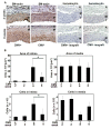
Figure 1
Figure 1
RCMV infection of aortic allografts enhances intimal hyperplasia and contributes to vessel remodelling. (A) The figure presents fragments of cross sections of RCMV-infected and uninfected allografts (controls) collected at 2 and 8 weeks after transplantation and stained for SM-α-actin (SM-actin) (Columns 1 and 2). Columns 3 and 4 show fragments of cross sections of the RCMV-infected and uninfected isograft controls at 2 and 8 weeks after transplantation to visualize that there was no intimal formation either in the infected or uninfected isografts. Blue, nuclei counterstained with hematoxylin. Brown, cells positive for SM-α-actin. I, intima, M, media, A, adventitia. (B) Bar charts show areas of intima and media presented as cross-sectional area (μm2) (upper panels) and the number of cells per cross section in the intima and media (lower panels) in RCMV-infected and uninfected allografts. At 2 weeks post transplantation the number of cells in the intima and media, and thickness of both layers were similar in infected and uninfected allografts. At 8 weeks, the intima was 3-fold thicker in infected vs. uninfected allografts and contained 2.5-fold more cells. The media of infected allografts had fewer cells at 8 week after transplantation. * p < 0.05 vs. control at 2 and 8 weeks, respectively.
Figure 2
Figure 2
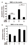
RCMV induces apoptosis of SMCs in the media layer of the aortic allografts. Bar charts show the Apoptotic Index of SMCs presented as the number of apoptotic SMCs per cross section in media (A) and intima (B) of infected and uninfected allografts (controls). Apoptotic SMCs were detected with double immunostaining for SM-α-actin and caspase 3. Both at 2 and 8 weeks after transplantation there was a significant increase of apoptotic SMCs in the media of infected allografts vs. uninfected controls. *p < 0.05 vs. control at 8 weeks. ***p < 0.001 vs. control at 2 weeks.
Figure 3
Figure 3
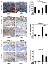
RCMV increases expression of matrix metalloproteinases and decreases collagen deposition in the aortic allografts. (A) Cross sections of RCMV-infected and uninfected allografts (controls) at 2 and 8 weeks post transplantation, stained with Masson's trichrome to identify collagen deposits. Blue, collagen; red, muscle fibers; black, nuclei; I, intima; M, media; A, adventitia. Bar chart shows the collagen deposits in the allografts of RCMV-infected and uninfected allografts at 2 and 8 weeks post transplantation presented as the size (μm2) of positive blue immunostained area per cross section. *p < 0.05 vs. uninfected controls 8 weeks after transplantation. At 8 weeks, infected allografts had significantly less collagen. (B) Cross sections of RCMV-infected and uninfected allografts (controls) stained to identify MMP-2. Brown indicates positivity for MMP-2. I, intima; M, media; A, adventitia. Bar chart shows percentage of positive areas for MMP-2 in infected and uninfected allografts at 2 and 8 weeks post transplantation determined as the percentage of positive area of the total cross section area. ***p < 0.001 vs. uninfected controls at 8 weeks. (C) Cross sections of RCMV-infected and uninfected allografts (controls) stained to identify MMP-9. Brown indicates positivity for MMP-9. I, intima; M, media; A, adventitia. Bar chart shows percentage of positive areas for MMP-9 in infected and uninfected allografts at 2 and 8 weeks post transplantation. Early after transplantation both MMP-2 and MMP-9 after confined mainly to the adventitia.
Figure 4
Figure 4
RCMV induces inflammation in the aortic allografts with increased infiltration of macrophages. (A) Cross sections of RCMV-infected allografts 2 and 8 weeks post transplantation stained to detect RCMV early/late antigen (R44-encoded protein). RCMV-infected cells were localized mainly in the adventitia and intima of the allograft. Brown, cells positive for RCMV. I, intima; M, media; A, adventitia. (B) Cross sections of RCMV-infected allografts and uninfected allografts (controls) at 2 and 8 weeks post transplantation stained for CD68-positive macrophages. Brown indicates positive staining for CD68 that appears mainly in the intima and adventitia. I, intima; M, media; A, adventitia. (C) Fragment of a cross section of RCMV-infected allograft double immunostained to detect RCMV-infected CD68-positive macrophages. 80% of infiltrating CD68-positive macrophages were RCMV infected. Green, CD68-positive macrophages; blue, RCMV; red, nuclei. (D) Cross sections of RCMV-infected allografts and uninfected controls at 2 and 8 weeks post transplantation stained for CD45-positive leukocytes. Brown indicates positive staining for CD45 that appears mainly in the intima and adventitia. I, intima; M, media; A, adventitia. (E) Cross sections of RCMV-infected allografts and uninfected controls at 2 and 8 weeks post transplantation stained for CD3-positive lymphocytes. Brown indicates positive staining for CD3 that appears mainly in the adventitia. I, intima; M, media; A, adventitia. (F) Bar charts show a number of CD68- and CD45-positive leukocytes in the media and intima of RCMV-infected and uninfected allografts (controls). Infiltration of CD68-positive macrophages into the intima was 4-fold higher and 2-fold higher in the media of infected allografts compared to uninfected controls. At 8 weeks, there was a massive infiltration of macrophages in the adventitia. ***p < 0.001 vs. uninfected control at 2 weeks in intima; **p < 0.01 vs. uninfected control at 8 weeks in intima; *p < 0.05 vs. uninfected control at 8 weeks in media.
Figure 5
Figure 5
RCMV increases expression of MCP-1 in the aortic allografts and cells from the infected adventitia migrate towards the intima. (A) Cross sections of the RCMV-infected and uninfected allografts (controls) at 2 and 8 weeks post transplantation stained for MCP-1. Green, MCP-1. Bar chart shows MCP-1 expression in the allografts, presented as positive area of the cross section (μm2). Both at 2 and 8 weeks after transplantation MCP-1 expression was increased in infected allografts (7-fold at 2 weeks and 3.5-fold at 8 weeks post transplantation). ***p < 0.001 vs. uninfected control at 2 and 8 weeks, respectively. I, intima; M, media; A, adventitia. (B) Cross section of the aortic allograft with transplanted fluorescently labeled isogenic adventitial graft. Grafts were collected 2 weeks post adventitia transplantation. Picture shows labeled blue adventitial cells in the allograft intima. Green, SM-actin; Blue, migrated adventitial cells; Red, nuclei.

Similar articles

Cited by

References

    1. Melnick JL, Petrie BL, Dreesman GR, Burek J, McCollum CH, DeBakey ME. Cytomegalovirus antigen within human arterial smooth muscle cells. Lancet. 1983;2(8351):644–7. doi: 10.1016/S0140-6736(83)92529-1. - DOI - PubMed
    1. Speir E, Modali R, Huang ES. et al.Potential role of human cytomegalovirus and p53 interaction in coronary restenosis. Science. 1994;265(5170):391–4. doi: 10.1126/science.8023160. - DOI - PubMed
    1. Vainas T, Stassen FR, Bruggeman CA. et al.Synergistic effect of Toll-like receptor 4 and CD14 polymorphisms on the total atherosclerosis burden in patients with peripheral arterial disease. J Vasc Surg. 2006;44:326–32. doi: 10.1016/j.jvs.2006.04.035. - DOI - PubMed
    1. Bentz GL, Yurochko AD. Human CMV infection of endothelial cells induces an angiogenic response through viral binding to EGF receptor and beta1 and beta3 integrins. Proc Natl Acad Sci USA. 2008;105(14):5531–6. doi: 10.1073/pnas.0800037105. - DOI - PMC - PubMed
    1. Streblow DN, Kreklywich CN, Andoh T. et al.The role of angiogenic and wound repair factors during CMV-accelerated transplant vascular sclerosis in rat cardiac transplants. Am J Transplant. 2008;8(2):277–87. doi: 10.1111/j.1600-6143.2007.02062.x. - DOI - PubMed

LinkOut - more resources