PTHrP treatment fails to rescue bone defects caused by Hedgehog pathway inhibition in young mice
- PMID: 21411723
- PMCID: PMC3576833
- DOI: 10.1177/0192623311399788
PTHrP treatment fails to rescue bone defects caused by Hedgehog pathway inhibition in young mice
Abstract
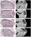
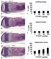
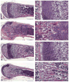
The advent of molecular targeted therapies offers the hope of therapeutic advance in the fight against cancer. However, this hope is tempered by recent findings that certain targeted therapies may have unique side effects. The Hedgehog (HH) pathway is a potential target for treatment of several cancers, including basal cell carcinoma and a subset of medulloblastoma. Recent clinical trials in adults have shown responses to HH pathway inhibition in both basal cell carcinoma and medulloblastoma. However, concerns have been raised about the use of HH pathway inhibitors in children because of the role the HH pathway plays in development. Indeed, young mice treated with the HH pathway inhibitor HhAntag developed severe bone defects, including premature differentiation of chondrocytes, thinning of cortical bone, and fusion of the growth plate. In an effort to lessen the severity of bone defects caused by HhAntag, we treated young mice simultaneously with HhAntag and parathyroid hormone-related protein (PTHrP), which functions downstream of Indian Hedgehog to maintain chondrocytes in a proliferative state. The results show that whereas treatment with PTHrP causes a significant increase in trabecular bone, it does not prevent fusion of the growth plate induced by HhAntag.
Figures


Similar articles
-
Transient inhibition of the Hedgehog pathway in young mice causes permanent defects in bone structure.Cancer Cell. 2008 Mar;13(3):249-60. doi: 10.1016/j.ccr.2008.01.027. Cancer Cell. 2008. PMID: 18328428
-
Regulation of chondrocyte terminal differentiation in the postembryonic growth plate: the role of the PTHrP-Indian hedgehog axis.Endocrinology. 2001 Sep;142(9):4131-40. doi: 10.1210/endo.142.9.8396. Endocrinology. 2001. PMID: 11517192
-
Partial rescue of postnatal growth plate abnormalities in Ihh mutants by expression of a constitutively active PTH/PTHrP receptor.Bone. 2010 Feb;46(2):472-8. doi: 10.1016/j.bone.2009.09.009. Epub 2009 Sep 15. Bone. 2010. PMID: 19761883 Free PMC article.
-
The parathyroid hormone-related protein and Indian hedgehog feedback loop in the growth plate.Novartis Found Symp. 2001;232:144-52; discussion 152-7. doi: 10.1002/0470846658.ch10. Novartis Found Symp. 2001. PMID: 11277077 Review.
-
[Cytokines in bone diseases. Genetic defects of PTH/PTHrP receptor in chondrodysplasia].Clin Calcium. 2010 Oct;20(10):1481-8. Clin Calcium. 2010. PMID: 20890029 Review. Japanese.
Cited by
-
Skeletal stem cells: origins, definitions, and functions in bone development and disease.Life Med. 2022 Dec 8;1(3):276-293. doi: 10.1093/lifemedi/lnac048. eCollection 2022 Dec. Life Med. 2022. PMID: 36811112 Free PMC article. Review.
-
The inhibitory roles of Ihh downregulation on chondrocyte growth and differentiation.Exp Ther Med. 2018 Jan;15(1):789-794. doi: 10.3892/etm.2017.5458. Epub 2017 Nov 7. Exp Ther Med. 2018. PMID: 29434683 Free PMC article.
-
Reproducibility of academic preclinical translational research: lessons from the development of Hedgehog pathway inhibitors to treat cancer.Open Biol. 2018 Aug;8(8):180098. doi: 10.1098/rsob.180098. Open Biol. 2018. PMID: 30068568 Free PMC article. Review.
-
FGFR3 Deficiency Causes Multiple Chondroma-like Lesions by Upregulating Hedgehog Signaling.PLoS Genet. 2015 Jun 19;11(6):e1005214. doi: 10.1371/journal.pgen.1005214. eCollection 2015 Jun. PLoS Genet. 2015. PMID: 26091072 Free PMC article.
References
-
- Adams SL, Cohen AJ, Lassova L. Integration of signaling pathways regulating chondrocyte differentiation during endochondral bone formation. J Cell Physiol. 2007;213:635–41. - PubMed
-
- Curran T, Ng JM. Cancer: Hedgehog’s other great trick. Nature. 2008;455:293–4. - PubMed
-
- Day TF, Guo X, Garrett-Beal L, Yang Y. Wnt/beta-catenin signaling in mesenchymal progenitors controls osteoblast and chondrocyte differentiation during vertebrate skeletogenesis. Developmental cell. 2005;8:739–50. - PubMed
MeSH terms
Substances
Grants and funding
LinkOut - more resources
Full Text Sources
Research Materials

