. 2011 May;31(10):2010-25.
doi: 10.1128/MCB.01006-10. Epub 2011 Mar 14.

PEST motif serine and tyrosine phosphorylation controls vascular endothelial growth factor receptor 2 stability and downregulation

Affiliations

PEST motif serine and tyrosine phosphorylation controls vascular endothelial growth factor receptor 2 stability and downregulation

Rosana D Meyer et al. Mol Cell Biol. 2011 May.

Abstract

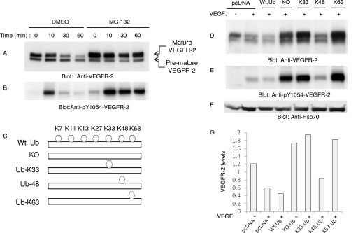
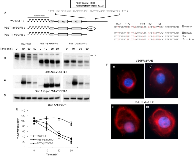
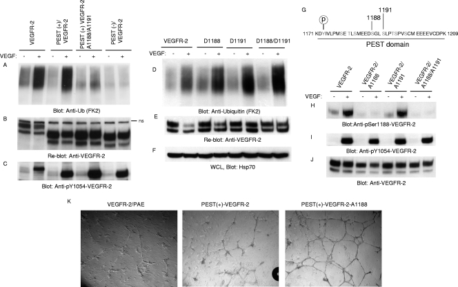
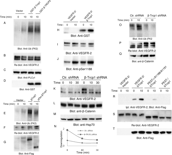
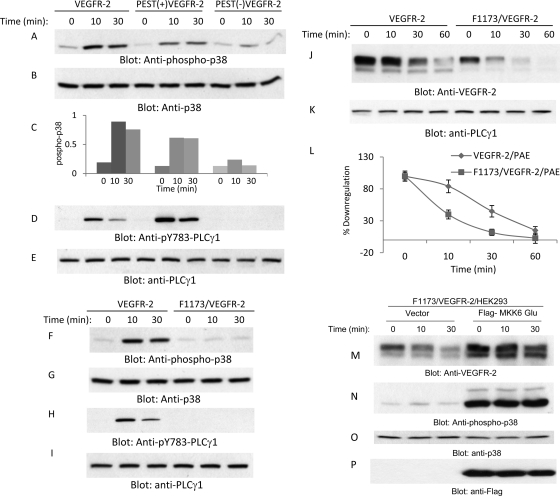
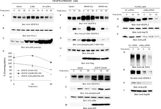
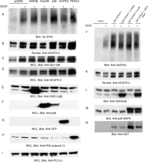
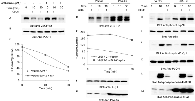
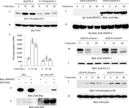
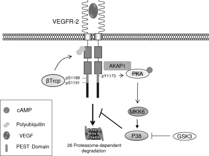
The internalization and degradation of vascular endothelial growth factor receptor 2 (VEGFR-2), a potent angiogenic receptor tyrosine kinase, is a central mechanism for the regulation of the coordinated action of VEGF in angiogenesis. Here, we show that VEGFR-2 is ubiquitinated in response to VEGF, and Lys 48-linked polyubiquitination controls its degradation via the 26S proteosome. The degradation and ubiquitination of VEGFR-2 is controlled by its PEST domain, and the phosphorylation of Ser1188/Ser1191 is required for the ubiquitination of VEGFR-2. F-box-containing β-Trcp1 ubiquitin E3 ligase is recruited to S1188/S1191 VEGFR-2 and mediates the ubiquitination and degradation of VEGFR-2. The PEST domain also controls the activation of p38 mitogen-activated protein kinase (MAPK) through phospho-Y1173. The activation of p38 stabilizes VEGFR-2, and its inactivation accelerates VEGFR-2 downregulation. The VEGFR-2-mediated activation of p38 is established through the protein kinase A (PKA)/MKK6 pathway. PKA is recruited to VEGFR-2 through AKAP1/AKAP149, and its phosphorylation requires Y1173 of VEGFR-2. The study has identified a unique mechanism in which VEGFR-2 stability and degradation is modulated. The PEST domain acts as a dual modulator of VEGFR-2; the phosphorylation of S1188/S1191 controls ubiquitination and degradation via β-Trcp1, where the phosphorylation of Y1173 through PKA/p38 MAPK controls the stability of VEGFR-2.


Figures

Fig. 1.
Fig. 1.
VEGFR-2 undergoes Lys 48-dependent proteasomal degradation. Serum-starved PAE cells expressing VEGFR-2 were preincubated with dimethylsulfoxide (DMSO) or with MG132 for 30 min, and then cells were left unstimulated (0) or were stimulated with VEGF for 10 and 30 min. Whole-cell lysates were immunoblotted with anti-VEGFR-2 antibody (A) or with anti-phospho-Y1054-VEGFR-2 antibody (B). HEK293 cells expressing VEGFR-2 were transfected with an empty vector (pcDNA 3.1His.Myc) or with ubiquitin constructs, including wild-type ubiquitin (Wt. Ub), a Lys mutant ubiquitin in which all of the lysines were mutated (KO), and ubiquitin constructs containing only Lys 33 (Ub-K33), Lys 48 (Ub-K48), or Lys63 (Ub-K63) in HEK293 cells. Cells were left unstimulated (−) or were stimulated with VEGF for 10 min (+), and whole-cell lysates were immunoblotted with anti-VEGFR-2 antibody (D), anti-phospho-Y1054-VEGFR-2 antibody (E), and anti-Hsp70 antibody for protein loading (F). (G) The quantification of the downregulation of VEGFR-2 by ubiquitin constructs.
Fig. 2.
Fig. 2.
PEST domain is required for ligand-mediated degradation of VEGFR-2. The schematic of VEGFR-2 and the presence of the putative PEST domain are shown. The PEST domain was predicted using the online program PESTFIND as outlined in Materials and Methods. The schematic of truncated VEGFR-2 encompassing PEST domain PEST(+)VEGFR-2 and PEST domain-deleted VEGFR-2, PEST(−)VEGFR-2, also are shown. (A) The PEST domain is conserved among mouse, human, bovine, and rat VEGFR-2. Wild-type VEGFR-2, PEST(+)VEGFR-2, and PEST(−)VEGFR-2 were expressed in PAE cells by a retroviral system, and their downregulation in response to VEGF in a time-dependent manner was measured. (B) Whole-cell lysates were subjected to Western blot analysis using anti-VEGFR-2 antibody. ns, nonspecific. The same cell lysates were blotted with anti-pY1054-VEGFR-2 antibody (C) and anti-PLCγ1 antibody (D). (E) The quantification of the downregulation of VEGFR-2, PEST(+)VEGFR-2, and PEST(−)VEGFR-2 in response to VEGF. The graph is an average from two independent experiments. (F) Confocal microscopy of VEGFR-2, PEST(+)VEGFR-2, and PEST(−)VEGFR-2 stimulated with VEGF for 10 min or left unstimulated (0).
Fig. 3.
Fig. 3.
Phosphorylation of serine 1188 is required for ubiquitination of VEGFR-2. (A) VEGFR-2, PEST(+)VEGFR-2, PEST(−)VEGFR-2, and PEST(+)VEGFR-2/A1188/A1191 (where Ser1188 and Ser1191 were mutated to Ala) were stimulated with VEGF for 10 min (+) or were left unstimulated (−), and cells were lysed, immunoprecipitated with anti-VEGFR-2 antibody, and immunoblotted with antiubiquitin (Anti-Ub; FK2) antibody. (B) The same membrane was reblotted with anti-VEGFR-2 antibody for protein levels. (C) Whole-cell lysates were blotted for phospho-Y1054-VEGFR-2. (D) PAE cells expressing VEGFR-2, D1188/VEGFR-2, D1191/VEGFR-2, and D1188/D1191/VEGFR-2 (Ser-to-Asp mutation) were stimulated as described for panel A and blotted with antiubiquitin antibody. (E) The same membrane was reblotted with anti-VEGFR-2 antibody for protein levels. (F) Whole-cell lysates (WCL) shown in panel D were blotted with anti-Hsp70 for internal protein levels. (G) Putative phosphorylation sites in the PEST domain are shown. PAE cells expressing wild-type VEGFR-2, VEGFR-2/A1188, VEGFR-2/A1188/A1191, and VEGFR-2/A1191 were stimulated with VEGF, and whole-cell lysates were blotted with anti-phospho-Ser1188-VEGFR-2 antibody (H), phospho-Y1054-VEGFR-2 antibody (F), and anti-VEGFR-2 antibody (I). (J) The same membrane was reblotted with anti-VEGFR-2 antibody for protein levels. (K) PAE cells expressing wild-type VEGFR-2, PEST(+)VEGFR-2, or PEST(+)VEGFR-2/A1188 (in which serine 1188 is mutated to alanine) were plated on the 24-well plates coated with Matrigel in the presence of VEGF, and pictures were taken under a light microscope equipped with a digital camera.
Fig. 4.
Fig. 4.
β-Trcp1 associates with VEGFR-2 and targets it for degradation. HEK293 cells expressing VEGFR-2 were transfected with an empty vector, GST-β-Trcp1, or with GST-β-Trcp2. (A) Cells were unstimulated (0) or stimulated with VEGF for 10 min, lysed, immunoprecipitated with anti-VEGFR-2 antibody, and blotted with antiubiquitin (Anti-Ub; FK2) antibody. (B) The same membrane was reblotted with anti-VEGFR-2. Whole-cell lysates from panel A were blotted with anti-PLCγ1 (C) and anti-GST antibody (D). (E) HEK293 cells coexpressing VEGFR-2 with empty vector, with β-Trcp1, or with F-box-deleted β-Trcp1(ΔFbx-β-Trcp1) were left unstimulated (0) or stimulated for 10 min (8) with VEGF, lysed, immunoprecipitated with anti-VEGFR-2 antibody, and blotted with antiubiquitin antibody. (F) The same membrane was reblotted for VEGFR-2. (G) Whole-cell lysates (WCL) from the same lysates also were blotted for β-Trcp1 using anti-Flag antibody. (H) HEK293 cells coexpressing VEGFR-2 with empty vector or with β-Trcp1 were left unstimulated (0) or were stimulated for 10 min (10) with VEGF, lysed, immunoprecipitated with anti-VEGFR-2 antibody, and blotted with anti-GST antibody. (I) The same membrane was reblotted for VEGFR-2 levels. (J) Whole-cell lysates from the same cell lysate were blotted for phospho-Ser1188 antibody. Primary endothelial cells, HUVEC cells expressing pSuper.puro or shRNA.β-Trcp1.pSuper.puro, were stimulated with VEGF for the indicated periods of time, and whole-cell lysates were blotted with anti-VEGFR-2 antibody (K), β-catenin (L), and Hsp70 (M). Ctr., control. (N) Quantification of downregulation of VEGFR-2 is shown. (O) HUVEC expressing pSuper.puro vector or shRNA.β-Trcp1.pSuper.puro were left unstimulated (0) or were stimulated with VEGF for 10 min, and cells were lysed and immunoprecipitated with anti-VEGFR-2 and blotted with antiubiquitin antibody. The same membrane was reblotted for VEGFR-2 (P). Whole-cell lysates from the same cell lysates were blotted for β-catenin (L) and Hsp70 (Q). (S) HEK293 cells coexpressing VEGFR-2 with empty vector, VEGFR-2 with β-Trcp1, PEST(+)Ser1188/Ser1191 mutant VEGFR-2 with β-Trcp1, or PEST(−)VEGFR-2 with β-Trcp1 were unstimulated or stimulated with VEGF, lysed, immunoprecipitated with anti-VEGFR-2 antibody, and blotted with anti-Flag antibody. (T) The same membrane was reblotted for VEGFR-2. Ipt, immunoprecipitation. (U) Whole-cell lysates from the same cell lysates were blotted for β-Trcp1 using anti-Flag antibody.
Fig. 5.
Fig. 5.
PEST domain controls the phosphorylation of p38 MAPK. Serum-starved PAE cells expressing VEGFR-2, PEST(+)VEGFR-2, and PEST(−)VEGFR-2 were stimulated with VEGF for the indicated periods of time, cells were lysed, and whole-cell lysates were subjected to Western blot analysis and blotted for phospho-p38 (A), total p38 (B), phospho-PLCγ1 (D), and total PLCγ1 (E). (C) Quantification of activation of p38. Serum-starved PAE cells expressing VEGFR-2 and F1173/VEGFR-2 were stimulated with VEGF for the indicated periods of time, cells were lysed, and whole-cell lysates were subjected to Western blot analysis and immunoblotted for phospho-p38 (F), total p38 (G), phospho-PLCγ1 (H), and total PLCγ1 (I). Serum-starved PAE cells expressing VEGFR-2 and F1173/VEGFR-2 were preincubated with cycloheximide for 90 min, and then cells were stimulated with VEGF for the indicated periods of time. Cells were lysed, and whole-cell lysates were immunoblotted for VEGFR-2 (J) and total PLCγ1 (K). (L) Quantification of VEGFR-2 protein levels from blot I is shown. The graph shows averages from two independent experiments. HEK293 cells coexpressing F1173/VEGFR-2 with an empty vector or constitutive active MKK6 (MKK6-Glu) were stimulated with VEGF for the indicated periods of time, and whole-cell lysates were immunoblotted for VEGFR-2 (M), phospho-p38 MAPK (pT180/pY182) (N), p38 MAPK (O), and MKK6 using anti-Flag antibody (P).
Fig. 6.
Fig. 6.
MKK6-dependent activation of p38 inhibits downregulation of VEGFR-2. HEK 293 cells coexpressing VEGFR-2 with an empty vector, VEGFR-2 with wild-type p38 MAPK, or VEGFR-2 with dominant-negative p38 (dn-P38) MAPK were preincubated with cycloheximide for 90 min and then stimulated with VEGF for the indicated periods of time. Whole-cell lysates were blotted for VEGFR-2 (A), PLCγ1 as a control for protein loading (B), and p38 (C). (D) The quantification of the downregulation of VEGFR-2 in response to ligand stimulation. Whole-cell lysates from HEK293 cells coexpressing VEGFR-2 either with an empty vector, constitutive active MKK6 (MKK6-Glu), or with dominant-negative MKK6 (MKK6-Ala) were immunoblotted for VEGFR-2 (E), phospho-VEGFR-2 (pY1054-VEGFR-2) (F), phospho-p38 MAPK (pT180/pY182) (G), p38 MAPK (H), and MKK6 using anti-Flag antibody (I). HEK293 cells expressing VEGFR-2 were transfected either with empty vector or with enhanced green fluorescent protein (EGFP)-tagged Cdc42. Serum-starved cells were stimulated with VEGF for the indicated times, and cells were lysed. Whole-cell lysates were blotted for VEGFR-2 (J), phospo-p38 (K), total p38 (L), and anti-GFP for Cdc42 expression (M). Cells also were treated with cycloheximide (20 mM for 90 min prior to stimulation with VEGF. HUVEC were transfected with control (Ctr.) siRNA or p38α siRNA after 24 h, and cells were starved overnight and stimulated with VEGF for the indicated periods of time. Cells were lysed, and whole-cell lysates were blotted with anti-VEGFR-2 antibody (N), anti-Hsp70 antibody (O), and anti-p38 antibody (P). (Q) Ubiquitination of VEGFR-2 in HUVEC in which p38α was silenced. (S) The same membrane was reblotted for VEGFR-2 levels. Whole-cell lysates were blotted for p38 (R) and Hsp90 (T) for protein loading.
Fig. 7.
Fig. 7.
p38 is not involved in the ubiquitination of VEGFR-2. HEK293 cells expressing VEGFR-2 were transfected with empty vector, GSK3β, dominant-negative p38 (Dn/p38), wild-type p38, dominant-negative PKA catalytic subunit C (Dn/PKA), and wild-type PKA catalytic subunit C [PKA(C)]. Cells were stimulated with VEGF for 10 min, lysed, immunoprecipitated with anti-VEGFR-2 antibody, and blotted with antiubiquitin (Ub; FK2) antibody (A). (B) The same membrane was reblotted for VEGFR-2 levels. Whole-cell lysates (WCL) from panel A were blotted with anti-phospho-Ser1188 antibody (C), anti-VEGFR-2 antibody (D), anti-GSK3 antibody (E), anti-p38 antibody (F), anti-GFP antibody to detect dominant-negative PKA (G), anti-PKA antibody (H), and anti-PLCγ1 antibody (I). HEK293 cells expressing VEGFR-2 were transfected with GST-tagged β-Trcp1 alone or GST-tagged β-Trcp1 with GSK3β, β-Trcp1 with p38, or β-Trcp1 with p38 and GSK3β. (J) Cells were stimulated with VEGF for 10 min, immunoprecipitated with anti-VEGFR-2 antibody, and blotted with anti-ubiquitin (FK2) antibody. (K) The same membrane was reblotted for VEGFR-2. (L to N) Whole-cell lysates from panel J were blotted for GSK3, p38, and β-Trcp1.
Fig. 8.
Fig. 8.
PKA pathway inhibits downregulation of VEGFR-2 and activates p38. PAE cells expressing wild-type VEGFR-2 were pretreated with forskolin (40 μM) and then stimulated with VEGF for the indicated time periods. Cells were treated with cycloheximide for 90 min to inhibit protein synthesis. Whole-cell lysates were blotted with anti-VEGFR-2 antibody (A) and with anti-PLCγ1 antibody for protein loading (B). (C) The quantification of VEGFR-2 downregulation in the presence or absence of forskolin is shown. Serum-starved HEK293 cells coexpressing VEGFR-2 either an empty vector or the catalytic active subunit (Cα) of PKA were pretreated with cycloheximide (CHX) for 90 min, and then cells were stimulated with VEGF for the indicated time periods. Whole-cell lysates were immunoblotted with anti-VEGFR-2 antibody (D), anti-PLCγ1 antibody (F), and anti-PKA antibody (G). (E) The quantification of the downregulation of VEGFR-2. The same cell lysates also were blotted for phospho-p38 (H), total p38 (I), phospho-PLCγ1 (J), total PLCγ1 (K), phospho-MAPK42/44 (L), and total PKA (M).
Fig. 9.
Fig. 9.
Tyrosine 1173 of VEGFR-2 is required for PKA phosphorylation but not for its association with VEGFR-2. PAE cells expressing VEGFR-2 or F1173/VEGFR-2 were stimulated with VEGF for the indicated periods of time. Cells were lysed, and whole-cell lysates (WCL) were blotted for phospho-PKA (A) and total PKA (B). (C) Quantification of the phosphorylation of PKA is shown. It represents averages from two experiments. HEK293 cells were transfected with GFP-tagged PKA catalytic subunit C alone or with Myc-tagged AKAP1. (D) Cells were lysed, immunoprecipitated (Ipt) with anti-GFP antibody, and blotted with anti-Myc antibody. (E) Whole-cell lysates also were blotted for anti-GFP. Serum-starved HEK293 cells coexpressing VEGFR-2 with empty vector or with c-myc-tagged AKAP1 were stimulated with VEGF for the indicated periods of time (F). Cells were lysed, and VEGFR-2 was immunoprecipitated with anti-VEGFR-2 antibody and immunoblotted with anti-c-myc antibody. (G) The same membrane was stripped and reblotted for VEGFR-2. Whole-cell lysates from the same cell groups were immunoblotted for VEGFR-2 (H), total PLCγ1 (I), phospho-p38 (J), and total p38 (K).
Fig. 10.
Fig. 10.
Proposed model for PEST-mediated downregulation of VEGFR-2. Upon ligand binding, the PEST domain of VEGFR-2 is phosphorylated on tyrosine and serine sites, including Y1173, S1188, and Ser1191. Ligand binding promotes β-Trcp1 association with VEGFR-2, which mediates the ubiquitination of VEGFR-2 through the Lys-48-dependent ubiquitin chain, leading to degradation by the 26S proteasome system. The activation of PKA by VEGFR-2 requires the AKAP1-mediated association of PKA with VEGFR-2, and its phosphorylation is mediated through Y1173 of VEGFR-2. The activation of PKA leads to the phosphorylation of p38. Activated p38 attenuates the downregulation of VEGFR-2.

Similar articles

Cited by

References

    1. Adams R. H., Alitalo K. 2007. Molecular regulation of angiogenesis and lymphangiogenesis. Nat. Rev. Mol. Cell Biol. 8:464–478 - PubMed
    1. Bhattacharya R. 2005. Regulatory role of dynamin-2 in VEGFR-2/KDR-mediated endothelial signaling. FASEB J. 19:1692–1694 - PubMed
    1. Bruns A. F. 2010. Ligand-stimulated VEGFR2 signaling is regulated by co-ordinated trafficking and proteolysis. Traffic 11:161–174 - PubMed
    1. Cleaver O., Melton D. A. 2003. Endothelial signaling during development. Nat. Med. 9:661–668 - PubMed
    1. Crusio K. M., King B., Reavie L. B., Aifantis I. 2010. The ubiquitous nature of cancer: the role of the SCF(Fbw7) complex in development and transformation. Oncogene 29:4865–4873 - PMC - PubMed

Publication types

MeSH terms

Substances