. 2011 Mar 14;208(3):605-15.
doi: 10.1084/jem.20101402. Epub 2011 Mar 7.

Herpesvirus telomeric repeats facilitate genomic integration into host telomeres and mobilization of viral DNA during reactivation

Affiliations

Herpesvirus telomeric repeats facilitate genomic integration into host telomeres and mobilization of viral DNA during reactivation

Benedikt B Kaufer et al. J Exp Med. .

Abstract

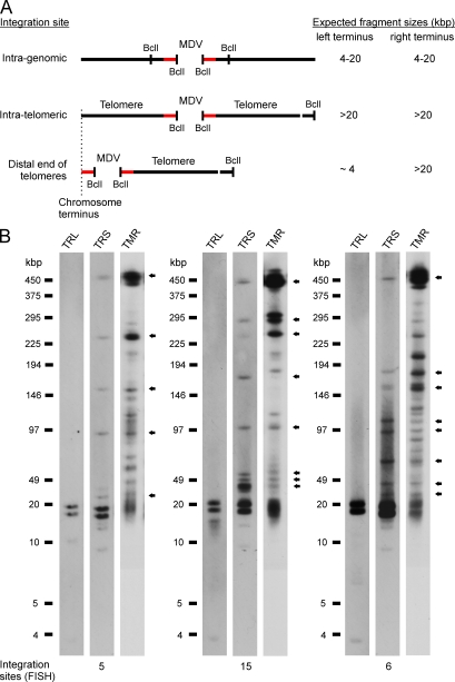
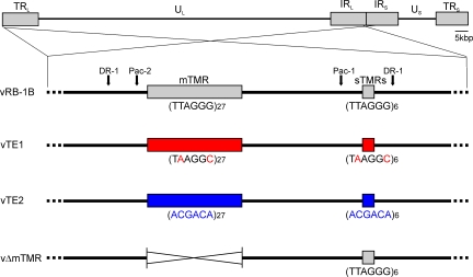
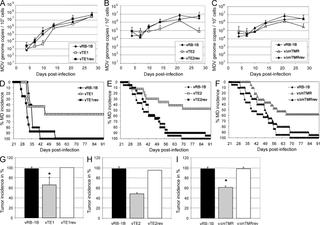
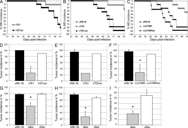
Some herpesviruses, particularly lymphotropic viruses such as Marek's disease virus (MDV) and human herpesvirus 6 (HHV-6), integrate their DNA into host chromosomes. MDV and HHV-6, among other herpesviruses, harbor telomeric repeats (TMRs) identical to host telomeres at either end of their linear genomes. Using MDV as a natural virus-host model, we show that herpesvirus TMRs facilitate viral genome integration into host telomeres and that integration is important for establishment of latency and lymphoma formation. Integration into host telomeres also aids in reactivation from the quiescent state of infection. Our results and the presence of TMRs in many herpesviruses suggest that integration mediated by viral TMRs is a conserved mechanism, which ensures faithful virus genome maintenance in host cells during cell division and allows efficient mobilization of dormant viral genomes. This finding is of particular importance as reactivation is critical for virus spread between susceptible individuals and is necessary for continued herpesvirus evolution and survival.


Figures

Figure 1.
Figure 1.
Identification of MDV integration sites. (A) Schematic representation of potential integration sites of the MDV genome within the host chromosome. Fragments resulting from digestion with BclI are depicted and expected fragment sizes are given. Terminal MDV genome fragments (red bars), telomeres, and BclI restriction sites are indicated. (B) PFGE analysis of LCL using BclI. PFGE patterns of three representative cell lines analyzed by Southern blotting, probing for the left MDV terminus (TRL), right MDV terminus (TRS), or telomere sequences (TMR), are shown. The 22.4- and 27.0-kbp fragments correspond to uncleaved termini and internal repeat fragments of the viral DNA. Arrows indicate colocalization of fragments that contain the TRS and high molecular mass telomere sequences. Results are representative of three independent experiments giving identical results. The number of integration sites detected by FISH is given at the bottom of the blot images.
Figure 2.
Figure 2.
MDV TMR mutants. Schematic representation of the MDV genome with a focus on viral a-like sequences containing the mTMR and sTMR regions present in the MDV genome. Recombinant BAC constructs in which the TMRs (TTAGGG)n were replaced by TE1 (TAAGGC)n or TE2 (ACGACA)n, as well as a BAC construct in which the mTMR region was deleted, are shown.
Figure 3.
Figure 3.
Mutation of herpesvirus TMRs mildly affects lytic replication but severely impairs disease and tumor development in vivo. (A–C) qPCR analysis of the viral ICP4 gene and host iNOS gene. Blood samples of animals infected with wild-type (vRB-1B), telomere mutant (TE1, TE2, and vΔmTMR), or revertant (TE1rev, TE2rev, and vΔmTMRrev) viruses were taken at 4, 7, 10, 14, 21, and 28 days after infection, and total DNA was extracted. Mean MDV genome copies per 1 × 10⁶ cells of eight infected chickens per group are shown with standard deviations (error bars) determined in one independent experiment for the indicated viruses. (D–F) Marek’s disease (MD) incidence in percentage of chickens infected by the intraabdominal route with wild-type (vRB-1B; D, n = 5; E, n = 19; F, n = 19), telomere mutant (TE1, n = 7; TE2, n = 17; vΔmTMR, n = 19), or revertant (TE1rev, n = 8; TE2rev, n = 20; vΔmTMRrev, n = 20) viruses was monitored during the indicated time period after infection. (G–I) Tumor incidence in P2a chickens infected with indicated viruses. Results are shown as mean tumor incidences in two (G), three (H), or four (I) independent experiments with standard deviations (error bars). The mean tumor incidences in chickens infected with vTE1 and vΔmTMR were significantly decreased compared with those infected with vRB-1B, which is indicated by asterisks (G, P = 0.021; I, P = 1.98 × 10⁻⁶). Each group contained between 5 and 20 animals with a mean group size of n = 13.6. vRB-1B, TTAGGG; TE1, TTAGGG→TAAGGC; TE2, TTAGGG→ACGACA; vΔmTMR, mTMR deletion.
Figure 4.
Figure 4.
Disease and tumor development are severely impaired in the absence of the viral TMRs in animals infected via the natural route of infection. (A–C) Marek’s disease (MD) incidence in the percentage determined for in-contact animals housed with chickens infected with vRB-1B (n = 9), vTE1 (n = 10), or vTE1rev (n = 10; A), vTE2 (n = 8) or vTE2rev (n = 9; B), and vΔmTMR (n = 8) or vΔmTMRrev (n = 8; C). Chickens were monitored during the indicated time period and MD was recorded after necropsy and gross pathological examination. (D and E) Tumor incidence in percentage of contact animals housed with animals infected with wild-type (vRB-1B), telomere mutant (TE1, TE2, and vΔmTMR), or revertant (TE1rev, TE2rev, and vΔmTMRrev) viruses as indicated. Results are shown as mean tumor incidences of two (D and E) or three (F) independent experiments with standard deviations (error bars). The mean tumor incidences in chickens infected with vΔmTMR (F) were significantly decreased compared with incidences in animals infected with vRB-1B as indicated by the asterisk (P = 0.011). Each group contained between 2 and 10 animals with a mean group size of n = 6.7. (G–I) Mean tumor incidence in highly susceptible P2a chickens infected by either the intraabdominal (G) or the natural (H) route of infection or in more resistant N2a chickens (I) with parental vRB-1B, vMut (vTE1, vTE2, or vΔmTMR), or vRev (vTE1rev, vTE2rev, or vΔmTMRrev) viruses. Results are shown as mean tumor incidences of nine (G), seven (H), or four (I) independent experiments with standard deviations (error bars). The mean group sizes were n = 13.6 (G), n = 6.7 (H), and n = 19.5 (I). A significant decrease of mean tumor incidence compared with vRB-1B (G, P = 1.26 × 10⁻⁸; H, P = 2.17 × 10⁻⁷) or vRev (G, P = 3.68 × 10⁻⁶; H, P = 7.72 × 10⁻⁵; I, P = 0.001) is indicated with an asterisk. vRB-1B, TTAGGG; TE1, TTAGGG→TAAGGC; TE2, TTAGGG→ACGACA; vΔmTMR, mTMR deletion.
Figure 5.
Figure 5.
Integration is severely impaired in the absence of the viral TMRs. (A) FISH analysis detecting MDV integration sites (anti-DIG FITC, green) in metaphase chromosomes (DAPI stain, blue) of representative cell lines of vRB-1B, vTE1, vTE2, and vΔmTMR. In the case of vTE1, vTE2, and vΔTMR, single integration sites are highlighted by arrows. (B) Mean number of integration sites in vRB-1B, mutant, and revertant cell lines. The number of analyzed cell lines and the frequencies of integration events are given. At least eight independent metaphase spread images were evaluated to determine the number of integration sites for each independent cell line.
Figure 6.
Figure 6.
Integration does not occur in host telomeres in the absence of viral TMRs. (A) Schematic representation and corresponding PFGE and Southern blot analysis of LCL DNA digested with SfiI. Fragment sizes generated by SfiI digestion of integrated and nonintegrated MDV genomes are depicted and sizes are given. The size of the linear MDV genome observed during lytic replication is indicated by an arrow. Results are representative of three independent Southern blot analyses. (B) Southern blotting of DNA of LCL derived from animals infected with vTE1 and digested with BclI. Potential intragenomic and telomeric integration sites are indicated. Results are representative of three independent Southern blot analyses. (C) Quantification of MDV copies in tumor cells. Results are shown as mean herpesvirus genome copies detected by the TRL probe relative to B2M in three independent experiments. The data are shown relative to LCL CU482 derived from a vRB-1B–infected chicken with standard deviations (error bars).
Figure 7.
Figure 7.
Reactivation is significantly impaired in the absence of viral TMRs. (A–C) Reactivation assay using LCL derived from animals infected with wild-type (vRB-1B), telomere mutant (TE1, TE2, and vΔmTMR), or revertant (TE1rev, TE2rev, and vΔmTMRrev) viruses as indicated. Representative images of virus reactivation in CECs are shown. Bars, 500 µm. Quantification of reactivation assays is shown as the mean (horizontal bars) number of plaques per 1 × 10⁶ tumor cells for each individual cell line (A–C, right), performed in triplicate for each of three independent experiments. Reactivation of lytic virus from vTE1-, vTE2-, and vΔmTMR-derived LCL was significantly reduced compared with that from vRB-1B–derived LCL, as indicated by the asterisks (A, P = 0.013; B, P = 0.039; C, P = 0.025). vRB-1B, TTAGGG; TE1, TTAGGG→TAAGGC; TE2, TTAGGG→ACGACA; vΔmTMR, mTMR deletion.

Similar articles

Cited by

References

    1. Arbuckle J.H., Medveczky M.M., Luka J., Hadley S.H., Luegmayr A., Ablashi D., Lund T.C., Tolar J., De Meirleir K., Montoya J.G., et al. 2010. The latent human herpesvirus-6A genome specifically integrates in telomeres of human chromosomes in vivo and in vitro. Proc. Natl. Acad. Sci. USA. 107:5563–5568 10.1073/pnas.0913586107 - DOI - PMC - PubMed
    1. Blasco M.A. 2005. Telomeres and human disease: ageing, cancer and beyond. Nat. Rev. Genet. 6:611–622 10.1038/nrg1656 - DOI - PubMed
    1. Brown A.C., Baigent S.J., Smith L.P., Chattoo J.P., Petherbridge L.J., Hawes P., Allday M.J., Nair V. 2006. Interaction of MEQ protein and C-terminal-binding protein is critical for induction of lymphomas by Marek’s disease virus. Proc. Natl. Acad. Sci. USA. 103:1687–1692 10.1073/pnas.0507595103 - DOI - PMC - PubMed
    1. Calnek B.W., Murthy K.K., Schat K.A. 1978. Establishment of Marek’s disease lymphoblastoid cell lines from transplantable versus primary lymphomas. Int. J. Cancer. 21:100–107 10.1002/ijc.2910210117 - DOI - PubMed
    1. Calnek B.W., Shek W.R., Schat K.A. 1981. Spontaneous and induced herpesvirus genome expression in Marek’s disease tumor cell lines. Infect. Immun. 34:483–491 - PMC - PubMed

Publication types

MeSH terms