. 2011 Apr 29;286(17):15171-81.
doi: 10.1074/jbc.M110.184374. Epub 2011 Mar 4.

G-actin participates in RNA polymerase II-dependent transcription elongation by recruiting positive transcription elongation factor b (P-TEFb)

Affiliations

G-actin participates in RNA polymerase II-dependent transcription elongation by recruiting positive transcription elongation factor b (P-TEFb)

Tianyang Qi et al. J Biol Chem. .

Abstract

Actin is a key regulator of RNA polymerase (Pol) II-dependent transcription. Positive transcription elongation factor b (P-TEFb), a Cdk9/cyclin T1 heterodimer, has been reported to play a critical role in transcription elongation. However, the relationship between actin and P-TEFb is still not clear. In this study, actin was found to interact with Cdk9, a catalytic subunit of P-TEFb, in elongation complexes. Using immunofluorescence and immunoprecipitation assays, Cdk9 was found to bind to G-actin through the conserved Thr-186 in the T-loop. Overexpression and in vitro kinase assays showed that G-actin promotes P-TEFb-dependent phosphorylation of the Pol II C-terminal domain. An in vitro transcription experiment revealed that the interaction between G-actin and Cdk9 stimulated Pol II transcription elongation. ChIP and immobilized template assays indicated that actin recruited Cdk9 to a transcriptional template in vivo and in vitro. Using cytokine IL-6-inducible p21 gene expression system, we revealed that actin recruited Cdk9 to endogenous gene. Moreover, overexpression of actin and Cdk9 increased histone H3 acetylation and acetylized histone H3 binding to a transcriptional template through the interaction with histone acetyltransferase, p300. Taken together, our results suggested that actin participates in transcription elongation by recruiting Cdk9 for phosphorylation of the Pol II C-terminal domain, and the actin-Cdk9 interaction promotes chromatin remodeling.


Figures

FIGURE 1.
FIGURE 1.
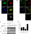
Actin binds P-TEFb in elongation complexes. A, diagram of the immobilized Ad20 and Ad22 DNA templates. The biotinylated 889-bp DNA transcription template named as Ad22 containing the AdMLP was linked to magnetic streptavidin beads. The Ad20 was a 490-bp DNA template identical to Ad22 but lacking the AdMLP. B, Western blot analysis of proteins released from the beads lacking DNA or linked to the Ad20 or Ad22 templates using the indicated antibodies. C, HeLa cells were stained with anti-actin antibody (a), anti-Cdk9 antibody (b), and Hoechst dye (c). The image (d) was a merge of a and b. Colocolization of actin and Cdk9 in d was analyzed by the confocal microscope software Olympus Fluoview (version 1.7c) to generate white dots in e. Scale bar, 10 μm. D, the immunoprecipitates were obtained from the NE of HeLa cells using an anti-actin antibody. Then, the immunoprecipitates were analyzed by immunoblotting (IB) in combination with antibodies against Pol II and Cdk9. E, HeLa cells were transfected with plasmids encoding HA-tagged actin or FLAG-tagged Cdk9. The NEs from the transfected HeLa cells were immunoprecipitated with either anti-HA or anti-FLAG antibody. The immunoprecipitates were then analyzed by Western blot with anti-Ser-2-phosphorylated Pol II (Pol II-Ser2), anti-Cdk9, or anti-actin antibodies, respectively.
FIGURE 2.
FIGURE 2.
Actin interacts with Thr-186 in the T-loop of transcriptionally active P-TEFb. A, HeLa cells were transfected with the indicated plasmids, expressing either FLAG-tagged wild-type or mutant Cdk9. The immunoprecipitates obtained with anti-FLAG antibody from the NEs of transfected HeLa cells were subjected to SDS-PAGE and analyzed by immunoblotting (IB) with the indicated antibodies. The proteins expressed in NEs were shown as a control in the right panel. B, HeLa cells transfected with HA-tagged actin were treated with actinomycin D (ActD) or the solvent DMSO (a negative control). Immunoprecipitates obtained with anti-HA antibody were detected with anti-Cdk9 antibody through Western blot (WB). The HA tag, Cdk9, and β-tubulin in the NEs were shown as controls for transfection and loading. C, HeLa cells were transfected with FLAG-tagged Cdk9. The lysates of NEs of the transfected HeLa cells were then immunoprecipitated with anti-FLAG antibody and analyzed by Western blot using an anti-actin antibody. FLAG tag, actin, and β-tubulin in the NEs were shown as controls for transfection and loading.
FIGURE 3.
FIGURE 3.
P-TEFb mainly interacts with monomeric actin. A and B, G- or F-actin was labeled with fluorescent DNase I conjugates or phallotoxin conjugates (a or f) respectively, Cdk9 was labeled with anti-Cdk9 antibody (b and g), and nuclei were stained with Hoechst 33342 (c and h). d was the merge of a and b, and i was the merge of f and g. d and i were analyzed by the confocal microscope software Olympus Fluoview (version 1.7c) to generate white dots in e and j, respectively. Scale bar, 10 μm. C, HeLa cells transfected with HA-tagged actin and a control vector were treated with jasplakinolide (Jas), cytochalasin D (CD), or 1% DMSO. Immunoprecipitates (IP) were obtained with anti-HA antibody from the NEs of transfected HeLa cells, and the amounts of Cdk9 were analyzed by Western blot (WB). HA tag, Cdk9, and β-tubulin expression in the NEs were shown as controls for transfection and loading. D, showing the amount of Cdk9 interacted with HA-actin after treatment with jasplakinolide, cytochalasin D, or 1% DMSO. The data shown are the mean ± S.D. from three independent experiments.
FIGURE 4.
FIGURE 4.
Interaction between G-actin and P-TEFb is critical for transcription elongation. A, nuclear extract from the HeLa cells transfected with indicated plasmids (top) were immunoblotted with antibody specific for Ser-2-phosphorylated Pol II CTD, and β-tubulin was used as a control. B, in vitro kinase reactions were performed with the indicated immunoprecipitation (IP) complexes of WT or mutant HA-actin and GST-CTD as a substrate. Phosphorylated GST-CTD was subjected to SDS-PAGE and detected with anti-Ser-2-phosphorylated Pol II CTD antibody. The HA tag and GST-CTD at the bottom were shown as controls. C, the depletion specificity of actin and Cdk9 was checked by Western blot using NEs from the transfected HeLa cells, and TFIIF, ELL, and NELF-A were used as negative controls. D, different combinations of WT or mutant actin, with or without P-TEFb, were introduced into the in vitro transcription system in the NEs of Cdk9 and actin-depleted HeLa cells. RNA transcripts derived from the transcription template Ad22 were analyzed as described under “Experimental Procedures.”
FIGURE 5.
FIGURE 5.
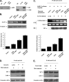
Actin recruits P-TEFb to the transcriptional template. A, schematic representation of the AdMLP-luciferase reporter gene template. B, HeLa cells were transfected with the AdMLP-luciferase reporter gene template, FLAG-tagged WT, or T186A-Cdk9. The ChIP assay was performed with anti-FLAG antibody. The promoter and coding regions of the AdMLP-luciferase reporter gene template were PCR-amplified. WT or mutant FLAG-tagged Cdk9 associated with the immunoprecipitated (IP) chromatin were used as controls. C and D (left panel), Western blot (WB) analysis of NEs in which endogenous actin or Cdk9 was immunodepleted under high salt conditions. TFIIF was used as a negative control. Right panel, the depleted NEs were incubated with immobilized Ad22 template under transcription conditions. The Cdk9 and actin retained by the template were detected with Western blot using TFIIF as a control.
FIGURE 6.
FIGURE 6.
Actin recruited Cdk9 to endogenous genes. A, HepG2 cells were stimulated for 2 h with IL-6 (20 ng/ml). Total RNA was isolated and expression of the p21 gene was analyzed by RT-PCR. In parallel, the recruitment of actin or Cdk9 to the p21 gene was analyzed by ChIP experiments. B, HepG2 cells in six-well plates were transiently transfected with 100 nm actin siRNA (siActin) or Cdk9 siRNA (siCDK9). 72 h after transfection, equivalent amounts of protein from the whole cell lysates were used for immunoblotting. β-Tubulin at bottom was shown as a control. C and D, siRNA transfection was performed as described in B. 48 h after siRNA transfection, cells were treated with IL-6 (20 ng/ml) for 2 h. ChIP assay was performed using anti-actin or anti-Cdk9 antibody. Actin and Cdk9 in NEs showed the efficiency of siRNA. WB, Western blot; IP, immunoprecipitation.
FIGURE 7.
FIGURE 7.
Actin-P-TEFb interaction enhances chromatin acetylation by activating p300. A, the immunoprecipitates were obtained from the NEs of HeLa cells using anti-actin antibody or anti-p300 antibody. Next, the immunoprecipitates were analyzed by immunoblotting in combination with antibodies against p300 or Cdk9. B, HeLa cells were transfected with the indicated expression plasmids. The whole cell lysates were subjected to Western blot (WB) analysis using anti-Ac-H3 antibody, and β-tubulin was shown as a control. Amount of acetylated histone 3 after transfection with indicated plasmids was quantified. The data shown are the mean ± S.D. from three independent experiments. C, the ChIP experiment was performed using anti-Ac-H3 antibody with the HeLa cells transfected with the AdMLP-luciferase reporter gene and indicated expression plasmids. The coding region of the AdMLP-luciferase gene was PCR-amplified. Relative fold for PCR product was quantified. The data shown are the mean ± S.D. from three independent experiments. D and E, HeLa cells were transfected with HA-tagged actin or FLAG-tagged Cdk9 and then treated with histone deacetylase inhibitor trichostatin A (TSA) (200 ng/ml) for 4 h. An immunoprecipitation assay was performed with anti-HA or anti-FLAG antibody, and the resulting immunoprecipitates (IP) were analyzed by immunoblotting with antibodies against Cdk9 or actin. β-Tubulin was used as a control.

Similar articles

Cited by

References

    1. Orphanides G., Reinberg D. (2002) Cell 108, 439–451 - PubMed
    1. Corden J. L. (1990) Trends Biochem. Sci 15, 383–387 - PubMed
    1. Dvir A., Conaway R. C., Conaway J. W. (1997) Proc. Natl. Acad. Sci. 94, 9006–9010 - PMC - PubMed
    1. O'Brien T., Hardin S., Greenleaf A., Lis J. T. (1994) Nature 370, 75–77 - PubMed
    1. Marshall N. F., Peng J., Xie Z., Price D. H. (1996) J. Biol. Chem. 271, 27176–27183 - PubMed

Publication types

MeSH terms

LinkOut - more resources