. 2011 May;85(10):4720-9.
doi: 10.1128/JVI.02032-10. Epub 2011 Mar 2.

Human adenovirus type 5 induces cell lysis through autophagy and autophagy-triggered caspase activity

Affiliations

Human adenovirus type 5 induces cell lysis through autophagy and autophagy-triggered caspase activity

Hong Jiang et al. J Virol. 2011 May.

Abstract

Oncolytic adenoviruses, such as Delta-24-RGD, are promising therapies for patients with brain tumor. Clinical trials have shown that the potency of these cancer-selective adenoviruses should be increased to optimize therapeutic efficacy. One potential strategy is to increase the efficiency of adenovirus-induced cell lysis, a mechanism that has not been clearly described. In this study, for the first time, we report that autophagy plays a role in adenovirus-induced cell lysis. At the late stage after adenovirus infection, numerous autophagic vacuoles accompany the disruption of cellular structure, leading to cell lysis. The virus induces a complete autophagic process from autophagosome initiation to its turnover through fusion with the lysosome although the formation of the autophagosome is sufficient for virally induced cell lysis. Importantly, downmodulation of autophagy genes (ATG5 or ATG10) rescues the infected cells from being lysed by the virus. Moreover, autophagy triggers caspase activity via the extrinsic FADD/caspase 8 pathway, which also contributes to adenovirus-mediated cell lysis. Therefore, our study implicates autophagy and caspase activation as part of the mechanism for cell lysis induced by adenovirus and suggests that manipulation of the process is a potential strategy to optimize clinical efficacy of oncolytic adenoviruses.


Figures
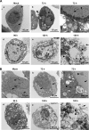
Fig. 1.
Fig. 1.
Representative electron micrographs show progressive development of autophagy and cell lysis in Delta-24-RGD-infected U-251 MG cells (A) and wild-type adenovirus 5 (Adwt)-infected MRC-5 cells (B). Cells were mock infected or infected with Delta-24-RGD or Adwt at a dose of 10 PFU/cell and examined at 72, 96, and 120 hours after infection, as indicated on the figure. Note that numerous mitochondria appear in mock-infected cells (arrows) but are barely seen in the cells after 96 h postinfection. Frame c in each panel is a close-up of a part of the cell in frame b showing vesicles in the cytoplasm (white arrows) and virions (arrowhead) clustered along or within an inclusion body (black arrow). Frame f in each panel is a close-up of a part of the cell in frame e revealing numerous vacuoles together with a discontinuous nuclear membrane (white arrow in panel A, frame f) and cellular membrane (black arrows) through which viral progeny (arrowheads) burst out of the disrupted cytoplasm.
Fig. 2.
Fig. 2.
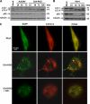
Adenovirus induces a complete autophagic flux. (A) U-87 MG cells were infected with replication-deficient adenoviral vector AdCMV (10 PFU/cell) and Delta-24-RGD (D24-RGD; 10 PFU/cell). (B) U-251 MG cells were infected with Adwt (10 PFU/cell). Cells were collected at 24, 48, and 72 h after infection. Bafilomycin A1 (BA1; 10 nM) was added to the cells 24 h after viral infection, and cells were collected 48 h later. Cell lysates were subjected to immunoblot analysis. Tubulin expression was used as a loading control. (C) Representative images of mCherry-EGFP-LC3 fluorescence in the U-87-MG cells. Cells were first transfected with plasmid expressing mCherry-EGFP-LC3. Twenty-four hours later, cells were infected with Delta-24-RGD at 50 PFU/cell in the absence or presence of 10 nM bafilomycin A1. After 48 h, live cells were observed under fluorescence microscopy.
Fig. 3.
Fig. 3.
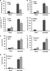
Effect of autophagy modulators on Delta-24-RGD-induced autophagy and cell lysis in U-87 MG cells. Cells were infected with Delta-24-RGD at 10 PFU/cell. Twenty-four hours later, cells were treated with 5 mM 3-methyladenine (3-MA; A, B, and C) or 10 nM bafilomycin A1 (BA1; D and E); alternatively, 3 h later cells were treated with 1 μM rapamycin (Rapa; F and G). Forty-eight hours after viral infection, cells were assayed for acidic vesicle organelles (AVO); alternatively, at 72 h postinfection, cells were assayed for cell lysis. P values are indicated by asterisks as follows: 0.001 (A), 0.008 (B), 0.0007 (C), 0.005 (D), 0.07 (E), 0.009 (F), and 0.02 (G). Note that cell lysis in panels B, E, and G was assayed with ethidium homodimer 1 staining, and cells in panel C were assayed for cell lysis with trypan blue staining. Three independent experiments were performed. Data are shown as means ± SD. D24-RGD, Delta-24-RGD. H2O, dimethyl sulfoxide (DMSO), or ethanol was used as a solvent for the 3-MA, BA1, or rapamycin, respectively.
Fig. 4.
Fig. 4.
Effect of autophagy modulators on Delta-24-RGD replication in U-87 MG cells. Cells were infected with Delta-24-RGD at 10 PFU/cell. Twenty-four hours later, cells were treated with 5 mM 3-methyladenine (3-MA) (A), or 3 h later cells were treated with 1 μM rapamycin (Rapa) (B). The titers of the viral progenies were determined 48 h after viral infection. Shown are the titers of viral progenies from 5 × 10⁴ cells in 1 ml infected with the virus. The experiments were performed once in triplicate. Means ± SD are shown (*, P > 0.05). H2O or ethanol was used as solvent for the 3-MA or rapamycin, respectively.
Fig. 5.
Fig. 5.
ATG5 knockout blocks adenovirus-induced autophagy and cell lysis in wild-type (wt) or ATG5 knockout (ATG5−/−) MEFs. (A) Cells were infected with Delta-24-RGD at the indicated doses (PFU/cell). After 72 h, cell lysates were collected for immunoblot analysis. Tubulin expression was used as a loading control. (B) Representative electron micrographs. Cells were infected with Delta-24-RGD at 50 PFU/cell for 72 h. Note that numerous vacuoles are induced by the virus in wt MEFs but only a few are induced in ATG5−/− MEFs. (C) Phase-contrast images of MEFs infected with Delta-24-RGD at 100 PFU/cell for 72 h. (D) Cell lysis caused by Delta-24-RGD in MEFs. Cells were infected with Delta-24-RGD at the indicated doses (PFU/cell). Seventy-two hours later, the cells were assayed for cell lysis. (E) Cells were infected with Adwt at 100 PFU/cell. Seventy-two hours later, the cells were assayed for cell lysis. Three independent experiments were performed. Data are shown as means ± SD. *, P = 0.004. M; mock; D24-RGD, Delta-24-RGD; UV-Adwt, UV-inactivated Adwt.
Fig. 6.
Fig. 6.
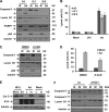
shRNA against ATG genes inhibits Delta-24-RGD-induced autophagy and cell lysis in U-87-MG cells. (A) ATG5 or ATG10 expression in U-87-MG (parental cells), cells transfected with control vector, and cells transfected with plasmid expressing shRNA against ATG5 (shATG5) or ATG10 (shATG10). Cell proteins were analyzed by immunoblotting. Actin expression was used as a loading control. (B) shRNA against ATG5 or ATG10 inhibits autophagy induction by Delta-24-RGD. Cells were infected with Delta-24-RGD at 10 PFU/cell. Forty-eight hours later, cell lysates were collected and analyzed by immunoblotting. Tubulin or actin was used as a loading control. (C) shRNA against ATG5 or ATG10 rescues cells from lysis by Delta-24-RGD. Cells were infected with Delta-24-RGD at 10 PFU/cell. Seventy-two hours later, cells were assayed for cell lysis. Three independent experiments were performed. Data are shown as means ± SD (*, P < 0.004). M; mock; Ad, Delta-24-RGD.
Fig. 7.
Fig. 7.
Autophagy-triggered caspase activity contributes to Delta-24-RGD-induced cell lysis. (A) Induction of autophagy and caspase activation by Delta-24-RGD. Human leukemia cell lines A3 (parental), I2.1 (FADD-null), and I9.2 (caspase 8-null) were infected with Delta-24-RGD at 100 PFU/cell. Seventy-two hours later, cell lysates were analyzed by immunoblotting. Tubulin expression was used as a loading control. (B) Cell lysis caused by Delta-24-RGD. Cells were treated as described for panel A and were assayed for cell death. Three independent experiments were performed. Data are shown as means ± SD (*, P < 0.05). (C) Inhibition of caspase activation by Z-VAD-Fmk. A3 cells were infected with Delta-24-RGD at 100 PFU/cell. Twenty-four hours later, 50 μM Z-VAD-Fmk was added to the cells. After 48 h, cell lysates were analyzed by immunoblotting. Tubulin expression was used as a loading control. (D) Z-VAD-Fmk rescues cells from lysis by Delta-24-RGD. A3 cells were treated as described for panel C. Cells were assayed for cell death. Three independent experiments were performed. Data are shown as means ± SD (*, P = 0.007). (E) Delta-24-RGD does not cause cytochrome c (Cyt C) release to the cytoplasm. A3 cells were infected with Delta-24-RGD at 100 PFU/cell. Seventy-two hours later, proteins from the cytosol (c) and mitochondria (m) were extracted from the cells and analyzed by immunoblotting. Tubulin expression was used as a loading control. (F) Atg5 is required for caspase activation. MEFs were infected with Delta-24-RGD at the indicated doses (PFU/cell). Seventy-two hours later, cell lysates were analyzed with immunoblotting. Tubulin expression was used as a loading control. M, mock; Ad, Delta-24-RGD. UVi, UV-inactivated Delta-24-RGD.

Similar articles

Cited by

References

    1. Abou El Hassan M. A., van der Meulen-Muileman I., Abbas S., Kruyt F. A. 2004. Conditionally replicating adenoviruses kill tumor cells via a basic apoptotic machinery-independent mechanism that resembles necrosis-like programmed cell death. J. Virol. 78:12243–12251 - PMC - PubMed
    1. Alonso M. M., et al. 2008. Delta-24-RGD in combination with RAD001 induces enhanced anti-glioma effect via autophagic cell death. Mol. Ther. 16:487–493 - PubMed
    1. Baehrecke E. H. 2005. Autophagy: dual roles in life and death? Nat. Rev. Mol. Cell Biol. 6:505–510 - PubMed
    1. Berk A. J. 2007. Adenoviridae: the viruses and their replication, 5th ed., vol. II Lippincott Williams & Wilkins, Philadelphia, PA
    1. Bodmer J. L., et al. 2000. TRAIL receptor-2 signals apoptosis through FADD and caspase-8. Nat. Cell Biol. 2:241–243 - PubMed

Publication types

LinkOut - more resources