. 2011 Apr 29;286(17):15458-72.
doi: 10.1074/jbc.M110.191924. Epub 2011 Mar 1.

Hrs recognizes a hydrophobic amino acid cluster in cytokine receptors during ubiquitin-independent endosomal sorting

Affiliations

Hrs recognizes a hydrophobic amino acid cluster in cytokine receptors during ubiquitin-independent endosomal sorting

Yuji Amano et al. J Biol Chem. .

Abstract

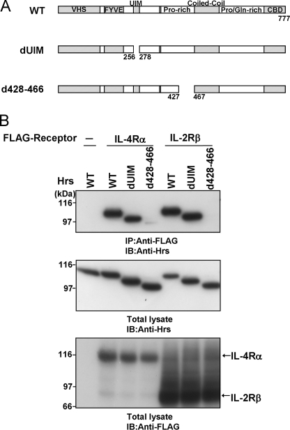
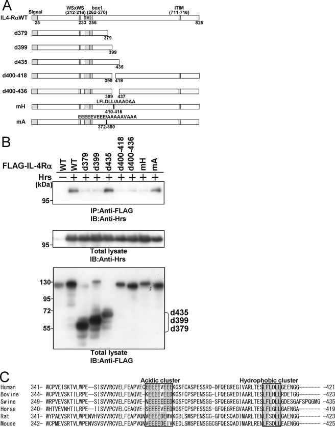
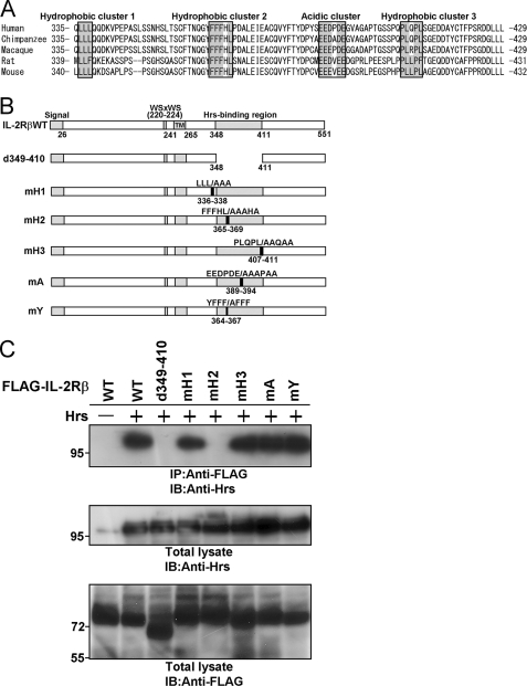
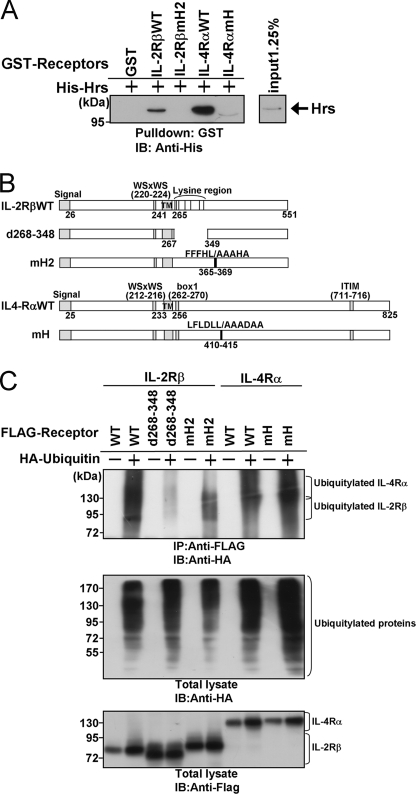
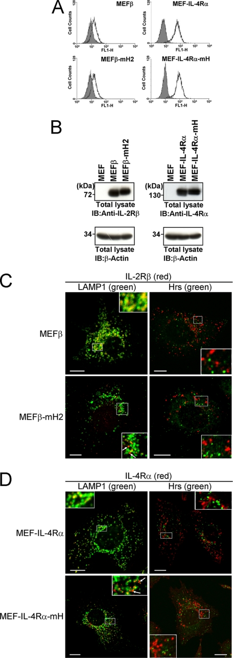
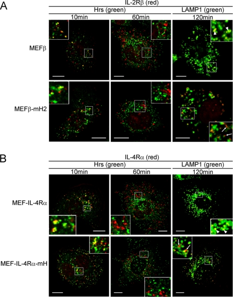
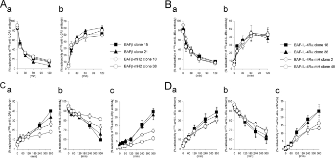
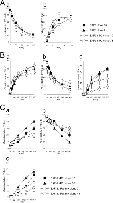
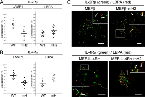
Hepatocyte growth factor-regulated tyrosine kinase substrate (Hrs) is a component of the ESCRT-0 protein complex that captures ubiquitylated cargo proteins and sorts them to the lysosomal pathway. Although Hrs acts as a key transporter for ubiquitin-dependent endosomal sorting, we previously reported that Hrs is also involved in ubiquitin-independent endosomal sorting of interleukin-2 receptor β (IL-2Rβ). Here, we show direct interactions between bacterially expressed Hrs and interleukin-4 receptor α (IL-4Rα), indicating that their binding is not required for ubiquitylation of the receptors, similar to the case for IL-2Rβ. Examinations of the Hrs binding regions of the receptors reveal that a hydrophobic amino acid cluster in both IL-2Rβ and IL-4Rα is essential for the binding. Whereas the wild-type receptors are delivered to LAMP1-positive late endosomes, mutant receptors lacking the hydrophobic amino acid cluster are sorted to lysobisphosphatidic acid-positive late endosomes rather than LAMP1-positive late endosomes. We also show that the degradation of these mutant receptors is attenuated. Accordingly, Hrs functions during ubiquitin-independent endosomal sorting of the receptors by recognizing the hydrophobic amino acid cluster. These findings suggest the existence of a group of cargo proteins that have this hydrophobic amino acid cluster as a ubiquitin-independent sorting signal.


Figures

FIGURE 1.
FIGURE 1.
The UIM domain of Hrs is dispensable for the interaction between Hrs and IL-4Rα. A, structures of wild-type Hrs and its mutants are shown. The Vps27-Hrs-STAM (VHS), Fab1-YGL023-Vps27-EEA1 (FYVE), ubiquitin interacting motif (UIM), proline (Pro)-rich region, coiled-coil, proline/glutamine (Pro/Gln)-rich region, and clathrin binding domain (CBD) are indicated. B, HEK293T cells (1 × 10⁶) were cotransfected with 3 μg of wild-type FLAG-IL-2Rβ, wild-type FLAG-IL-4Rα, or empty vector and 3 μg of wild-type Hrs or its mutants. Aliquots (400 μg) of the cell lysates were immunoprecipitated with an anti-FLAG monoclonal antibody and immunoblotted with an anti-Hrs monoclonal antibody (top panel). The expression levels of Hrs and FLAG-tagged receptors in total lysates of the transfected HEK293T cells were examined by immunoblotting with an anti-Hrs monoclonal antibody and anti-FLAG monoclonal antibody, respectively. Total lysate, aliquots (10 μg) of the lysates were immunoblotted with an anti-Hrs antibody (middle panel) or anti-FLAG monoclonal antibody (lower panel). IP, immunoprecipitation; IB, immunoblotting.
FIGURE 2.
FIGURE 2.
A hydrophobic amino acid cluster in the cytoplasmic tail of IL-4Rα is required for Hrs binding. A, structures of wild-type IL-4Rα and its mutants are shown. The signal sequence, WSXWS (tryptophan, serine, any amino acid, tryptophan, serine) motif, transmembrane region (TM), box1 motif, and immunoreceptor tyrosine-based inhibitory motif (ITIM) are indicated. B, HEK293T cells (1 × 10⁶) were cotransfected with 3 μg of FLAG-tagged wild-type IL-4Rα or its mutants and 3 μg of wild-type Hrs or empty vector. Aliquots (400 μg) of the cell lysates were immunoprecipitated with an anti-FLAG monoclonal antibody and immunoblotted with an anti-Hrs monoclonal antibody (top panel). The expression levels of Hrs and IL-4Rα were examined by immunoblotting with an anti-Hrs monoclonal antibody and anti-FLAG monoclonal antibody, respectively. Total lysate, aliquots (10 μg) of the lysates were immunoblotted with an anti-Hrs monoclonal antibody (middle panel) or anti-FLAG monoclonal antibody (lower panel). IP, immunoprecipitation; IB, immunoblotting. C, multiple alignment of the Hrs binding regions of human, bovine, swine, horse, rat, and mouse IL-4Rα is shown. The regions defined as acidic or hydrophobic clusters are boxed.
FIGURE 3.
FIGURE 3.
A hydrophobic amino acid cluster in the cytoplasmic tail of IL-2Rβ is required for Hrs binding. A, multiple alignment of the Hrs binding regions of human, chimpanzee, macaque, rat, and mouse IL-2Rβ is shown. The regions defined as acidic and hydrophobic clusters are boxed. B, shown are structures of wild-type IL-2Rβ and its mutants. The signal sequence, WSXWS motif, transmembrane region (TM), and Hrs binding region are indicated. C, HEK293T cells (1 × 10⁶) were cotransfected with 3 μg of FLAG-tagged wild-type IL-2Rβ or its mutants and 3 μg of wild-type Hrs or empty vector. Aliquots (400 μg) of the cell lysates were immunoprecipitated with an anti-FLAG monoclonal antibody and immunoblotted with an anti-Hrs monoclonal antibody (top panel). The expression levels of Hrs and IL-2Rβ were examined by immunoblotting with an anti-Hrs monoclonal antibody and anti-FLAG monoclonal antibody, respectively. Total lysate, aliquots (10 μg) of the lysates were immunoblotted with an anti-Hrs monoclonal antibody (middle panel) or anti-FLAG monoclonal antibody (lower panel). IP, immunoprecipitation; IB, immunoblotting.
FIGURE 4.
FIGURE 4.
Hrs directly associates with the cytokine receptors IL-2Rβ and IL-4Rα by recognizing the hydrophobic amino acid cluster in a ubiquitin-independent manner. A, glutathione-Sepharose beads containing immobilized GST, GST-fused cytoplasmic tail fragment of IL-2Rβ (GST-IL-2RβcyWT or GST-IL-2RβcymH2), or GST-fused cytoplasmic tail fragment of IL-4Rα (GST-IL-4RαcyWT or GST-IL-4RαcymH) were incubated with His-tagged Hrs. The bound proteins were separated by SDS-PAGE and analyzed by immunoblotting with an anti-His tag antibody. The input control of His-Hrs is shown in the right panel. B, structures of the IL-2Rβ and IL-4Rα mutants used in the receptor ubiquitylation assays are shown. C, HEK293T cells were cotransfected with 2 μg of HA-ubiquitin or empty vector and 2 μg of FLAG-IL-2Rβ, FLAG-IL-2Rβd268–348, FLAG-IL-2RβmH2, FLAG-IL-4Rα, or FLAG-IL-4RαmH. Aliquots (200 μg) of the cell lysates were immunoprecipitated with an anti-FLAG monoclonal antibody and immunoblotted with an anti-HA monoclonal antibody (top panel). The expression levels of the receptors (IL-2Rβ and IL-4Rα) and ubiquitylated total proteins were examined by immunoblotting with an anti-FLAG monoclonal antibody and anti-HA monoclonal antibody, respectively. Total lysate, aliquots (10 μg) of the lysates were immunoblotted with an anti-HA antibody (middle panel) or anti-FLAG antibody (lower panel). IP, immunoprecipitation; IB, immunoblotting.
FIGURE 5.
FIGURE 5.
Effects of the hydrophobic amino acid clusters in IL-2Rβ and IL-4Rα on their late-endosomal localizations. A, the IL-2Rβ and IL-4Rα expression levels on the surface of MEF transfectants were examined by flow cytometry. MEFβ, MEFβ-mH2, MEF-IL-4Rα, and MEF-IL-4Rα-mH cells were incubated with an anti-IL-2Rβ monoclonal antibody (TU11) or anti-IL-4Rα monoclonal antibody (MAB230) followed by a FITC-conjugated secondary antibody. B, the expression levels of IL-2Rβ and IL-4Rα in the indicated cells were examined by immunoblotting. Aliquots (15 μg) of the total lysates were immunoblotted with an anti-IL-2Rβ antibody (C20) or anti-IL-4Rα antibody (C20) (upper panels) or with an anti-β-actin antibody (lower panels). C and D, fluorescence images were observed using a confocal laser microscope. The indicated cells were grown on coverslips, fixed, and double-labeled with an anti-IL-2Rβ antibody (TU11) or anti-IL-4Rα antibody (MAB230) and an anti-LAMP1 monoclonal antibody or anti-Hrs antibody. Subsequently, the cells were incubated with fluorescently labeled secondary antibodies. Fairly large amounts of IL-2RβmH2 and IL-4RαmH are not sorted to LAMP1-positive compartments (arrows). Scale bars, 10 μm. IB, immunoblotting.
FIGURE 6.
FIGURE 6.
Kinetics of the endosomal localizations of IL-2Rβ and IL-4Rα. A and B, the indicated MEF transfectants were grown on coverslips, and the cell surface receptors were bound by an anti-IL-2Rβ antibody (TU11) or anti-IL-4Rα antibody (MAB230) at 0 °C followed by treatment with the chemical cross-linker DTSSP. The cells were cultured at 37 °C, fixed at the indicated times, and incubated with an anti-Hrs antibody or anti-LAMP1 monoclonal antibody. Fluorescence labeling was carried out for IL-2Rβ (red), IL-4Rα (red), Hrs (green), and LAMP1 (green). Large proportions of IL-2Rβ and IL-4Rα are delivered to LAMP1-positive compartments (arrowheads). In contrast, fairly large amounts of IL-2RβmH2 and IL-4RαmH are not delivered to LAMP1-positive compartments (arrows). Scale bars, 10 μm.
FIGURE 7.
FIGURE 7.
Internalization and degradation of IL-2Rβ and IL-4Rα in BAF-B03 transfectants. A and B, shown is internalization of IL-2Rβ and IL-4Rα in the transfectants. BAF transfectants were incubated with 125I-anti-IL-2Rβ antibody (TU11) or 125I-anti-IL-4Rα antibody (MAB230) at 0 °C. The cells were then cultured at 37 °C and harvested at the indicated times. The radioactivities of the cell surface-bound acid-removable fractions (a) and intracellular acid-unremovable fractions (b) were counted. C and D, degradation of IL-2Rβ and IL-4Rα in the transfectants is shown. BAF transfectants were incubated with 125I-anti-IL-2Rβ antibody or 125I-anti-IL-4Rα antibody at 0 °C followed by treatment with the chemical cross-linker DTSSP. The cells were then cultured at 37 °C and harvested at the indicated times. The radioactivities of the culture supernatants (a), cell precipitate fractions (b), and TCA-soluble fractions of the culture supernatants (c) were counted. The values represent the means ± S.E. of three separate experiments.
FIGURE 8.
FIGURE 8.
Internalization and degradation of 125I-IL-2 and 125I-IL-4 in BAF-B03 transfectants. A, internalization of 125I-IL-2 in the transfectants is shown. BAF transfectants were incubated with 350 pm 125I-IL-2 at 0 °C. The cells were then cultured at 37 °C and harvested at the indicated times. The radioactivities of the cell surface-bound acid-removable fractions (a) and intracellular acid-unremovable fractions (b) were counted. B, degradation of 125I-IL-2 in the transfectants is shown. BAF transfectants were incubated with 350 pm 125I-IL-2 and then cultured for the indicated times. The radioactivities of the culture supernatants (a), cell precipitate fractions (b), and TCA-soluble fractions of the culture supernatants (c) were counted. C, degradation of 125I-IL-4 in the transfectants is shown. BAF transfectants were incubated with 250 pm 125I-IL-4, treated with the cross-linker DTSSP, and then cultured for the indicated times. The radioactivities of the culture supernatants (a), cell precipitate fractions (b), and TCA-soluble fractions of the culture supernatants (c) were counted. The values represent the means ± S.E. of three separate experiments.
FIGURE 9.
FIGURE 9.
Localizations of IL-2Rβ and IL-4Rα mutants lacking the hydrophobic amino acid cluster to LBPA-positive compartments. MEF transfectants were grown on coverslips, fixed, and double-labeled with an anti-IL-2Rβ antibody (C20) or anti-IL-4Rα antibody (C20) and an anti-LAMP1 monoclonal antibody or anti-LBPA monoclonal antibody. Fluorescence images of the MEF transfectants (n ≥ 10 cells) were captured using a confocal laser microscope. The percentages of the IL-2Rβ-positive (A) and IL-4Rα-positive (B) pixel areas that were colocalized with LAMP1 (left) and LBPA (right) were analyzed. C, the IL-2Rβ-positive and IL-4Rα-positive pixel areas that were colocalized with LBPA (arrowheads) and not with LBPA (arrows) are indicated. Fluorescence labeling was carried out for IL-2Rβ (green), IL-4Rα (green), and LBPA (red). Scale bars, 10 μm.

Similar articles

Cited by

References

    1. Bonifacino J. S., Traub L. M. (2003) Annu. Rev. Biochem. 72, 395–447 - PubMed
    1. Di Fiore P. P., Polo S., Hofmann K. (2003) Nat. Rev. Mol. Cell. Biol. 4, 491–497 - PubMed
    1. Hicke L., Schubert H. L., Hill C. P. (2005) Nat. Rev. Mol. Cell. Biol. 6, 610–621 - PubMed
    1. Katzmann D. J., Babst M., Emr S. D. (2001) Cell 106, 145–155 - PubMed
    1. Alam S. L., Sun J., Payne M., Welch B. D., Blake B. K., Davis D. R., Meyer H. H., Emr S. D., Sundquist W. I. (2004) EMBO J. 23, 1411–1421 - PMC - PubMed

Publication types

Substances

LinkOut - more resources