. 2011 Oct;469(10):2773-84.
doi: 10.1007/s11999-011-1826-x.

Engineered cartilage maturation regulates cytokine production and interleukin-1β response

Affiliations

Engineered cartilage maturation regulates cytokine production and interleukin-1β response

Silvia Francioli et al. Clin Orthop Relat Res. 2011 Oct.

Abstract

Background: Because the injured joint has an actively inflammatory environment, the survival and repair potential of cartilage grafts may be influenced by inflammatory processes. Understanding the interactions of those processes with the graft may lead to concepts for pharmacologic or surgical solutions allowing improved cartilage repair.

Questions/purposes: We asked whether the maturation level of cartilaginous tissues generated in vitro by expanded human articular chondrocytes (HACs) modulate (1) the spontaneous production of cytokines and (2) the response to interleukin (IL)-1β.

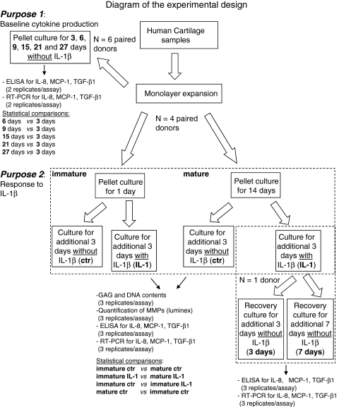
Methods: Twelve pellets/donor prepared with monolayer-expanded HACs (n = 6 donors) were evaluated at six different culture times for mRNA expression (n = 72) and spontaneous baseline release of monocyte chemoattractant protein (MCP)-1, IL-8, and transforming growth factor (TGF)-β1 (n = 72). We cultured 24 pellets/donor from each of four donors for 1 or 14 days (defined as immature and mature, respectively) and exposed the pellets to IL-1β for 3 days. MCP-1, IL-8, TGF-β1, and metalloprotease (MMP)-1 and MMP-13 were quantified in pellets and culture supernatants.

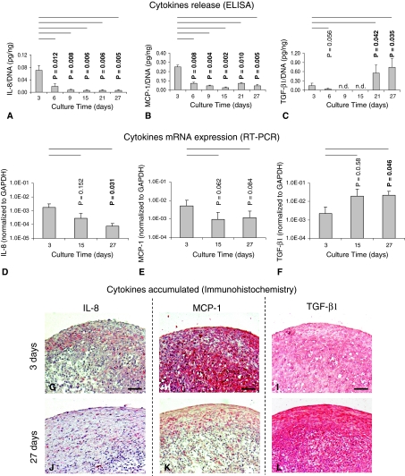
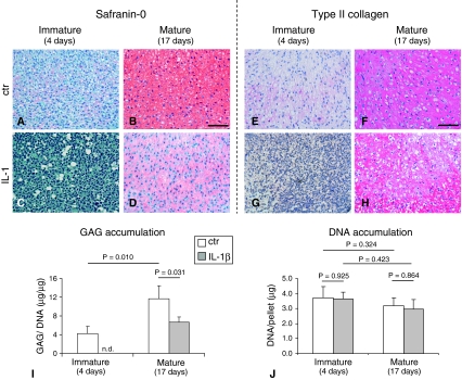
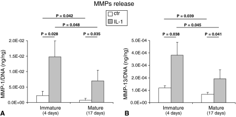
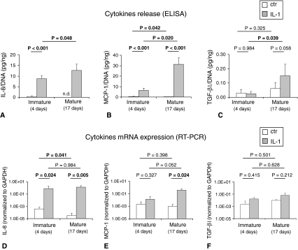
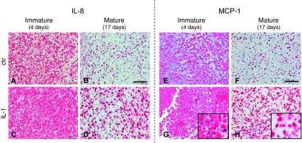
Results: By increasing culture time, the spontaneous release of IL-8 and MCP-1 decreased (12.0- and 5.5-fold, respectively), whereas that of TGF-β1 increased (5.4-fold). As compared with immature pellets, mature pellets responded to IL-1β by releasing lower amounts of MMP-1 (2.9-fold) and MMP-13 (1.7-fold) and increased levels of IL-8, MCP-1, and TGF-β1 (1.5-, 5.0-, and 7.5-fold, respectively). IL-8 and MCP-1 promptly returned to baseline on withdrawal of IL-1β.

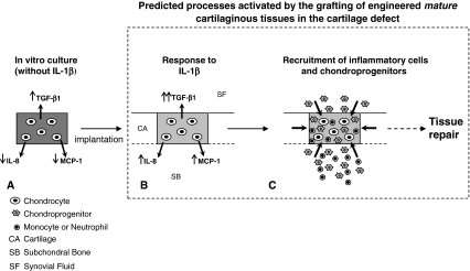
Conclusions: Our observations suggest more mature cartilaginous tissues are more resistant to IL-1β exposure and can activate chemokines required to initiate tissue repair processes.

Clinical relevance: The implantation of more mature cartilaginous tissues might provide superior graft survival and improve/accelerate cartilage repair.


Figures

Fig. 1
Fig. 1
Diagram of the experimental design. HACs harvested from six donors were expanded in monolayer and then cultured in pellets. For Purpose 1, six donor primaries were used. The total number of pellets (per donor) was 24, four for each culture time: two were analyzed by real-time RT-PCR (total number of replicates: 12) and the others by histology/immunohistochemistry (total number of replicates: 12, not included in this scheme). ELISA quantification of MCP-1, TGF-β1, and IL-8 was instead performed in the same supernatants harvested from two pellets (total number of replicates: 12). For Purpose 2, four donor primaries were used. The total number of pellets generated per donor was 48, nine for each group: three were analyzed by RT-PCR, (total number of replicates: 12) three by biochemical test (GAGs, DNA, total number of replicates: 12), and three by histology/immunohistochemistry (not included in this scheme, total number of replicates: 12). ELISA and Luminex analyses were performed in the same supernatants harvested from three pellets (total number of replicates: 12). Differences in the cytokine expression and release, MMP release, GAG and DNA contents between the indicated groups (in bold) were assessed using the nonparametric two-tailed Wilcoxon test. Pellets generated from one donor were also exposed to 3 or 7 days of recovery culture after IL-1 removal. ctr = control (without IL-1β).
Fig. 2A–L
Fig. 2A–L
Images show the quantification of IL-8, MCP-1, and TGF-β1 produced/accumulated by HACs at different time of pellet culture. Amounts of (A) IL-8, (B) MCP-1, and (C) TGF-β1 proteins are shown, normalized to the DNA contents of the corresponding pellets, released in the supernatants (six donors, 12 specimens). Real-time RT-PCR analysis of the expressions of (D) IL-8, (E) MCP-1, and (F) TGF-β1 mRNA by chondrocytes cultured in pellets at the different times (four donors, 12 specimens) is shown. Values are expressed as mean ± SD of measurements. Probability values (p) versus the 3-day groups are reported on the top of each bar. n.d. = not detectable. Immunohistologic assessment of representative pellets cultured for (GI) 3 days and (JL) 27 days is shown (staining with antibodies against [G, J] IL-8, [H, K] MCP-1, and [I, L] TGF-β1; original magnification, ×10). Scale bars = 100 μm. Results indicate a decrease of IL-8 and MCP-1 and an increase of TGF-β1 spontaneous production with increasing maturation extent.
Fig. 3A–J
Fig. 3A–J
Images illustrate (AD) histologic and (EH) immunohistologic assessment of representative pellets maintained in culture for a total of (A, C, E, G) 4 days (immature) or (B, D, F, H) 17 days (mature) in which the last 3 days were (A, B, E, F) without IL-1β (ctr) or (C, D, G, H) with IL-1β ([AD] safranin O and [EH] Type II collagen staining; original magnification, ×2). Scale bars = 200 μm. (I) Sulfated GAG content normalized to the amount of DNA and (J) DNA content of mature and immature pellets are shown. Values are expressed as mean ± SD of measurements (four donors, 12 specimens). n.d. = not detectable. Probability values (p) (mature versus mature [same culture condition] and ctr versus IL-1 [same culture time]) are reported on the top of the bars. Results indicate a superior IL-1β-mediated cartilage loss by immature pellets.
Fig. 4A–B
Fig. 4A–B
Graphs show the quantification of released MMPs by HACs. Amounts of (A) MMP-1 and (B) MMP-13 proteins are shown, normalized to the DNA contents of the corresponding pellets, released in the supernatants of pellets maintained in culture for a total of 4 days (immature) or 17 days (mature) in which the last 3 days with IL-1β or without IL-1β (ctr). Values are expressed as mean ± SD of measurements (four donors, 12 specimens). Probability values (p) (mature versus mature [same culture condition] and ctr versus IL-1β [same culture time]) are reported on the top of the bars. Results indicate a superior IL-1β-mediated MMPs production by immature pellets.
Fig. 5A–F
Fig. 5A–F
Graphs show the quantification of IL-8, MCP-1, and TGF-β1 produced by HACs. Amounts of (A) IL-8, (B) MCP-1, and (C) TGF-β1 are shown, normalized to the DNA contents of the corresponding pellets, released in the supernatants of tissues maintained in culture for a total of 4 days (immature) or 17 days (mature) in which the last 3 days were with IL-1β or without IL-1β (ctr). Real-time RT-PCR analysis of the expression of (D) IL-8, (E) MCP-1, and (F) TGF-β1 mRNA by chondrocytes cultured in pellets at the different times is shown. Values are expressed as mean ± SD of measurements (four donors, 12 specimens). n.d. = not detectable. Probability values (p) (mature versus mature [same culture condition] and ctr versus IL-1β [same culture time]) are reported on the top of the bars. Results indicate a superior IL-1β-mediated production of IL-8, MCP-1, and TGF-β1 by immature pellets.
Fig. 6A–H
Fig. 6A–H
Images illustrate the accumulation of (AD) IL-8 and (EH) MCP-1 in the tissues. Immunohistologic assessment of representative pellets maintained in culture for a total of (A, C, E, G) 4 days (immature) or (B, D, F, H) 17 days (mature) in which the last 3 days were (A, B, E, F) without IL-1β (ctr) or (C, D, G, H) with IL-1β is shown (staining with antibodies against [AD] IL-8 and [EH] TGF-β1; original magnification, ×20 for pictures and ×40 for inserts). Scale bars = 50 μm.
Fig. 7A–C
Fig. 7A–C
Schematic view of the hypothesis and results is shown. (A) During the in vitro maturation (in the absence of IL-1β), chondrocyte-based constructs accumulate a large amount of cartilaginous matrix (dark gray) and express/release lower amounts of IL-8 and MCP-1 and higher amounts of TGF-β1. (B) When implanted in the cartilage defect, mature grafts respond to IL-1β by losing some of the accumulated cartilage matrix (light gray) and enhancing the expression/release of IL-8 and MCP-1 in a transient manner and of TGF-β1. (C) IL-8 and MCP-1 would attract neutrophils, monocytes, and chondroprogenitors from the surrounding tissues. Although the former cells would induce tissue remodeling, the chondroprogenitors, especially in the presence of elevated concentration of TGF-β1, would contribute to the deposition of extracellular matrix. Taken together, the contribution of the different recruited cell types would improve/accelerate cartilage repair processes (see dashed arrow), and the transient nature of the enhanced release of IL-8 and MCP-1 would not induce chronic inflammation processes.

Similar articles

Cited by

References

    1. Aulthouse AL, Beck M, Griffey E, Sanford J, Arden K, Machado MA, Horton WA. Expression of the human chondrocyte phenotype in vitro. In Vitro Cell Dev Biol. 1989;25:659–668. doi: 10.1007/BF02623638. - DOI - PubMed
    1. Baici A, Lang A, Horler D, Knopfel M. Cathepsin B as a marker of the dedifferentiated chondrocyte phenotype. Ann Rheum Dis. 1988;47:684–691. doi: 10.1136/ard.47.8.684. - DOI - PMC - PubMed
    1. Barbero A, Grogan SP, Schaefer D, Heberer M, Mainil-Varlet P, Martin I. Age related changes in human articular chondrocyte yield, proliferation and post-expansion chondrogenic capacity. Osteoarthritis Cartilage. 2004;12:476–484. doi: 10.1016/j.joca.2004.02.010. - DOI - PubMed
    1. Bedi A, Feeley BT, Williams RJ., 3rd Management of articular cartilage defects of the knee. J Bone Joint Surg Am. 2010;92:994–1009. doi: 10.2106/JBJS.I.00895. - DOI - PubMed
    1. Behrens P, Bitter T, Kurz B, Russlies M. Matrix-associated autologous chondrocyte transplantation/implantation (MACT/MACI)—5-year follow-up. Knee. 2006;13:194–202. doi: 10.1016/j.knee.2006.02.012. - DOI - PubMed

Publication types

MeSH terms