. 2011 Feb 15;6(2):e17248.
doi: 10.1371/journal.pone.0017248.

Drosophila S2 cells are non-permissive for vaccinia virus DNA replication following entry via low pH-dependent endocytosis and early transcription

Affiliations

Drosophila S2 cells are non-permissive for vaccinia virus DNA replication following entry via low pH-dependent endocytosis and early transcription

Zain Bengali et al. PLoS One. .

Abstract

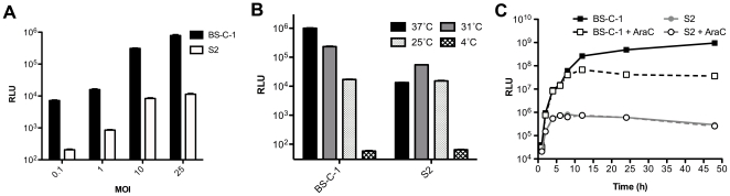
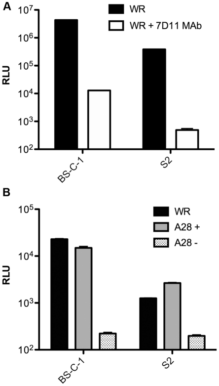
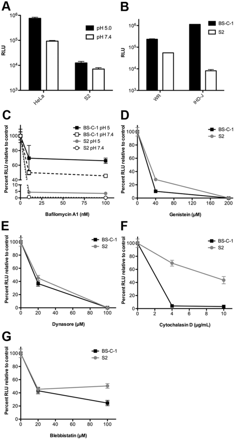
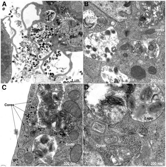
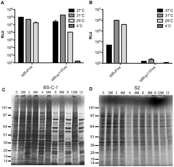
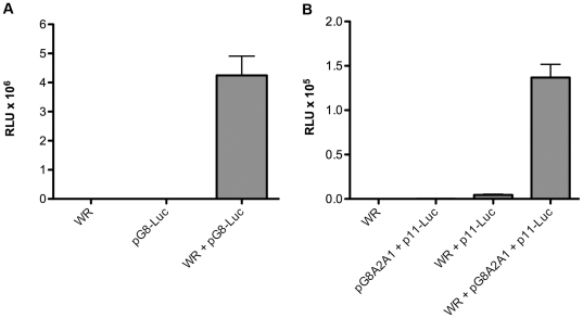
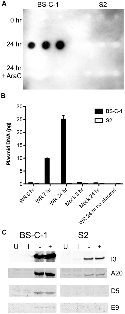
Vaccinia virus (VACV), a member of the chordopox subfamily of the Poxviridae, abortively infects insect cells. We have investigated VACV infection of Drosophila S2 cells, which are useful for protein expression and genome-wide RNAi screening. Biochemical and electron microscopic analyses indicated that VACV entry into Drosophila S2 cells depended on the VACV multiprotein entry-fusion complex but appeared to occur exclusively by a low pH-dependent endocytic mechanism, in contrast to both neutral and low pH entry pathways used in mammalian cells. Deep RNA sequencing revealed that the entire VACV early transcriptome, comprising 118 open reading frames, was robustly expressed but neither intermediate nor late mRNAs were made. Nor was viral late protein synthesis or inhibition of host protein synthesis detected by pulse-labeling with radioactive amino acids. Some reduction in viral early proteins was noted by Western blotting. Nevertheless, synthesis of the multitude of early proteins needed for intermediate gene expression was demonstrated by transfection of a plasmid containing a reporter gene regulated by an intermediate promoter. In addition, expression of a reporter gene with a late promoter was achieved by cotransfection of intermediate genes encoding the late transcription factors. The requirement for transfection of DNA templates for intermediate and late gene expression indicated a defect in viral genome replication in VACV-infected S2 cells, which was confirmed by direct analysis. Furthermore, VACV-infected S2 cells did not support the replication of a transfected plasmid, which occurs in mammalian cells and is dependent on all known viral replication proteins, indicating a primary restriction of DNA synthesis.


Conflict of interest statement

Competing Interests: The authors have declared that no competing interests exist.

Figures

Figure 1
Figure 1. Entry of VACV in Drosophila S2 cells.
A) BS-C-1 and S2 cells were incubated with purified WRvFire virions at 4°C at neutral pH for 1 h at the indicated MOI to allow attachment, washed, and then incubated for 1 h at 37°C for BS-C-1 cells and 25°C for S2 cells. Cells were lysed and Luc activity measured and plotted as relative light units (RLU). B) Cells were infected as in panel A with a MOI of 2 PFU per cell and after virus attachment the plates were incubated at the indicated temperatures for 2 h and then assayed for Luc. C) Cells were infected with an MOI of 1 PFU per cell as in panel A. After attachment, plates were incubated at 31°C and Luc assays were made over a 48 h period. Note that the solid and dashed lines representing Luc activity from S2 cells in the presence and absence of AraC are practically superimposed. Standard error bars were plotted in all three panels but are too close to discern in some places.
Figure 2
Figure 2. VACV entry into S2 cells is dependent on components of the EFC.
A) Purified WRvFire virions (WR) (MOI of 10 PFU per cell) were incubated with or without the L1 MAb 7D11 (20 µg/ml) for 30 min at room temperature and then added to BS-C-1 or S2 cells. After attachment, the cells were incubated at 31°C for 1 h and assayed for Luc. B) BS-C-1 and S2 cells were incubated with purified WRvFire virions (WR) or A28ivFire virions (MOI of 10 PFU per cell) containing (+) or lacking (-) the A28 protein. After attachment, the cells were incubated for 1 h and assayed for Luc. Standard error bars were plotted in both panels but are too close to discern in some places.
Figure 3
Figure 3. Effects of pH and chemical inhibitors on VACV entry.
A) HeLa and S2 cells were incubated with purified WRvFire virions at 4°C for 1 h at a MOI of 2 PFU per cell. After attachment, cells were washed and incubated with pH 5 or pH 7.4 buffer for 3 min at 37°C. Cells were then washed and incubated at 31°C at neutral pH for 2 h and Luc assayed. B) BS-C-1 and S2 cells were infected with WRvFire or IHD-JvFire virions at neutral pH and Luc assayed as in panel A. C) BS-C-1 and S2 cells were infected as above except cells were pre-treated with bafilomycin A1 for 1 h at 31°C and then pre-chilled to 4°C prior to virion attachment, followed by wash and pH treatment. Inhibitor was maintained in the media throughout the infection. BS-C-1 and S2 cells were pretreated with: D) genistein; E) dynasore; F) cytochalasin; G) blebbistatin followed by wash and incubation at 31°C for 2 h. Standard error bars were plotted in all panels but are too close to discern in some places.
Figure 4
Figure 4. Transmission electron microscopy of VACV infected S2 cells.
Purified VACV virions were spinoculated onto cells for 1 h at 4°C at a MOI of 150 PFU per cell. The cells were then incubated at 31°C. A) Low magnification of virions associated with cells and in endosomes at 1 h after infection. B–D) Higher magnifications showing virions in vesicles and cores in the cytoplasm at 1 h after infection. V, virions; Endo, endosome. A size marker is present at the lower right corner of each panel.
Figure 5
Figure 5. Quantification of virions in endosomes and cores in cytoplasm.
Data are from the same experiment used to obtain images in Fig. 4. The numbers of virus particles were counted in single sections of 90 different cells and the totals plotted: A) MVs in vesicles; B) cores in cytoplasm.
Figure 6
Figure 6. Relative amount of VACV mRNA in infected S2 cells.
Total polyadenylated RNA was extracted at various times after infection and subjected to deep RNA sequencing. The sequences were divided into those that mapped to the Drosophila and VACV genomes. The percentage of the sequence hits to VACV genome relative to the total mapped reads at progressive time after infection is shown.
Figure 7
Figure 7. VACV genome-wide transcriptome maps of S2 cells.
A) The number of viral sequence reads per nucleotide was determined as indicated in the legend to Fig. 6 and displayed over the entire VACV genome with early and post-transcriptional ORFs in green and red, respectively. The counts above the line map to the upper (rightward) strand and counts below the line map to the lower (leftward) strand of the VACV genome. The highest read counts are off-scale in the 2- to 12-h samples for display purposes. The counts were normalized by the total reads of the samples and those duplicated because of their location within the inverted terminal repetition were divided by 2. The HindIII restriction map of the VACV genome is shown at the bottom for reference purposes. B) The HindIII D region from the 6 h time point in panel A has been enlarged.
Figure 8
Figure 8. Late gene expression.
BS-C-1 (A) and S2 (B) cells were infected at an MOI of 1 PFU per cell with a recombinant VACV with Luc regulated under the VACV early/late synthetic promoter (WRvFire) or the p11 late promoter (WRvp11Fire) at indicated temperatures. Luc activity was measured at 16 h after infection. Standard error bars were plotted in both panels but are too close to discern in some places. BS-C-1 (C) and S2 (D) cells were infected with VACV at an MOI of 20 PFU per cell and pulse-labeled with 35S-labeled amino acids for 30 min intervals at the times indicated and analyzed by sodium dodecyl sulfate-polyacrylamide gel electrophoresis and autoradiography. The masses of marker proteins in kDA are indicated on the left. M denotes mock-infected cells.
Figure 9
Figure 9. Expression of reporter genes regulated by intermediate and late promoters in transfected plasmids.
A) Intermediate expression. S2 cells were mock infected or infected with VACV strain WR at a MOI of 1 PFU per cell and mock transfected or transfected with a plasmid containing the Luc ORF regulated by the G8R intermediate promoter. Luc activity was measured at 16 h. B) S2 cells were infected with VACV strain WR and transfected with a plasmid containing the Luc ORF regulated by the late p11 promoter and cotransfected or not with a second plasmid containing the three late transcription factor genes (G8R, A1L, A2L) regulated by intermediate promoters. Luc activity was measured at 16 h. Standard error bars were plotted in both panels but are too close to discern in some places.
Figure 10
Figure 10. DNA replication.
A) VACV genome replication. BS-C-1 and S2 cells were infected with VACV strain WR (MOI of 5 PFU/cell) in the presence or absence of AraC. At 0 and 24 h, the amount of VACV DNA was determined in triplicate by slot blot hybridization using digoxigenin-dUTP labeled F17R probe. B) Plasmid replication. BS-C-1 and S2 cells were infected with VACV strain WR (MOI of 3 PFU/cell) and then transfected with a plasmid. At 0, 7 and 24 h, plasmid sequences were quantified by real-time PCR. Mock infected cells and incubations without plasmids were used as controls. Standard error bars were plotted but are too close to discern in some places. C) Western blotting of VACV DNA replication proteins. BS-C-1 and S2 cells were infected with VACV (MOI of 20 PFU/cell), incubated after overnight at 31°C in the presence (+) and absence (-) of AraC analyzed by SDS-polyacrylamide gel electrophoresis and Western blotting with antibody to the I3, A20, D5 and E9 proteins. Uninfected cells (U) and cells harvested after inoculation (I) were used as controls.

Similar articles

Cited by

References

    1. Moss B. Poxviridae: the viruses and their replication. In: Knipe DM, Howley PM, editors. Fields Virology. Philadelphia: Lippincott Williams & Wilkins; 2007. pp. 2905–2946.
    1. Upton C, Slack S, Hunter AL, Ehlers A, Roper RL. Poxvirus orthologous clusters: toward defining the minimum essential poxvirus genome. J Virol. 2003;77:7590–7600. - PMC - PubMed
    1. Langridge WH. Detection of Amsacta moorei entomopoxvirus and vaccinia virus proteins in cell cultures restrictive for poxvirus multiplication. J Invertebr Pathol. 1983;42:77–82. - PubMed
    1. Li Y, Hall RL, Moyer RW. Transient, non-lethal expression of genes in vertebrate cells by recombinant entemopoxviruses. J Virol. 1997;71:9557–9562. - PMC - PubMed
    1. Li Y, Yuan S, Moyer RW. The non-permissive infection of insect (Gypsy moth) LD-652 cells by vaccinia virus. Virology. 1998;248:74–82. - PubMed

Publication types

LinkOut - more resources