

Multimodal wide-field two-photon excitation imaging: characterization of the technique for in vivo applications
- PMID: 21339880
- PMCID: PMC3038450
- DOI: 10.1364/BOE.2.000356
Multimodal wide-field two-photon excitation imaging: characterization of the technique for in vivo applications
Abstract
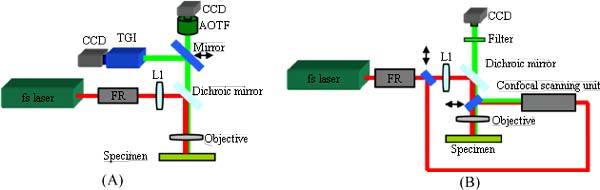
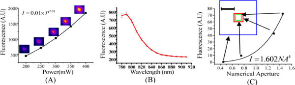
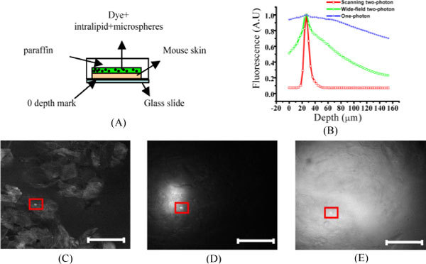
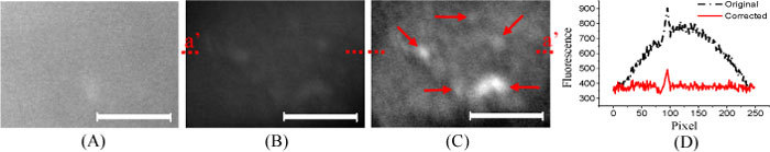
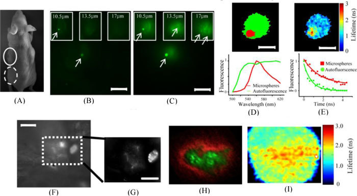
We report fast, non-scanning, wide-field two-photon fluorescence excitation with spectral and lifetime detection for in vivo biomedical applications. We determined the optical characteristics of the technique, developed a Gaussian flat-field correction method to reduce artifacts resulting from non-uniform excitation such that contrast is enhanced, and showed that it can be used for ex vivo and in vivo cellular-level imaging. Two applications were demonstrated: (i) ex vivo measurements of beta-amyloid plaques in retinas of transgenic mice, and (ii) in vivo imaging of sulfonated gallium(III) corroles injected into tumors. We demonstrate that wide-field two photon fluorescence excitation with flat-field correction provides more penetration depth as well as better contrast and axial resolution than the corresponding one-photon wide field excitation for the same dye. Importantly, when this technique is used together with spectral and fluorescence lifetime detection modules, it offers improved discrimination between fluorescence from molecules of interest and autofluorescence, with higher sensitivity and specificity for in vivo applications.
Keywords: (110.4234) Imaging systems; (180.4315) Microscopy.
Figures
Similar articles
-
Multimode Optical Imaging for Translational Chemotherapy: In Vivo Tumor Detection and Delineation by Targeted Gallium Corroles.Proc SPIE Int Soc Opt Eng. 2011 Feb 28;7902:79020F. doi: 10.1117/12.877780. Proc SPIE Int Soc Opt Eng. 2011. PMID: 26412924 Free PMC article.
-
An excitation wavelength-scanning spectral imaging system for preclinical imaging.Rev Sci Instrum. 2008 Feb;79(2 Pt 1):023707. doi: 10.1063/1.2885043. Rev Sci Instrum. 2008. PMID: 18315305
-
Aggregation-Induced Emission Luminogen with Near-Infrared-II Excitation and Near-Infrared-I Emission for Ultradeep Intravital Two-Photon Microscopy.ACS Nano. 2018 Aug 28;12(8):7936-7945. doi: 10.1021/acsnano.8b02452. Epub 2018 Aug 1. ACS Nano. 2018. PMID: 30059201
-
Non-invasive imaging of skin physiology and percutaneous penetration using fluorescence spectral and lifetime imaging with multiphoton and confocal microscopy.Eur J Pharm Biopharm. 2011 Apr;77(3):469-88. doi: 10.1016/j.ejpb.2010.12.023. Epub 2011 Jan 21. Eur J Pharm Biopharm. 2011. PMID: 21256962 Review.
-
Two-photon microscopy of cells and tissue.Circ Res. 2004 Dec 10;95(12):1154-66. doi: 10.1161/01.RES.0000150593.30324.42. Circ Res. 2004. PMID: 15591237 Review.
Cited by
-
Widefield Two-Photon Excitation without Scanning: Live Cell Microscopy with High Time Resolution and Low Photo-Bleaching.PLoS One. 2016 Jan 29;11(1):e0147115. doi: 10.1371/journal.pone.0147115. eCollection 2016. PLoS One. 2016. PMID: 26824845 Free PMC article.
-
Chemotherapy targeting by DNA capture in viral protein particles.Nanomedicine (Lond). 2012 Mar;7(3):335-52. doi: 10.2217/nnm.11.104. Nanomedicine (Lond). 2012. PMID: 22385197 Free PMC article.
-
Fast Optical Sectioning for Widefield Fluorescence Mesoscopy with the Mesolens based on HiLo Microscopy.Sci Rep. 2018 Nov 2;8(1):16259. doi: 10.1038/s41598-018-34516-2. Sci Rep. 2018. PMID: 30390029 Free PMC article.
-
Photoexcitation of tumor-targeted corroles induces singlet oxygen-mediated augmentation of cytotoxicity.J Control Release. 2012 Nov 10;163(3):368-73. doi: 10.1016/j.jconrel.2012.09.015. Epub 2012 Oct 4. J Control Release. 2012. PMID: 23041277 Free PMC article.
-
Wide-field multiphoton imaging of cellular dynamics in thick tissue by temporal focusing and patterned illumination.Biomed Opt Express. 2011 Feb 25;2(3):696-704. doi: 10.1364/BOE.2.000696. Biomed Opt Express. 2011. PMID: 21412473 Free PMC article.