. 2011 Apr 29;286(17):14903-12.
doi: 10.1074/jbc.M110.211656. Epub 2011 Feb 18.

S-adenosyl-L-methionine induces compaction of nascent peptide chain inside the ribosomal exit tunnel upon translation arrest in the Arabidopsis CGS1 gene

Affiliations

S-adenosyl-L-methionine induces compaction of nascent peptide chain inside the ribosomal exit tunnel upon translation arrest in the Arabidopsis CGS1 gene

Noriyuki Onoue et al. J Biol Chem. .

Abstract

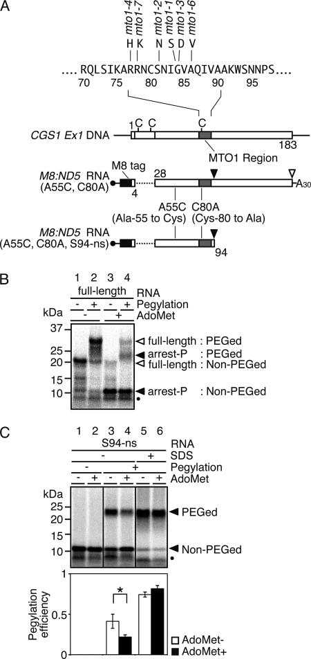
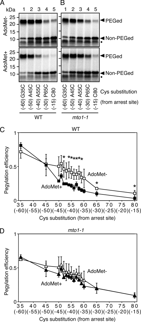
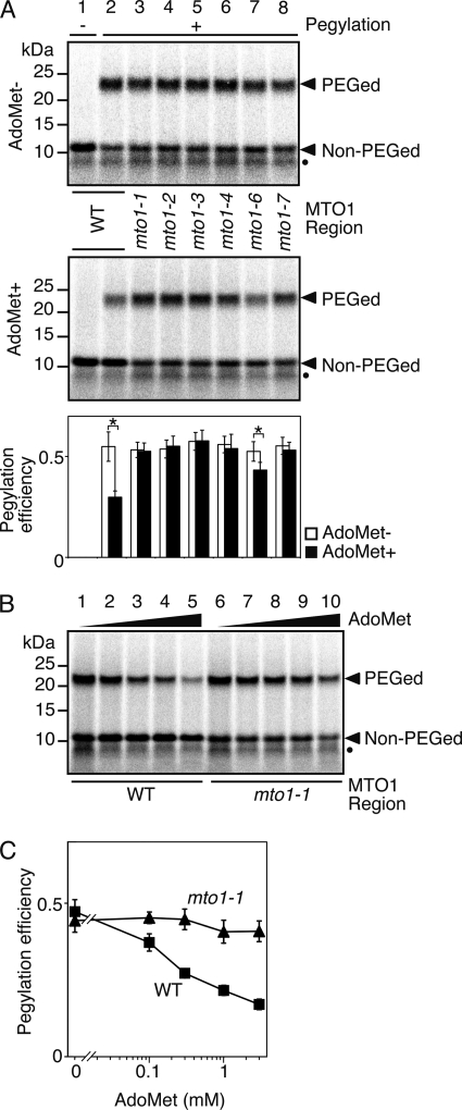
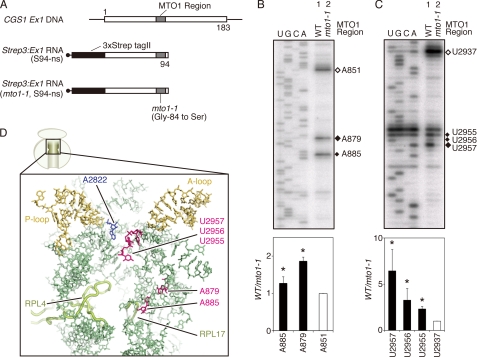
Expression of the Arabidopsis CGS1 gene, encoding the first committed enzyme of methionine biosynthesis, is feedback-regulated in response to S-adenosyl-L-methionine (AdoMet) at the mRNA level. This regulation is first preceded by temporal arrest of CGS1 translation elongation at the Ser-94 codon. AdoMet is specifically required for this translation arrest, although the mechanism by which AdoMet acts with the CGS1 nascent peptide remained elusive. We report here that the nascent peptide of CGS1 is induced to form a compact conformation within the exit tunnel of the arrested ribosome in an AdoMet-dependent manner. Cysteine residues introduced into CGS1 nascent peptide showed reduced ability to react with polyethyleneglycol maleimide in the presence of AdoMet, consistent with a shift into the ribosomal exit tunnel. Methylation protection and UV cross-link assays of 28 S rRNA revealed that induced compaction of nascent peptide is associated with specific changes in methylation protection and UV cross-link patterns in the exit tunnel wall. A 14-residue stretch of amino acid sequence, termed the MTO1 region, has been shown to act in cis for CGS1 translation arrest and mRNA degradation. This regulation is lost in the presence of mto1 mutations, which cause single amino acid alterations within MTO1. In this study, both the induced peptide compaction and exit tunnel change were found to be disrupted by mto1 mutations. These results suggest that the MTO1 region participates in the AdoMet-induced arrest of CGS1 translation by mediating changes of the nascent peptide and the exit tunnel wall.


Figures

FIGURE 1.
FIGURE 1.
Pegylation assay of CGS1 nascent peptide. A, schematic representation of CGS1 exon 1 DNA (CGS1 Ex1 DNA), the “full-length” CGS1 exon 1 RNA construct M8:ND5(A55C, C80A), and the nonstop RNA construct M8:ND5(A55C, C80A, S94-ns). Positions of endogenous cysteine codons are marked with vertical bars and labeled with C. Gray boxes indicate the MTO1 region, and the black box indicates the M8 tag sequence. Amino acid sequence around the MTO1 region and mto1 mutant alleles are shown above the top construct. Positions of introduced amino acid substitutions are indicated. B, pegylation of translation products carrying a single cysteine residue at the 55th position in the “full-length” CGS1 exon 1 RNA. M8:ND5(A55C, C80A) RNA was translated for 30 min in the absence (lanes 1 and 2) or presence (lanes 3 and 4) of 1 mm AdoMet. Translation products were labeled with [35S]methionine. RNCs were isolated by ultracentrifugation and subjected to pegylation reaction (Pegylation +; lanes 2 and 4) or translation-only reaction (Pegylation −; lanes 1 and 3). Samples were separated by SDS-PAGE followed by detection of the radioactive signals. Positions of 32-kDa pegylated full-length (full-length: PEGed), 20-kDa unpegylated (full-length: Non-PEGed), 23-kDa pegylated (arrest-P: PEGed), and 11-kDa unpegylated (arrest-P: Non-PEGed) products are indicated. Unpegylated arrested peptide (10 kDa) produced by a secondary arrested ribosome is marked with a black dot. A representative result of triplicate experiments is shown. C, pegylation of translation arrest products carrying a single cysteine residue at the 55th position in the Ser-94 nonstop RNA. Upper panel, M8:ND5(A55C, C80A, S94-ns) RNA was translated for 30 min in the absence (lanes 1, 3, and 5) or presence (lanes 2, 4, and 6) of 1 mm AdoMet. Translation products were labeled with [35S]methionine. RNCs were isolated by ultracentrifugation and subjected to pegylation reaction (lanes 3–6) or translation-only reaction (Pegylation −; lanes 1 and 2). In lanes 5 and 6, RNCs were treated with 1% SDS for 20 min at 25 °C prior to the pegylation reaction. Samples were separated by SDS-PAGE followed by detection of the radioactive signals. Positions of the 23-kDa pegylated (PEGed) and 11-kDa unpegylated (Non-PEGed) products are indicated. Unpegylated arrested peptide (10 kDa) produced by a secondary arrested ribosome is marked with a black dot. A representative result of triplicate experiments is shown. Lower panel, radioactive signals of the pegylated and unpegylated bands were quantified, and the pegylation efficiency was calculated (see “Experimental Procedures”). Average ± S.D. of at least three independent experiments are shown. Significant difference in pegylation efficiency between the presence and absence of AdoMet is indicated by an asterisk (p < 0.05 by t test).
FIGURE 2.
FIGURE 2.
Cysteine-scanning and pegylation assay of CGS1 nascent peptide. A and B, pegylation assay of wild-type MTO1 (A) and mto1-1 mutant (B) versions of M8:ND5(G35C, C80A, S94-ns) (lane 1), M8:ND5(A45C, C80A, S94-ns) (lane 2), M8:ND5(A55C, C80A, S94-ns) (lane 3), M8:ND5(P65C, C80A, S94-ns) (lane 4), and M8:ND5(S94-ns) (lane 5) RNAs in the absence (upper panel) and presence (lower panel) of 1 mm AdoMet was carried out as described for Fig. 1C. Representative results of triplicate experiments are shown. Positions of the 23-kDa pegylated (PEGed) and 11-kDa unpegylated (Non-PEGed) products are indicated. Unpegylated arrested peptide (10 kDa) produced by a secondary arrested ribosome is marked with a black dot. The single cysteine position is indicated below each lane, and the distance of this cysteine from the arrest site (Ser-94) is indicated in parentheses. C and D, pegylation experiments were carried out using a series of cysteine substitution constructs of wild-type MTO1 (C) and mto1-1 mutant (D) versions. The radioactive signals of the pegylated and unpegylated bands were quantified. Pegylation efficiencies were calculated as in Fig. 1C, and average ± S.D. of at least three independent experiments are shown. Significant difference in pegylation efficiency between the absence and presence of AdoMet are marked with asterisks (p < 0.05 by t test).
FIGURE 3.
FIGURE 3.
Correlation between regulatory function of the MTO1 region and pegylation efficiency. A, effects of different alleles of mto1 mutations were tested. Pegylation assays of wild-type MTO1 (lane 2), mto1-1 (lane 3), mto1-2 (lane 4), mto1-3 (lane 5), mto1-4 (lane 6), mto1-6 (lane 7), and mto1-7 (lane 8) mutant versions of M8:ND5(S58C, C80A, S94-ns) RNA in the absence (upper panel) and presence (middle panel) of 1 mm AdoMet (representative result of quadruplicate experiments) and quantification of radioactive signals (lower panel) were carried out as described for Fig. 1C. A translation-only reaction using wild-type MTO1 version is shown in lane 1. Significant difference in pegylation efficiency between the presence and absence of AdoMet are indicated by asterisks (p < 0.05 by t test). Positions of the 23-kDa pegylated (PEGed) and 11-kDa unpegylated (Non-PEGed) products are indicated. Unpegylated arrested peptide (10 kDa) produced by a secondary arrested ribosome is marked with a black dot. B, pegylation assay at different AdoMet concentrations was carried out as in Fig. 1C using wild-type MTO1 (lanes 1–5) and mto1-1 mutant (lanes 6–10) versions of M8:ND5(S58C, C80A, S94-ns) RNAs. The samples were translated for 30 min with 0 (lanes 1 and 6), 0.1 mm (lanes 2 and 7), 0.3 mm (lanes 3 and 8), 1 mm (lanes 4 and 9), and 3 mm (lanes 5 and 10) AdoMet. The 23-kDa pegylated (PEGed) and 11-kDa unpegylated (Non-PEGed) products are indicated. A representative result of triplicate experiments is shown. Unpegylated arrested peptide (10 kDa) produced by a secondary arrested ribosome is marked with a black dot. C, radioactive signals in B were quantified for the wild-type MTO1 (B, lanes 1–5) and mto1-1 mutant (B, lanes 6–10) versions. The pegylation efficiency was calculated as in Fig. 1C, and average ± S.D. of three independent experiments are shown.
FIGURE 4.
FIGURE 4.
Methylation protection and UV cross-link experiments of 28 S rRNA. A, schematic representation of Strep3:Ex1(S94-ns) and Strep3:Ex1(mto1-1, S94-ns) RNAs. Gray boxes indicate the MTO1 region, and the black boxes indicate the three consecutive Strep-tag II sequence. B, upper panel, methylation protection assay of arrested ribosome. Wild-type MTO1 (lane 1) and mto1-1 (lane 2) versions of Strep3:Ex1(S94-ns) RNAs were translated in WGE for 30 min in the presence of 1 mm AdoMet. RNCs were subjected to methylation reaction using DMS. Total RNA was extracted and used for primer extension analysis using 32P-labeled oligonucleotides complementary to wheat 28 S rRNA. Primer extension signals were detected after separation on a sequence gel. Filled diamonds mark the nucleotides in which a methylation difference was detected between lane 1 (WT) and lane 2 (mto1-1), and an open diamond marks the position of A851 that was taken as no difference between the two lanes. Sequence ladders were synthesized using the same oligonucleotides as primer and are shown in the rRNA sequence. A representative result of triplicate experiments is shown. Lower panel, radioactive signals of the bands marked in the upper panel were quantified. Relative methylation levels were calculated as lane 1 (WT)/lane 2 (mto1-1) and normalized with that for A851. Average ± S.D. of three independent experiments are shown. Significant difference in methylation between lanes 1 (WT) and lane 2 (mto1-1) are indicated by asterisks (p < 0.05 by t test). C, upper panel, UV cross-link assay of arrested ribosome. Wild-type MTO1 (lane 1) and mto1-1 (lane 2) versions of Strep3:Ex1(S94-ns) RNAs were translated in WGE as in B. RNCs were irradiated with UV, and primer extension analysis was carried out as in B. Filled diamonds mark the nucleotides in which a difference in UV cross-link was detected between lanes 1 (WT) and lane 2 (mto1-1), and an open diamond marks the position of U2937 that was taken as no difference between the two lanes. Sequence ladders were synthesized as in B. A representative result of triplicate experiments is shown. Lower panel, radioactive signals of the bands marked in the upper panel were quantified. Relative levels of UV cross-link were calculated as lane 1 (WT)/lane 2 (mto1-1) and normalized with that for U2937. Average ± S.D. of three independent experiments are shown. Significant difference in UV cross-link levels between lane 1 (WT) and lane 2 (mto1-1) are indicated by asterisks (p < 0.05 by t test). D, wheat 60 S subunit showing components in the upper half region of the exit tunnel, including A2822 (EcA2451) in the PTase center (blue) and ribosomal proteins L4 and L17 that constitutes the constriction region (light green). Nucleotides A879, A885, U2955, U2956, and U2957 are shown in magenta; the A-loop and P-loop are marked in ocher; and other 28 S rRNA nucleotides are shown in pale green. Protein Data Bank codes 3IZ9 and 3IZR for Triticum aestivum 28 S rRNA and ribosomal proteins (14, 54), and PyMol software are used.

Similar articles

Cited by

References

    1. Kim J., Leustek T. (1996) Plant Mol. Biol. 32, 1117–1124 - PubMed
    1. Matthews B. F. (1999) in Plant Amino Acids: Biochemistry and Biotechnology (Singh B. K. ed) pp. 205–225, Marcel Dekker, Inc., New York
    1. Chiba Y., Ishikawa M., Kijima F., Tyson R. H., Kim J., Yamamoto A., Nambara E., Leustek T., Wallsgrove R. M., Naito S. (1999) Science 286, 1371–1374 - PubMed
    1. Chiba Y., Sakurai R., Yoshino M., Ominato K., Ishikawa M., Onouchi H., Naito S. (2003) Proc. Natl. Acad. Sci. U.S.A. 100, 10225–10230 - PMC - PubMed
    1. Inaba K., Fujiwara T., Hayashi H., Chino M., Komeda Y., Naito S. (1994) Plant Physiol. 104, 881–887 - PMC - PubMed

Publication types

LinkOut - more resources