. 2011 Aug;18(8):1337-45.
doi: 10.1038/cdd.2011.8. Epub 2011 Feb 18.

Induction of reaper ortholog mx in mosquito midgut cells following baculovirus infection

Affiliations

Induction of reaper ortholog mx in mosquito midgut cells following baculovirus infection

B Liu et al. Cell Death Differ. 2011 Aug.

Abstract

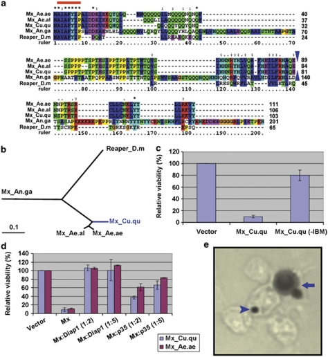
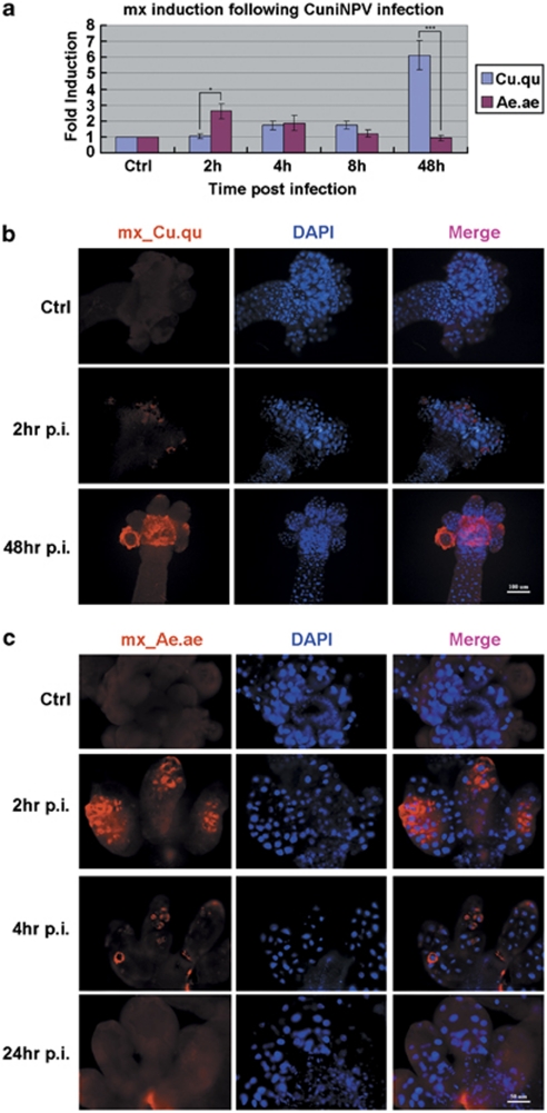
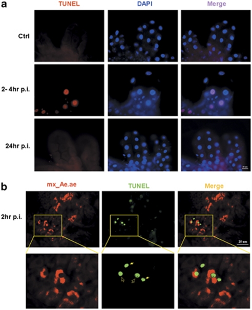
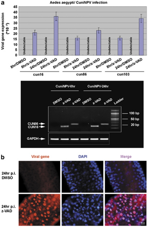
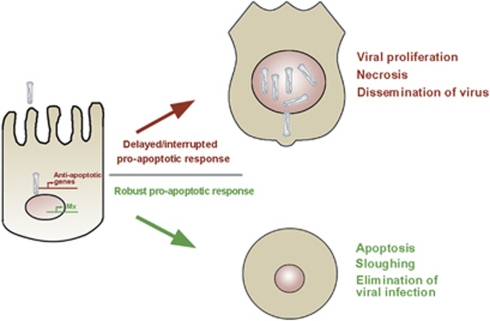
Many vertebrate and insect viruses possess antiapoptotic genes that are required for their infectivity. This led to the hypothesis that apoptosis is an innate immunoresponse important for limiting virus infections. The role of apoptosis may be especially important in insect antiviral defense because of the lack of adaptive immunity. However, the cellular mechanism that elicits apoptosis in response to viral infection in insects has not been determined. Using an in vivo infection system with the mosquito baculovirus CuniNPV (Culex nigripalpus nucleopolyhedrovirus), we demonstrated that michelob_x (mx), the mosquito ortholog of Drosophila proapoptotic gene reaper, is specifically induced in larval midgut cells following viral infection. Interestingly, the dynamics of mx induction corresponds with the outcome of the infection. In the permissive mosquito C. quinquefasciatus, a slow induction of mx failed to induce prompt apoptosis, and the infected cells eventually undergo necrosis with heavy loads of encapsulated viruses. In contrast, in the refractory mosquito Aedes aegypti, a rapid induction of mx within 30 min p.i. is followed by apoptosis within 2-6 h p.i., suggesting a possible role for apoptosis in limiting viral infection. When the execution of apoptosis was delayed by caspase inhibitors, viral gene expression became detectable in the A. aegypti larvae.


Figures

Figure 1
Figure 1
Gene cloning and characterization of mx_Cu.qu. (a) Alignment of protein sequences of Mx orthologs in mosquitoes and Reaper from D. melanogaster. The blue triangles denote the relative position of the intron in Mx sequences (there is no intron in Reaper). The three Mx orthologs from Culicinae share considerable similarity besides the IBM (red line). However, they share little similarity with Mx from Anopheles or the Drosophila Reaper. (b) Distance tree of these orthologs. (c) Expression of Mx_Cu.qu kills C6/36 cells. However, when amino acids 2–4 are removed (−IBM), the protein has little, if any, proapoptotic activity. (d) Mx_Cu.qu-induced cell death is almost completely blocked by co-transfection of Diap1 and significantly inhibited by co-transfection of p35. Numbers in parentheses denote the ratio of respective genes. (e) C6/36 cells killed by expression of mx_Cu.qu or mx_Ae.ae show typical apoptotic morphology including cell fragmentation (arrow) and phagocytosis by neighboring cells (arrow head). Cells co-transfected with pIE-lacZ and pIE-mx_Cu.qu were fixed 20 h later and processed for X-Gal staining
Figure 2
Figure 2
Induction of mx_Cu.qu and mx_Ae.ae following CuniNPV infection. (a) The level of mx_Cu.qu and mx_Ae.ae in pooled larvae (15–20 for each time points) following CuniNPV infection of C. quinquefasciatus and A. aegypti, respectively. Expression level was first normalized with house-keeping genes before calculating the fold induction. Data are presented as mean±S.D. of at least three independent experiments. (*P-value<0.05 and ***P-value<0.001.) (b) Expression of mx_Cu.qu in the midguts of CuniNPV-infected or control (Ctrl) C. quinquefasciatus larvae was monitored with FISH. While there is little induction of mx_Cu.qu at early infection (2 h p.i.), there is very high level of mx_Cu.qu in infected cells at 24 and 48 h p.i. (c) Expression of mx_Ae.ae in the midguts of Ctrl or CuniNPV-infected A .aegypti larvae was monitored with FISH. There is a rapid induction of mx_Ae.ae in cells at the gastric caeca at 2 h p.i. The number of mx_Ae.ae-positive cells returned to the background level by 8 h p.i. For each time point, at least 50 midguts were analyzed in two to three independent experiments, and 80–100% examined guts presented the features as represented in b and c
Figure 3
Figure 3
Necrosis in CuniNPV-infected C. quinquefasciatus larvae. (a) Cells infected with CuniNPV at 48 h p.i. were identified with a pool of fluorescein-labeled cRNA probes against CuniNPV genes, cun24, cun75, and cun85. Virus-infected cells at this time demonstrated hypertrophied nuclei (arrow) compared with uninfected cells (arrow head). Note that there is clear separation between cells that were positive for viral gene expression versus those that were negative (dashed line). More than 100 midguts were examined in several independent experiments; all cells positive for viral gene expression at this time point have hypertrophied nuclei (additional images in Supplementary Figure S2). (b) Cells with hypertrophied nuclei have compromised cell membrane integrity. Live larvae at 48 h p.i. were exposed to 1 μg/ml PI in culture media for 10 min before the midguts were dissected out, fixed with paraformaldehyde, and counterstained using DAPI. All the cells with hypertrophied nuclei (arrow) are also permeable to PI, indicating necrotic cell death. In contrast, cells in the anterior midgut (arrow head) have normal nuclei and intact membrane. More than 40 midguts were observed in three independent experiments, and essentially all of them showed the described association between nuclei morphology and permeability to PI
Figure 4
Figure 4
Rapid apoptosis following the mx_Ae.ae induction in A. aegypti larvae exposed to CuniNPV. (a) TUNEL assay in A. aegypti midgut. At 2–4 h after CuniNPV infection (2–4 h p.i.), significant increase of TUNEL-positive cells can be detected in over 80% of the examined midguts. In contrast, few TUNEL-positive cells were detectable in the control animal (Ctrl) or the midgut of infected animal at late time points (24 h p.i.). (b) Colocalization analysis indicated that TUNEL-positive cells are the ones that express mx_Ae.ae. Early-stage TUNEL-positive cells (open arrow), indicated by relatively normal nuclei morphology and intact cytoplasm, have high levels of FISH signal for mx_Ae.ae. The mx_Ae.ae signal is not visible in later-stage TUNEL-positive cells with significantly condensed nuclei (solid arrow), which is likely because of the shrinkage of cytoplasm and/or degradation of macromolecules at this stage. Some mx-expressing cells are not TUNEL positive, which is likely because of the lag between mx expression and the activation of caspase-dependent DNase
Figure 5
Figure 5
Suppressing apoptosis in A. aegypti leads to expressions of viral genes. (a) Suppressing apoptosis allowed the expression of ie viral genes. None of the three ie CuniNPV genes, cun16, cun86, and cun103, could be detectable by Q-PCR in A. aegypti larvae exposed to CuniNPV and treated with DMSO only. In contrast, all the three genes could be detected in A. aegypti larvae treated with capsase inhibitors z-VAD-FMK (z-VAD). Bottom panels are gel pictures of the Q-PCR products. (b) FISH was performed with a pool of cRNA probes against CuniNPV genes, cun16, cun86, and cun103. Top panel, no viral gene could be detected in the midgut of any of the DMSO-treated larvae (over 100 examined) at 24 h after CuniNPV infection. In contrast, viral gene expression was detectable in ∼20% of the midguts dissected from caspase inhibitor-treated larvae
Figure 6
Figure 6
The race to apoptosis through the mosquito reaper. This is a schematic presentation of our findings. On CuniNPV infection, there is a competing ‘race' between the host cell to express cellular proapoptotic gene(s) to eliminate the infected cell and the virus to express early genes to block apoptosis and initiate proliferation. A prompt induction of mx and apoptosis leads to elimination of the infected cell before the viral genes are detectable in the refractory mosquito A. aegypti. A delay in this process, either due to insufficient proapoptotic response or interference of caspase inhibitors, could subjugate the cell under viral control and render the organism susceptible to the virus

Similar articles

Cited by

References

    1. Benedict CA, Norris PS, Ware CF. To kill or be killed: viral evasion of apoptosis. Nat Immunol. 2002;3:1013–1018. - PubMed
    1. Galluzzi L, Brenner C, Morselli E, Touat Z, Kroemer G. Viral control of mitochondrial apoptosis. PLoS Pathog. 2008;4:e1000018. - PMC - PubMed
    1. Thomson BJ. Viruses and apoptosis. Int J Exp Pathol. 2001;82:65–76. - PMC - PubMed
    1. Clem RJ, Miller LK. Control of programmed cell death by the baculovirus genes p35 and iap. Mol Cell Biol. 1994;14:5212–5222. - PMC - PubMed
    1. Crook NE, Clem RJ, Miller LK. An apoptosis-inhibiting baculovirus gene with a zinc finger-like motif. J Virol. 1993;67:2168–2174. - PMC - PubMed

Publication types

MeSH terms