. 2011 Apr;31(8):1624-36.
doi: 10.1128/MCB.00470-10. Epub 2011 Feb 7.

Identification of PITX1 as a TERT suppressor gene located on human chromosome 5

Affiliations

Identification of PITX1 as a TERT suppressor gene located on human chromosome 5

Dong-Lai Qi et al. Mol Cell Biol. 2011 Apr.

Abstract

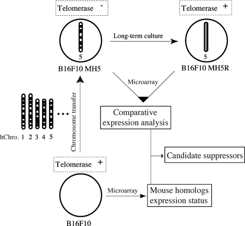
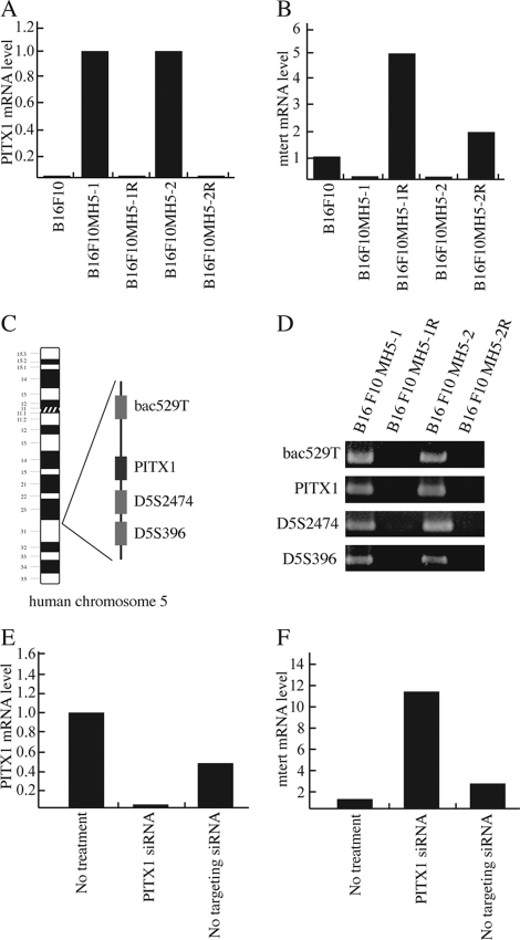
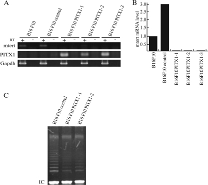
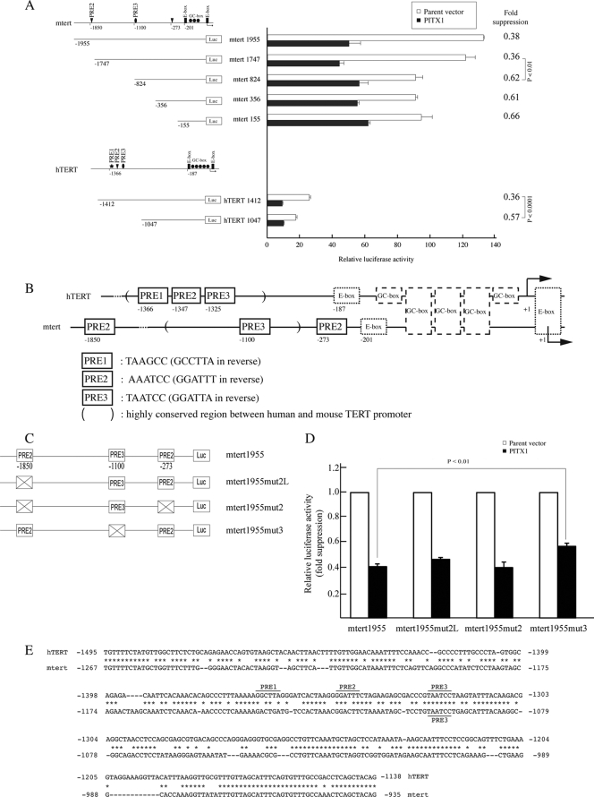
Telomerase, a ribonucleoprotein enzyme that maintains telomere length, is crucial for cellular immortalization and cancer progression. Telomerase activity is attributed primarily to the expression of telomerase reverse transcriptase (TERT). Using microcell-mediated chromosome transfer (MMCT) into the mouse melanoma cell line B16F10, we previously found that human chromosome 5 carries a gene, or genes, that can negatively regulate TERT expression (H. Kugoh, K. Shigenami, K. Funaki, J. Barrett, and M. Oshimura, Genes Chromosome Cancer 36:37-47, 2003). To identify the gene responsible for the regulation of TERT transcription, we performed cDNA microarray analysis using parental B16F10 cells, telomerase-negative B16F10 microcell hybrids with a human chromosome 5 (B16F10MH5), and its revertant clones (MH5R) with reactivated telomerase. Here, we report the identification of PITX1, whose expression leads to the downregulation of mouse tert (mtert) transcription, as a TERT suppressor gene. Additionally, both human TERT (hTERT) and mouse TERT (mtert) promoter activity can be suppressed by PITX1. We show that three and one binding site within the hTERT and mtert promoters, respectively, that express a unique conserved region are responsible for the transcriptional activation of TERT. Furthermore, we showed that PITX1 binds to the TERT promoter both in vitro and in vivo. Thus, PITX1 suppresses TERT transcription through direct binding to the TERT promoter, which ultimately regulates telomerase activity.


Figures

Fig. 1.
Fig. 1.
Outline of the strategy used to identify negative regulators of telomerase. Normal human chromosomes were previously individually transferred into B16F10 cells. Only the introduction of human chromosome 5 (MH5) inhibited telomerase activity, which subsequently was reactivated during long-term culture (MH5R). Genes that were differentially expressed from human chromosome 5 between MH5 and MH5R were identified by cDNA microarray. Mouse homologs of these genes, which exhibited reduced expression in B16F10 cells, ultimately were chosen as candidate genes for further analysis.
Fig. 2.
Fig. 2.
Downregulation of mtert expression in MH5 clones is associated with PITX1 expression. (A and B) Analysis of the mRNA expression of PITX1 and mtert in microcell hybrids with human chromosome 5 (MH5) was performed using qRT-PCR. GAPDH was used as an internal control for each sample. MH5-1 and -2 showed strong repression of mtert expression, which was associated with the expression of PITX1. In contrast, revertant clones (MH5-1R and -2R) with telomerase activity showed reduced PITX1 transcription. (C) Schematic diagram showing the location of the genomic loci of PITX1 and flanking STS markers on human chromosome 5. (D) The chromosomal regions neighboring PITX1 were not detected by genomic PCR in MH5R clones. (E and F) Depletion of PITX1 by siRNA induces reactivation of mtert transcription in both MH5-1 and -2 clones in B16F10.
Fig. 3.
Fig. 3.
Expression of PITX1 inhibited mtert mRNA expression and telomerase activity in B16F10 cells. (A and B) RNA was prepared from B16F10 cells transfected with a PITX1 expression vector or with control vector (pcDNA3.1) and was analyzed for mtert and PITX1 mRNA expression using RT-PCR and qRT-PCR. (C) PITX1 transfection resulted in depleted telomerase activity as assessed using the TeloChaser kit. IC is the internal control provided in the kit.
Fig. 4.
Fig. 4.
Luciferase assay of various truncated hTERT and mtert promoters in PITX1- and control vector-transfected B16F10 cells. (A) The schematic drawing (left) shows the position of the truncation site of each of the reporter plasmids. The firefly luciferase activity was standardized using Renilla reniformis luciferase activity from cotransfected pGL4.70. The promoter region that contains PREs significantly inhibits, and is therefore crucial for, TERT transcriptional activity in both humans and mice. Error bars represent standard deviations from three experiments. Statistical analysis (t test) using SigmaPlot 2000 and indicating a significant difference is shown (P values). (B) Comparison of transcription factor binding sequences in the hTERT and mtert promoters. The proximal promoter region of hTERT is partially similar to that of mtert, containing both E and GC boxes. The transcription start site is indicated by an arrow. Candidate PITX1 binding sequences are indicated as PITX1 response elements (PRE1, PRE2, and PRE3). (C) Schematic diagram showing the luciferase reporter plasmids that encode wild-type and PRE3-mutated versions of a fragment of the mtert promoter region. The crossed box represents mutation. (D) PITX1 expression vectors were cotransfected with the mtert reporter plasmids shown in panel C and mtert 824, which lacks PRE3, and promoter activity was assayed using a luciferase assay. The firefly luciferase activity was standardized using Renilla reniformis luciferase activity from cotransfected pGL4.70. The transfection of control vector was used as a suppression control to normalize the effect of PITX1 transfection. The transcriptional activity of mtert was suppressed by the wild-type reporter compared to that of the mutated reporters. Error bars represent the standard deviations from three experiments. Statistical analysis (t test) using SigmaPlot 2000 and indicating a significant difference is shown (P values). (E) The sequences within the TERT promoter conserved between human and mouse. Three and one PRE sites of the hTERT and mTERT promoters lie within this conserved region.
Fig. 5.
Fig. 5.
Suppression of the transcriptional activity of the hTERT promoter by PITX1. (A) PITX1 expression vectors were cotransfected into the human melanoma cell line A2058 with reporter plasmids containing a wild-type (hTERT1412) or a truncated (hTERT1047) hTERT promoter region in which the PRE site in the hTERT promoter region is eliminated. The resulting firefly luciferase activity was standardized using the Renilla reniformis luciferase activity from cotransfected pGL4.70. The transcriptional activity in hTERT1412 indicated suppression effects that were approximately 2-fold greater than those of hTERT1047. Error bars represent the standard deviations from three experiments. Statistical analysis (t test) using SigmaPlot 2000 and indicating a significant difference is shown (P values). (B) Schematic diagram showing the luciferase reporter plasmids that carry wild and mutated versions of the mtert promoter region. The crossed box represents mutation. (C) PITX1 expression vectors were cotransfected with reporter plasmids containing the wild-type or PRE-mutated versions of the hTERT promoter. The transcriptional activity of hTERT was suppressed by the wild-type reporter compared to that of the mutated versions. Error bars represent the standard deviations from three experiments. Statistical analysis (t test) using SigmaPlot 2000 and indicating a significant difference is shown (P values).
Fig. 6.
Fig. 6.
PITX1 directly binds to the hTERT promoter in vitro. (A) Western blotting to confirm the overexpression of PITX1 protein in B16F10-transfected cells. (B and C) EMSA was performed using the human PITX1 protein and a radiolabeled oligonucleotide probe designed to detect the binding of PITX1 to PRE1 (B) or PRE2 (C). The open circles indicate background signals. P indicates probe binding to PITX1. F, free probe; wt, wild type; mut, mutation; −, no competitor. (D) A supershift assay using the PITX1 antibody or control IgG. Triangles indicate increasing amounts of antibody.
Fig. 7.
Fig. 7.
PITX1 directly binds to both the hTERT and the mtert promoter in vivo. A ChIP assay was carried out using the anti-PITX1 antibody to verify the binding of PITX1 to the hTERT and mtert promoters in human A2058, mouse B16F10 cells, and the B16F10MH5 clone. PITX1 expressed in human- and mouse-transfected cells and B16F10MH5 clone was cross-linked to DNA using an anti-PITX1 antibody. Rabbit immunoglobulin (IgG) was used as a negative control. Input represents PCR of the hTERT promoter DNA before immunoprecipitation.
Fig. 8.
Fig. 8.
Detection of PITX1 protein by immunohistochemistry in gastric mucosa and adenocarcinoma. (A) PITX1-positive cells exist in surface mucous cells and fundic glands. (B) In contrast, PITX1-positive cells were not observed in carcinoma lesions.

Similar articles

Cited by

References

    1. Artandi S., et al. 2002. Constitutive telomerase expression promotes mammary carcinomas in aging mice. Proc. Natl. Acad. Sci. U. S. A. 99:8191–8196 - PMC - PubMed
    1. Blasco M. 2005. Telomeres and human disease: ageing, cancer and beyond. Nat. Rev. Genet. 6:611–622 - PubMed
    1. Cavalli L. R., Riggins R. B., Wang A., Clarke R., Haddad B. R. 2010. Frequent loss of heterozygosity at the interferon regulatory factor-1 gene locus in breast cancer. Breast Cancer Res. Treat. 121:227–231 - PMC - PubMed
    1. Chen Y., et al. 2007. Decreased PITX1 homeobox gene expression in human lung cancer. Lung Cancer 55:287–294 - PubMed
    1. Chen Y. N., Chen H., Xu Y., Zhang X., Luo Y. 2008. Expression of pituitary homeobox 1 gene in human gastric carcinogenesis and its clinicopathological significance. World J. Gastroenterol. 14:292–297 - PMC - PubMed

Publication types