. 2011 Apr 1;286(13):11756-64.
doi: 10.1074/jbc.M110.192773. Epub 2011 Feb 1.

Mapping the interactions between Escherichia coli TolQ transmembrane segments

Affiliations

Mapping the interactions between Escherichia coli TolQ transmembrane segments

Xiang Y-Z Zhang et al. J Biol Chem. .

Abstract

The tolQRAB-pal operon is conserved in Gram-negative genomes. The TolQRA proteins of Escherichia coli form an inner membrane complex in which TolQR uses the proton-motive force to regulate TolA conformation and the in vivo interaction of TolA C-terminal region with the outer membrane Pal lipoprotein. The stoichiometry of the TolQ, TolR, and TolA has been estimated and suggests that 4-6 TolQ molecules are associated in the complex, thus involving interactions between the transmembrane helices (TMHs) of TolQ, TolR, and TolA. It has been proposed that an ion channel forms at the interface between two TolQ and one TolR TMHs involving the TolR-Asp(23), TolQ-Thr(145), and TolQ-Thr(178) residues. To define the organization of the three TMHs of TolQ, we constructed epitope-tagged versions of TolQ. Immunodetection of in vivo and in vitro chemically cross-linked TolQ proteins showed that TolQ exists as multimers in the complex. To understand how TolQ multimerizes, we initiated a cysteine-scanning study. Results of single and tandem cysteine substitution suggest a dynamic model of helix interactions in which the hairpin formed by the two last TMHs of TolQ change conformation, whereas the first TMH of TolQ forms intramolecular interactions.


Figures

FIGURE 1.
FIGURE 1.
TolQ forms multimers. A, in vivo formaldehyde (FA) cross-linking of TPS13 (tolQR) cells producing the indicated Tol proteins (TolQR, TolQ, and TolR; TolQHAR, TolQHA, and TolR; TolQHA, TolQHA, and no TolR). 0.4 × 10⁸ cells treated (+) or not (−) by the chemical cross-linked formaldehyde were loaded on 12.5% acrylamide SDS-PAGE gel, and TolQHA and TolQHA-containing complexes were immunodetected using anti-HA mAb. The ∼43-, ∼52-kDa, a and b complexes are indicated. B, ∼0.5 μg of the purified TolQ8H protein was treated (+) or not (−) with formaldehyde, loaded on a 12.5% acrylamide SDS-PAGE gel, and immunodetected using anti-His5 mAb. The monomeric (Q) and dimeric (Q2) forms are indicated. C, TPS13 (tolQR) cells producing TolQHA (TPS13 pOK-QHA, lane 1), TolQ8H (TPS13 pQ8R, lane 2), or both proteins simultaneously (TPS13 pQ8R pOK-QHA, lane 3) treated with formaldehyde were solubilized. The TolQHA proteins were immunoprecipitated from detergent-solubilized membrane extracts using the anti-HA mAb, and the TolQHA and TolQ8H proteins were immunodetected using anti-HA (upper panel) and anti-His (lower panel) mAbs. Prestained molecular mass markers (in kDa) are indicated.
FIGURE 2.
FIGURE 2.
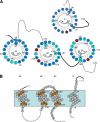
Summary of the TolQ cysteine substitution phenotypes. A, TolQ TMHs residues shown on a helical wheel projection from the periplasmic to the cytoplasmic side. The color code summarizes the result of the phenotypic analyses of the cysteine substitutions (Table 1): WT phenotype (blue, colicin-sensitive and DOC-resistant), tol phenotype (red, colicin-resistant and DOC-sensitive), and discriminative phenotype (cyan, colicin- and DOC-sensitive). The residues used in the tandem cysteine scanning and Ile154 are numbered. Residues numbered on the outside of the wheel locate at the periplasmic side of the TMH, whereas residues numbered on the inside locate at the cytoplasmic side. Periplasmic and cytoplasmic segments are indicated by unbroken and dashed lines, respectively. The phenotypic analysis of TolR TMH cysteine substitutions (30) is also reported using the same color code. B, schematic lateral view of TolQ and TolR TMHs in which the residues used for the tandem cysteine mutagenesis are indicated by orange balls. The residues locating at the boundaries of each TMH are numbered.
FIGURE 3.
FIGURE 3.
TolQ dimerizes through TMH2-TMH2 contacts. A, 0.4 × 10⁸ Tuner (DE3) pET-Q cells producing the indicated TolQ8H variant were incubated (lower panel) or not (upper panel) with 0.3 mm CuoP before treatment with the thiol-blocking agent NEM. Heat-denatured samples were loaded on a non-reducing 12% acrylamide SDS-PAGE gel and analyzed by Western blot immunodetections using anti-His5 mAb. Prestained molecular mass markers (in kDa) and TolQ monomers (Q) and dimers (Q2) are indicated. B, about 3 μg of n-dodecyl-β-d-maltoside purified TolQ8H157C treated (+) or not (−) with the reducing agent β-mercaptoethanol (red) was loaded on a 12.5% acrylamide SDS-PAGE gel and analyzed by Coomassie Blue staining.
FIGURE 4.
FIGURE 4.
Tandem cysteine scanning of TolQ TMHs. A, summary of the residues present at the cytoplasmic (cyto) and periplasmic (peri) extremities of the TolQ TMHs targeted for the tandem cysteine scanning. All tandem combinations at the cytoplasmic or periplasmic side were tested for their phenotypes (Table 2) and their ability to form disulfide bridges (see supplemental Fig. S4). B, representative profiles obtained for TolQ double cysteine mutants. Membrane extracts from 0.4 × 10⁸ TPS13 (tolQR) cells producing TolR and the WT TolQHA protein (class i, lane 1) or the TolQHA-G157C (class ii, lane 2), -G157C/P169C (class iii, lane 3), or -Q37C/I186C (class iv, lane 4) cysteine mutants were loaded on a 12% SDS-PAGE gel and analyzed by Western blot immunodetections using anti-HA mAb (upper panel, non-reducing conditions (ox); lower panel, β-mercaptoethanol-treated samples (red)). The TolQ monomer and multimers are indicated by arrows on the left. The asterisk indicates a TolQ mobility shift associated with an intramolecular disulfide bond. C, 0.4 × 10⁸ Tuner (DE3) cells producing the TolQ8H-G157C/P169C double mutant, treated (+) or not (−) with the oxidative agent CuoP, were heat-denatured in Laemmli buffer and analyzed by non-reducing 12% acrylamide SDS-PAGE gel and Western blot immunodetection using anti-His5 mAb. Prestained molecular mass markers (in kDa) are indicated on the left.
FIGURE 5.
FIGURE 5.
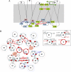
Schematic model of the TolQ and TolR TMH interactions. A, putative model of TolQ TMH arrangement emphasizing side-specific contacts. Two adjacent TolQ molecules are depicted. TMH1, -2, and -3 are indicated. Residues of TMH1 (light pink), TMH2 (light green), and TMH3 (light blue) involved in intermolecular (symbolized by red sticks) and intramolecular (symbolized by the blue sticks) interactions are indicated. Putative TMH rearrangements are indicated by green arrows. B, putative model of TolQ-TolR TMHs organization. TolQ-TolR residues in contact are shown in a model containing six TolQ molecules, two TolR molecules, and two putative ionic channels. TolQ TMH organization defined by genetic suppressive data was kept identical in each TolQ molecule. Balls represent cysteine-substituted positions involved in disulfide bond formation (large balls, residues at the periplasmic side of TMHs; small balls, residues at the cytoplasmic side of TMHs). Disulfide interactions are symbolized by red (intermolecular) or blue (intramolecular) sticks, whereas suppressor pairs are indicated by black sticks (unbroken, periplasmic side; dotted, cytoplasmic side). Numbers above the sticks indicate the following TMH intermolecular interactions: i) suppressors, (1) TolR-D23A/TolQ-A185D; (2) TolR-M33Q/TolQ-T145A; (3) TolQ-H151E/TolQ-E173Q; (4) TolQ-G144A/TolQ-A184G; (5) TolQ-A177V/TolQ-L19P; ii) cysteine substitutions, (6) TolQ-Gly157/TolQ-Pro169; (7) TolQ-Gly157/TolQ-Gly170; (8) TolQ-Ala155/TolQ-Pro169; (9) TolQ-Tyr139/TolQ-Ala185; (10) TolQ-Tyr139/TolQ-Ile186; (11) TolQ-Ile140/TolQ-Ala185; (12) TolR-Leu22/TolR-Leu22; (arrow) TolR-Val24/TolR-Val24; (157) TolQ-Gly157/TolQ-Gly157; (154) TolQ-Gly154/TolQ-Gly154. The intramolecular TMH1-TMH2 and TMH1-TMH3 interactions indicated by blue sticks are shown without numbering (TolQ-Ile36/TolQ-Pro138/Tyr139/Ile140 and TolQ-Gln37/TolQ-Ale185/Ile186). The different TMHs of the TolQ and TolR proteins are shown in the inset with residues shown to be involved in dimer formation in this work (represented by gray balls). Polar (red balls) and small lateral chain residues (white balls) characterized in previous studies (18, 24) are also indicated.

Similar articles

Cited by

References

    1. Lloubès R., Cascales E., Walburger A., Bouveret E., Lazdunski C., Bernadac A., Journet L. (2001) Res. Microbiol. 152, 523–529 - PubMed
    1. Lazzaroni J. C., Germon P., Ray M. C., Vianney A. (1999) FEMS Microbiol. Lett 177, 191–197 - PubMed
    1. Gerding M. A., Ogata Y., Pecora N. D., Niki H., de Boer P. A. (2007) Mol. Microbiol. 63, 1008–1025 - PMC - PubMed
    1. Anwari K., Poggio S., Perry A., Gatsos X., Ramarathinam S. H., Williamson N. A., Noinaj N., Buchanan S., Gabriel K., Purcell A. W., Jacobs-Wagner C., Lithgow T. (2010) PLoS ONE 5, e8619. - PMC - PubMed
    1. Yeh Y. C., Comolli L. R., Downing K. H., Shapiro L., McAdams H. H. (2010) J. Bacteriol. 192, 4847–4858 - PMC - PubMed

Publication types

MeSH terms

LinkOut - more resources