. 2011 Feb 7;192(3):401-15.
doi: 10.1083/jcb.201006011. Epub 2011 Jan 31.

Replication protein A safeguards genome integrity by controlling NER incision events

Affiliations

Replication protein A safeguards genome integrity by controlling NER incision events

René M Overmeer et al. J Cell Biol. .

Abstract

Single-stranded DNA gaps that might arise by futile repair processes can lead to mutagenic events and challenge genome integrity. Nucleotide excision repair (NER) is an evolutionarily conserved repair mechanism, essential for removal of helix-distorting DNA lesions. In the currently prevailing model, NER operates through coordinated assembly of repair factors into pre- and post-incision complexes; however, its regulation in vivo is poorly understood. Notably, the transition from dual incision to repair synthesis should be rigidly synchronized as it might lead to accumulation of unprocessed repair intermediates. We monitored NER regulatory events in vivo using sequential UV irradiations. Under conditions that allow incision yet prevent completion of repair synthesis or ligation, preincision factors can reassociate with new damage sites. In contrast, replication protein A remains at the incomplete NER sites and regulates a feedback loop from completion of DNA repair synthesis to subsequent damage recognition, independently of ATR signaling. Our data reveal an important function for replication protein A in averting further generation of DNA strand breaks that could lead to mutagenic and recombinogenic events.


Figures

Figure 1.
Figure 1.
Impairment of DNA repair synthesis and ligation inhibits repair independently of ATR. (A) Removal of 6-4PP in time was measured in the presence (■) and absence (▲) of inhibitors after global UV irradiation (30 J/m2) in NHF (blue lines) and ATR-deficient cells (orange lines); NHF cells were treated with L67 for 4 h before irradiation (•). Cells were stained with a 6-4PP–specific antibody and lesion removal was measured by immunofluorescent staining and quantification of at least three independent experiments with over 40 cells per point. (B) Seckle syndrome cells (ATR) show impaired γ-H2AX staining at sites of UV damage compared with NHF. Cells were irradiated with 30 J/m2 1 h before fixation and were processed for immunofluorescent staining with antibodies against XPB and γ-H2AX. (C) NHFs, treated and nontreated with L67 for 4 h before local UV irradiation (30 J/m2), were stained for XPB and PCNA localization.
Figure 2.
Figure 2.
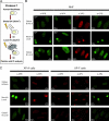
Prolonged accumulation of pre- and post-incision factors at NER sites in the presence of replication inhibitors. (A) Fluorescent immunostaining of XPB, RPA, Polδ, and PCNA localization at damage sites in confluent NHF after local UV irradiation (30 J/m2) at time points as indicated. “Merge” refers to the combined image of DAPI and PCNA staining. (B) Immunolocalization of XPB, RPA, Polδ, and PCNA in NHF in the presence of HU and AraC at different repair times after 30 J/m2 local UV irradiation. (C) Immunolocalization of XPB in confluent XP-A cells at different repair times after 30 J/m2 local UV irradiation. (D) XPC and 6-4PP were visualized by immunostaining after local UV irradiation; cells were irradiated with 30 J/m2 UV and incubated for either 30 min or 20 h.
Figure 3.
Figure 3.
Preincision factors remain associated to initial repair sites in the absence of incision. (A) Schematic representation of Protocol 1. Cells were globally UV irradiated (30 J/m2) in the absence of HU and AraC, recovered for 1 h, and were subsequently irradiated with the same dose of local UV. (B) Release of XPB, XPC, and XPA from repair complexes is dependent on functional incision. Confluent NHF were treated according to Protocol 1 (A) and immunostained with the indicated antibodies. Exposure times within experiments are equal. (C) Immunolocalization of XPB, XPC, and XPA antibodies in XP-A and XP-F cells treated according to Protocol 1 (A).
Figure 4.
Figure 4.
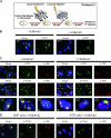
Impairment of DNA synthesis prevents recruitment of RPA and post-incision factors to de novo UV damage. For clarity, representative cells (numbered boxes) are enlarged and depicted below. (A) Schematic representation of competition experiments according to Protocol 3. Cells were treated or mock-treated with DNA synthesis inhibitors 30 min before the first local (8 µm) UV irradiation, which was followed (after 30 min) by the second local (3 µm) irradiation. Cells that have been hit twice by UV irradiation contain both large and small spots. (B) Competition experiments (Protocol 3) confirm dynamic association of XPA with damage sites in the presence and absence of DNA synthesis inhibitors in confluent NHFs. (C) Confluent NHFs treated as in B and co-stained with RPA, PCNA, and XPB antibodies. (D) Confluent NHFs and Seckle syndrome cells (ATR) were treated according to Protocol 3 in the presence of HU and AraC, followed by immunofluorescent staining of PCNA and XPB. For clarity, small spots are indicated with arrows.
Figure 5.
Figure 5.
Inhibition of DNA repair synthesis or ligation prevents novel incision events and leads to prolonged accumulation of post-incision factors. (A) Quiescent NHFs were irradiated according to Protocol 3. Incision events are visualized by γ-H2AX staining; counterstaining for XPB revealed areas of damage induction. Γ-H2AX accumulation increases in time, thus the intensity is lower at the second UV spots when compared with the initially induced damage. Also, incubation with inhibitors increases γ-H2AX accumulation, therefore microscopic exposure time for γH2AX is threefold shorter when cells are irradiated in the presence of inhibitors. (B) NHFs were irradiated according to Protocol 3 in the presence of L67 inhibitor and stained for PCNA, XPB, and γ-H2AX. For clarity, small spots are indicated with arrows. (C) NHFs were locally irradiated in the presence or absence of inhibitors, fixed 1 h later, and stained for XPA or XPB in combination with PCNA, Polδ, or γH2AX. Average intensity inside local spots was measured and normalized to normal conditions (no inhibitors). Error bars represent the SEM values of >40 nuclei per experiment; out of at least three independent experiments.
Figure 6.
Figure 6.
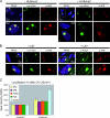
Single 5′ incision leads to γ-H2AX phosphorylation independent of DNA synthesis. (A) Catalytically dead XP-G and XP-F cells were stained for γ-H2AX 1 h after UV or mock irradiation with 30 J/m2. Average intensity per nucleus was quantified and normalized to mock treatment. (B) Catalytically dead XP-G and XP-F were locally irradiated with 30 J/m2 in the presence or absence of inhibitors. 1 h later, cells were fixed and stained for XPB and γ-H2AX.
Figure 7.
Figure 7.
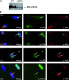
RPA is prerequisite for the functional assembly of NER subcomplexes. (A) Western blot analysis of equal amounts of whole cell lysates prepared from cells treated or nontreated (NT) with siRNA against RPA p70. (B) Cells treated with siRNA against RPA p70 were locally UV irradiated (30 J/m2), fixed 1 h later, and stained with XPA, XPB, Polδ, and γ-H2AX antibodies. Absence of RPA was verified with RPA costaining (arrows).
Figure 8.
Figure 8.
DNA synthesis inhibitors sequester RPA to sites of incomplete NER repair synthesis. Confluent NHFs were (mock) irradiated with 20 J/m2 in the presence or absence of inhibitors, let to recover, and cross-linked 40 min later. ChIP was performed with XPB (A) and XRCC1 (B) specific antibodies and Western blot analysis of the coprecipitating proteins was performed with antibodies as indicated.
Figure 9.
Figure 9.
Schematic depiction of regulation of NER-mediated incision events in vivo. Upon lesion recognition by UV-DDB and XPC-hHR23B, local opening of DNA by TFIIH provides access to the core GG-NER machinery, i.e., XPA, RPA, XPG, and XPF/ERCC1. RPA binds to the undamaged single-stranded DNA stabilizing the complex. Subsequently, incision is followed by the release of core NER factors, which are then free to associate with other damages with the exception of RPA, which remains bound to the repair site, probably on the undamaged single-stranded DNA. The later stages of repair are performed by RFC stable loading PCNA onto the incised DNA, the recruitment of DNA polymerases polδ/polε/polκ and XRCC1-Lig3/Lig1 to fill in and ligate the gap, respectively. After ligation, post-incision factors and RPA are able to dissociate. RPA can then stabilize the otherwise abortive preincision complexes, enabling the initiation of new NER events.

Similar articles

Cited by

References

    1. Aboussekhra A., Wood R.D. 1995. Detection of nucleotide excision repair incisions in human fibroblasts by immunostaining for PCNA. Exp. Cell Res. 221:326–332 10.1006/excr.1995.1382 - DOI - PubMed
    1. Aboussekhra A., Biggerstaff M., Shivji M.K.K., Vilpo J.A., Moncollin V., Podust V.N., Protić M., Hübscher U., Egly J.M., Wood R.D. 1995. Mammalian DNA nucleotide excision repair reconstituted with purified protein components. Cell. 80:859–868 10.1016/0092-8674(95)90289-9 - DOI - PubMed
    1. Araújo S.J., Tirode F., Coin F., Pospiech H., Syväoja J.E., Stucki M., Hübscher U., Egly J.M., Wood R.D. 2000. Nucleotide excision repair of DNA with recombinant human proteins: definition of the minimal set of factors, active forms of TFIIH, and modulation by CAK. Genes Dev. 14:349–359 - PMC - PubMed
    1. Auclair Y., Rouget R., Affar B., Drobetsky E.A. 2008. ATR kinase is required for global genomic nucleotide excision repair exclusively during S phase in human cells. Proc. Natl. Acad. Sci. USA. 105:17896–17901 10.1073/pnas.0801585105 - DOI - PMC - PubMed
    1. Barr S.M., Leung C.G., Chang E.E., Cimprich K.A. 2003. ATR kinase activity regulates the intranuclear translocation of ATR and RPA following ionizing radiation. Curr. Biol. 13:1047–1051 10.1016/S0960-9822(03)00376-2 - DOI - PubMed

Publication types