. 2011 Apr;85(7):3570-83.
doi: 10.1128/JVI.01295-10. Epub 2011 Jan 26.

LEDGF dominant interference proteins demonstrate prenuclear exposure of HIV-1 integrase and synergize with LEDGF depletion to destroy viral infectivity

Affiliations

LEDGF dominant interference proteins demonstrate prenuclear exposure of HIV-1 integrase and synergize with LEDGF depletion to destroy viral infectivity

Anne M Meehan et al. J Virol. 2011 Apr.

Abstract

Target cell overexpression of the integrase binding domain (IBD) of LEDGF/p75 (LEDGF) inhibits HIV-1 replication. The mechanism and protein structure requirements for this dominant interference are unclear. More generally, how and when HIV-1 uncoating occurs postentry is poorly defined, and it is unknown whether integrase within the evolving viral core becomes accessible to cellular proteins prior to nuclear entry. We used LEDGF dominant interference to address the latter question while characterizing determinants of IBD antiviral activity. Fusions of green fluorescent protein (GFP) with multiple C-terminal segments of LEDGF inhibited HIV-1 replication substantially, but minimal chimeras of either polarity (GFP-IBD or IBD-GFP) were most effective. Combining GFP-IBD expression with LEDGF depletion was profoundly antiviral. CD4(+) T cell lines were rendered virtually uninfectable, with single-cycle HIV-1 infectivity reduced 4 logs and high-input (multiplicity of infection = 5.0) replication completely blocked. We restricted GFP-IBD to specific intracellular locations and found that antiviral activity was preserved when the protein was confined to the cytoplasm or directed to the nuclear envelope. The life cycle block triggered by the cytoplasm-restricted protein manifested after nuclear entry, at the level of integration. We conclude that integrase within the viral core becomes accessible to host cell protein interaction in the cytoplasm. LEDGF dominant interference and depletion impair HIV-1 integration at distinct postentry stages. GFP-IBD may trigger premature or improper integrase oligomerization.


Figures

FIG. 1.
FIG. 1.
GFP-LEDGF fusion proteins. The domains of LEDGF are indicated. The PWWP domain and the AT hooks are dominant chromatin binding elements (7, 30, 32, 50). The C-terminal domain (amino acids 325 to 530) contains the IBD (amino acids 347 to 429). In the present study “LEDGF” refers to the 75-kDa splice variant (i.e., LEDGF/p75) of the gene PSIP1. The gene also encodes an IBD-lacking splice variant LEDGF/p52, which has a different 8-amino-acid C-terminal domain and does not interact with integrase. CR, charged region; IBD, integrase binding domain; NDE, N-terminal domain ensemble involved in chromatin binding; NLS, nuclear localization signal.
FIG. 2.
FIG. 2.
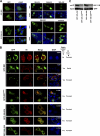
Subcellular localization of IBD proteins, with or without IN. (A) Cells were fixed at 48 h after transfection, stained with DAPI, and imaged with a confocal microscope. These transfections were done in LEDGF-deficient 293T cells (L1340 cells, Table 2), but similar results were seen in 293T, HeLa, and Cos7 cells (data not shown). Dashed line circles indicate cells in mitosis. Fusions terminating at LEDGF residue 432 are mainly nuclear, while those terminating at amino acid 530 have greater cytoplasmic localization. (B) GFP-LEDGF fusion proteins were coexpressed with HIV-1 IN in the presence or absence of endogenous LEDGF. Cells used are noted at right (white font). The morphologically distinctive inclusions seen in COS7 cells were also seen in HeLa cells (data not shown). As described in Table 2, L1340 (or “L”) cells have a stable LEDGF knockdown (31). LH4 cells are L1340 cells that contain a stably integrated HIV-1 IN expression plasmid (28, 40). IN is unstable in the absence of an IBD protein, so that very low levels are present at steady state in LH4 cells, too low to be detected at this confocal gain (28). Therefore, IN seen in these LH4 cells images is that which is stabilized by the GFP-IBD interaction (40). Wild-type LEDGF re-expression in LH4 cells (not shown here) results in nuclear (chromatin-bound) IN-LEDGF complexes (40). (C) Coimmunoprecipitation of HIV-1 IN with GFP-LEDGF fusion proteins. Immunoblotting for Myc epitope-tagged HIV-1 IN was performed after immunoprecipitation of indicated GFP-LEDGF fusion proteins from LH4 cells.
FIG. 3.
FIG. 3.
Fusion proteins inhibit lentiviral infection. (A) Western blotting of MT4 cell lysates with antibody to GFP. Tubulin was immunoblotted as a loading control. (B) HIV-1luc challenge of stable MT4 cells. WT/D366N pairs are indicated. Values were normalized to cell number or protein. Error bars are between duplicates in each experiment. All challenge experiments were performed at least twice with similar results. In panels B to D, cell lysates were collected 5 to 7 days after infection. (C) FIV-luc infection. (D) MLV-luc infection. (E) Alu-PCR assessment of integration. Values are normalized to GAPDH standard. (F) 2-LTR circles, analyzed at 22 h posttransduction with HIV-1luc. Values are normalized to MtDNA as a loading control.
FIG. 4.
FIG. 4.
GFP-IBD inhibits HIV-1 infection in multiple T cell lines and primary cells. (A) SupT1 and MT4 cell lines that stably express GFP-IBD or IBD-GFP were challenged with HIV-1luc, and luciferase was analyzed at 5 days. The macrophages were transduced as described in Materials and Methods and challenged with HIV-1luc, and expression was analyzed at 7 days. (B) Alu-PCR-based assay of integration in SupT1 GFP-IBD cell lines challenged with HIV-1luc.
FIG. 5.
FIG. 5.
Combined dominant interference and knockdown. (A and B) Subcellular fractions of SupT1 (A) and MT4 cells (B) stably transduced with lentiviral vectors expressing LEDGF-targeted shRNA (p75shRNA) with or without GFP-IBD were analyzed for residual LEDGF expression using anti-LEDGF antibody. S1, Triton X-100-extractable, non-chromatin-bound fraction. S2, Triton X-100-resistant, chromatin-bound fraction releasable with salt and DNase treatment. See Fig. 3 in reference for assay details and validation. (C) LEDGF mRNA levels in stable cell lines, assessed by real-time quantitative RT-PCR. The primers span the exons 14/15 junction and do not detect GFP-IBD. The results were normalized to copies of cyclophilin A mRNA. (D) SupT1 and MT4 cell lines stably expressing GFP-IBD and the LEDGF mRNA-targeting shRNA were challenged with HIV-1luc, and the luciferase activity was analyzed after 5 days. The fold inhibition values for the combined modalities in the three graphs from right to left are 2,860, 18,600, and 8,730, respectively. (E) SupT1 cells expressing both GFP-IBD and LEDGF shRNA were transduced with a retroviral vector encoding neoR and p75syn7, an LEDGF cDNA insensitive to the shRNA. Cells were selected in G418. Western blotting of subcellular fractions confirmed re-expression of LEDGF. (F and G) LEDGF re-expression partially restores HIV-1 infection in cells expressing GFP-IBD and LEDGF shRNA. A luciferase assay (F) and an Alu-PCR integration assay (G) were performed in SupT1 cell lines. (H and I) SupT1 (H) and MT4 (I) cells stably transduced with the indicated expression elements were challenged with HIV-1. Supernatant p24 antigen was determined at the various time points shown.
FIG. 6.
FIG. 6.
GFP-199-530 does not inhibit HIV-1 infection. SupT1 cells stably expressing GFP-199-530 or GFP-199-530D366N were challenged with either HIV-1luc (A) or replication-competent HIV-1 (B).
FIG. 7.
FIG. 7.
NETC-GFP-IBD inhibits HIV-1 infection. (A) NETC-GFP-IBD and NETC-GFP-IBDD366N display a nuclear envelope localization pattern. (B) NETC-GFP-IBD colocalizes with HIV-1 IN at the nuclear envelope, in contrast to NETC-GFP-IBDD366N, which does not. Cells were transiently transfected with the indicated constructs, and immunofluorescence analyses were performed as described in Materials and Methods. Untransfected LH4 cells are shown in the top panel for comparison. Note that IN is poorly visualized in LH4 cells in the presence of NETC-GFP-IBDD366N because it is not protected from degradation (28); the confocal gain is much higher in the control LH4 panel at the top than in the images of the bottom two rows, enabling the low level of IN to be seen in these LEDGF-deficient cells. (C) NETC-GFP-IBD and NETC-GFP-IBDD366N were stably expressed in MT4 with lentiviral vectors. The lines were challenged with HIV-1luc, and the luciferase activity was analyzed at 5 days.
FIG. 8.
FIG. 8.
Characterization of a cytoplasmically restricted DI protein. (A) LEDGF-deficient 293T cells (L1340 cells [31]) were transfected with PK-GFP-IBD. Cells were fixed, and stained with DAPI at 48 h after transfection, and imaged with a confocal microscope. (B) MT4 cells stably expressing PK-GFP-IBD were centrifuged onto slides and imaged as described in Materials and Methods. (C) PK-GFP-IBD and Myc-epitope-tagged IN expression plasmids were transiently transfected into L1340 cells. Cells were fixed 48 h later and stained with anti-Myc. (D) Proliferative properties of MT4 cell lines derived by lentiviral vector transduction. (The “A” cell line is shown in the photomicrographs of this figure). (E) PK-GFP-IBD does not shuttle. PK-, Rev-CFP-, or PK-GFP-IBD-transfected L1340 cells or a stable CD4+ T cell line (MT4) expressing PK-GFP-IBD were each treated with leptomycin B to assess shuttling via the CRM1 pathway. The positive control in the experiment was Rev-CFP, which was imaged in the green channel to distinguish it from DAPI. In the presence of leptomycin B, Rev-CFP becomes completely nucleolar. In contrast, pyruvate kinase (PK; detected with an Alexa Green-conjugated antibody to the N-terminal Myc epitope tag) and PK-GFP-IBD remain completely cytoplasmic under the same conditions. The areas marked by dashed line boxes are enlarged at right.
FIG. 9.
FIG. 9.
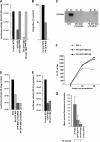
Inhibition of HIV-1 infection by PK-GFP-IBD. (A) MT4 cell lines stably expressing GFP-IBD, GFP-IBDD366N, PK-GFP-IBD, and PK-GFP-IBDD366N,5E were challenged with HIV-1luc. (B) Alu-PCR based integration assay of HIV-1luc in MT4 PK-GFP-IBD cells. (C) Western blot of subcellular fractions of MT4 cells expressing either GFP-IBD+LEDGFshRNA or PK-GFP-IBD+LEDGFshRNA. LEDGF is markedly decreased in both cell lines. See the Fig. 5A legend for assay details. (D) MT4 cells stably expressing PK-GFP-IBD, a LEDGF-targeted shRNA and GFP-IBD, or a LEDGF-targeted shRNA and PK-GFP-IBD were challenged with HIV-1luc. (E) LAI luciferase challenge of indicated cell lines. Luciferase was analyzed 4 days after infection. (F) Time course of HIV-1luc 2-LTR circle formation in the indicated cell lines. (G) Mean nuclear fluorescence intensity versus DI activity. The MFI was obtained for 10 control MT4 cells, and 20 each of GFP-IBD, GFP-IBDLOW, and PK-GFP-IBD cells (see Fig. S1A to C in the supplemental material). The legend at the bottom indicates whether the cell line produces antiviral dominant interference versus MT4 control cells.

Similar articles

Cited by

References

    1. Bao, K. K., et al. 2003. Functional oligomeric state of avian sarcoma virus integrase. J. Biol. Chem. 278:1323-1327. - PubMed
    1. Bartholomeeusen, K., et al. 2007. Differential interaction of HIV-1 integrase and JPO2 with the C terminus of LEDGF/p75. J. Mol. Biol. 372:407-421. - PubMed
    1. Bartholomeeusen, K., et al. 2009. Lens epithelium-derived growth factor/p75 interacts with the transposase-derived DDE domain of pogZ. J. Biol. Chem. 284:11467-11477. - PMC - PubMed
    1. Busschots, K., et al. 2005. The interaction of LEDGF/p75 with integrase is lentivirus-specific and promotes DNA binding. J. Biol. Chem. 280:17841-17847. - PubMed
    1. Cherepanov, P. 2007. LEDGF/p75 interacts with divergent lentiviral integrases and modulates their enzymatic activity in vitro. Nucleic Acids Res. 35:113-124. - PMC - PubMed

Publication types

MeSH terms

Substances