. 2011 Jan 24;192(2):295-306.
doi: 10.1083/jcb.201007018.

Bro1 binding to Snf7 regulates ESCRT-III membrane scission activity in yeast

Affiliations

Bro1 binding to Snf7 regulates ESCRT-III membrane scission activity in yeast

Megan Wemmer et al. J Cell Biol. .

Abstract

Endosomal sorting complexes required for transport (ESCRTs) promote the invagination of vesicles into the lumen of endosomes, the budding of enveloped viruses, and the separation of cells during cytokinesis. These processes share a topologically similar membrane scission event facilitated by ESCRT-III assembly at the cytosolic surface of the membrane. The Snf7 subunit of ESCRT-III in yeast binds directly to an auxiliary protein, Bro1. Like ESCRT-III, Bro1 is required for the formation of intralumenal vesicles at endosomes, but its role in membrane scission is unknown. We show that overexpression of Bro1 or its N-terminal Bro1 domain that binds Snf7 enhances the stability of ESCRT-III by inhibiting Vps4-mediated disassembly in vivo and in vitro. This stabilization effect correlates with a reduced frequency in the detachment of intralumenal vesicles as observed by electron tomography, implicating Bro1 as a regulator of ESCRT-III disassembly and membrane scission activity.


Figures
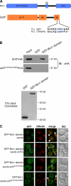
Figure 1.
Figure 1.
The Bro1 domain binds a MIM1-like motif in Snf7. (A) Schematic diagrams of Bro1 and Snf7, including the amino acid sequence alignment of MIM1-like motifs from yeast Snf7 and human CHMP4a. (B) In vitro pull-downs of purified GST or GST-Bro1 domain incubated with bacterial lysates expressing Snf7-HA or Snf7L231A/L234A-HA. GST and GST-Bro1 domain inputs were loaded at 10% of their total used in each pull-down. IB, immunoblotting. (C) Fluorescence and DIC microscopy of vps4Δ, vps4Δ snf7Δ, and vps4Δ snf7L231/234A cells expressing GFP-Bro1 domain or GFP-Bro1. Arrowheads indicate where GFP does (closed) or does not (open) colocalize with FM4-64–labeled class E compartments. Bars, 2 µm.
Figure 2.
Figure 2.
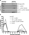
Overexpression of Bro1 or the Bro1 domain stabilizes ESCRT-III polymers at endosomes. (A) Western blot analysis of detergent-solubilized yeast membranes resolved by isopycnic density gradient centrifugation. Inclusion of 1% Triton X-100 throughout the gradients yielded identical sedimentation profiles. (B) Quantitation of triplicate gradients from A.
Figure 3.
Figure 3.
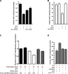
Bro1 binding to Snf7 inhibits in vitro disassembly of ESCRT-III. (A) 0.1 µM purified Vps4, ATP, and an ATP regeneration system were incubated with membranes isolated from vps4Δ yeast cells; overexpression of full-length Bro1 or the Bro1 domain (BOD) is indicated. The amount of membrane-associated Snf7 remaining after incubation at 30°C for 10 min was determined by quantitative Western blot analysis. P < 0.001 for both samples. (B) In vitro ESCRT-III release performed in the presence or absence of ATP, 0.1 µM Vps4, and 0.1 µM GST-Bro1 domain (GST-BOD). (C) In vitro ESCRT-III release performed as in B except that the reaction was allowed to proceed for 5 min in the absence of GST-BOD or for 20 min in the absence or presence of 1 µM GST-BOD that was added either at the same time as purified Vps4 or 5 min after the addition of Vps4. (D) In vitro ESCRT-III release performed with membranes from yeast expressing wild-type Snf7 or the Snf7Vps2MIM1 chimera in the presence or absence of ATP, 0.1 µM Vps4, and 0.1 µM GST-BOD. Error bars show the standard deviation.
Figure 4.
Figure 4.
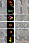
Endosome morphologies in cells overexpressing Bro1 or the Bro1 domain. (A–F) 2D cross sections and 3D models from 200-nm-thick section electron tomograms. Spherical endosomal limiting membranes are depicted in yellow, whereas flattened class E compartments are depicted in various colors to discriminate discrete membranes. Lumenal vesicles are red. V, vacuole. Bars, 100 nm or 200 nm as noted. (G–L) Gallery of tomographic slices of wild-type cells with and without overexpression of Bro1 or the Bro1 domain. Freely detached ILVs are traced in red, whereas ILV budding profiles and the limiting endosomal membrane are traced in yellow. In some cases, the continuity of ILV budding profiles with the limiting endosomal membrane is out of plane in the tomographic slice but evident in the 3D reconstruction. Bars, 50 nm.
Figure 5.
Figure 5.
Quantitation of endosome morphology and vesicle budding profiles. Quantitation of the mean number of ILV budding profiles observed per MVB (A) versus the frequency distribution of the number of ILV budding profiles observed per MVB (B) observed in tomograms. Quantitation of the relative percentage of limiting versus lumenal membrane surface areas (C) and the mean ILV diameters (D) observed in tomograms. (E) The percentage of the limiting membrane incorporated into ILV budding profiles was measured for each strain. Note that no ILV budding profiles were seen for snf7Δ cells, but five freely detached ILVs were observed at the periphery of a class E compartment in a single tomogram of snf7Δ cells (Fig. S5); the rarity of this occurrence is reflected by the observation that the membrane surface area of these ILVs comprised 0.27% of the total endosomal membrane surface area in this strain. Tomograms for 12, 14, and 17 MVBs were prepared and modeled for wild-type cells, cells overexpressing the Bro1 domain, and cells overexpressing full-length Bro1, respectively. Four individual tomograms were prepared and modeled for snf7L231A/L234A cells, which contained a total of 33 MVB, VTE, and cisternal structures. Three individual tomograms were generated and modeled for snf7Δ cells, each containing E compartments that were composed of multiple cisternal stacks and occasionally also spherical endosomal membranes. Methods used for the preparation of the tomograms that were used to generate the measurements shown in this experiment are described in detail in the Materials and methods section.
Figure 6.
Figure 6.
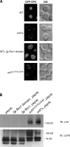
Cargo sorting and deubiquitination. (A) Fluorescence and DIC microscopy of cells expressing GFP-CPS. Bars, 2 µm. (B) Western blot analysis of anti-CPS immunoprecipitates. IB, immunoblotting.

Similar articles

Cited by

References

    1. Agromayor M., Martin-Serrano J. 2006. Interaction of AMSH with ESCRT-III and deubiquitination of endosomal cargo. J. Biol. Chem. 281:23083–23091 10.1074/jbc.M513803200 - DOI - PubMed
    1. Azmi I., Davies B., Dimaano C., Payne J., Eckert D., Babst M., Katzmann D.J. 2006. Recycling of ESCRTs by the AAA-ATPase Vps4 is regulated by a conserved VSL region in Vta1. J. Cell Biol. 172:705–717 10.1083/jcb.200508166 - DOI - PMC - PubMed
    1. Azmi I.F., Davies B.A., Xiao J., Babst M., Xu Z., Katzmann D.J. 2008. ESCRT-III family members stimulate Vps4 ATPase activity directly or via Vta1. Dev. Cell. 14:50–61 10.1016/j.devcel.2007.10.021 - DOI - PubMed
    1. Babst M., Sato T.K., Banta L.M., Emr S.D. 1997. Endosomal transport function in yeast requires a novel AAA-type ATPase, Vps4p. EMBO J. 16:1820–1831 10.1093/emboj/16.8.1820 - DOI - PMC - PubMed
    1. Babst M., Wendland B., Estepa E.J., Emr S.D. 1998. The Vps4p AAA ATPase regulates membrane association of a Vps protein complex required for normal endosome function. EMBO J. 17:2982–2993 10.1093/emboj/17.11.2982 - DOI - PMC - PubMed

Publication types

MeSH terms

Substances