Review
. 2011:6:395-423.
doi: 10.1146/annurev.pathol.4.110807.092150.

A glimpse of various pathogenetic mechanisms of diabetic nephropathy

Affiliations
Review

A glimpse of various pathogenetic mechanisms of diabetic nephropathy

Yashpal S Kanwar et al. Annu Rev Pathol. 2011.

Abstract

Diabetic nephropathy is a well-known complication of diabetes and is a leading cause of chronic renal failure in the Western world. It is characterized by the accumulation of extracellular matrix in the glomerular and tubulointerstitial compartments and by the thickening and hyalinization of intrarenal vasculature. The various cellular events and signaling pathways activated during diabetic nephropathy may be similar in different cell types. Such cellular events include excessive channeling of glucose intermediaries into various metabolic pathways with generation of advanced glycation products, activation of protein kinase C, increased expression of transforming growth factor β and GTP-binding proteins, and generation of reactive oxygen species. In addition to these metabolic and biochemical derangements, changes in the intraglomerular hemodynamics, modulated in part by local activation of the renin-angiotensin system, compound the hyperglycemia-induced injury. Events involving various intersecting pathways occur in most cell types of the kidney.


Figures

Figure 1
Figure 1
Light photomicrographs illustrating various stages of developing glomerular lesions and tubulointerstitial disease in diabetic nephropathy. (a) A normal glomerulus. (b) Thickened basement membranes (arrowheads) and expanded mesangial regions (asterisks). (c) The nodular appearance of the mesangial regions characteristic of Kimmelstiel-Wilson lesions (asterisks). (d) The tubulointerstitial lesions include thickened tubular basement membrane (TBM), hyalinization of afferent arteriole (ART), and fibrosis of the interstitium (INT). Abbreviations: c, capillary lumen; En, endothelial cell; Ep, visceral epithelial cell (podocyte); Me, mesangium; US, urinary space.
Figure 2
Figure 2
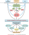
An overview of different signaling events induced by exposure of renal cells to high glucose concentrations, with resulting altered expression of various genes and cellular abnormalities leading to diabetic nephropathy. The schematic drawing also highlights the hypothetical cross talk between AGE-RAGE (advanced glycation end product–receptor for AGE) and the renin-angiotensin system (RAS) and the reciprocal-cyclical modulation of the interactions among AGEs, reactive oxygen species (ROS), and protein kinase C (PKC), with ROS as the central mediator. Abbreviations: Ang II, angiotensin II; AP-1, activator protein 1; AT1, Ang II receptor; ECM, extracellular matrix; GLUT, glucose transporter; JAK-STAT, Janus kinase–signal transducer and activator of transcription; MAPK, mitogen-activated protein kinase; MCP-1, monocyte chemoattractant protein 1; NF-κB; nuclear factor κB; RNS, reactive nitrogen species; TGF-β, transforming growth factor β; VEGF, vascular endothelial growth factor.
Figure 3
Figure 3
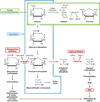
Schematics of different pathways for glucose metabolism that lead to the activation of protein kinase C (PKC) and transforming growth factor β(TGF-β). In addition to undergoing glycolysis, excess glucose is channeled into the polyol and hexosamine pathways, which results in increased lipid synthesis and the generation of diacylglycerol (DAG). This leads to the activation of PKC and TGF-β and, consequently, increased extracellular matrix (ECM) synthesis. Abbreviation: GFAT, glutamine:fructose-6-phosphate-aminotransferase.
Figure 4
Figure 4
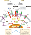
Schematic drawing depicting the generation of advanced glycation end products (AGEs) and downstream events. AGEs are formed by condensation of a sugar (R) such as glucose with a reactive ε-amino (NH2) group of the protein; this process is followed by the formation of a Schiff base, Amadori rearrangement, and a complex series of reactions. AGE:RAGE (receptor for AGE) interactions lead to the generation of reactive oxygen species (ROS) and the activation of protein kinase C (PKC), transforming growth factor β (TGF-β), mitogenactivated protein kinase (MAPK), and transcription factors such as nuclear factor κB (NF-κB), leading to increased synthesis of the extracellular matrix (ECM). Diabetic injury is further amplified by the feedback loops of angiotensin II (Ang II) and cross-linking of de novo synthesized excess ECM proteins with sugars. Abbreviations: AT1, Ang II receptor; TGF-βR, TGF-β receptor.
Figure 5
Figure 5
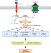
Sequence of events following AGE:RAGE (advanced glycation end products:receptor for advanced glycation end products) interactions and excess glucose entry into the cell via glucose transporter (GLUT). Diacylglycerol (DAG) and activated phospholipase C (PLC) increase the expression and activity of protein kinase C (PKC), which modulates the expression of a wide variety of genes that adversely affect glomerular pathophysiology, thereby leading to increased vascular permeability, mesangial expansion, hyperfiltration, and proteinuria. Abbreviations: Ang II, angiotensin II; e-NOS, endothelial nitric oxide synthase; ET-1, endothelin 1; IP3, inositol trisphosphate; PAI-1, plasminogen activator inhibitor 1; PIP2, phosphatidylinositol 4,5-bisphosphate; ROS, reactive oxygen species; TGF-β, transforming growth factor β; VEGF, vascular endothelial growth factor.
Figure 6
Figure 6
Summary of events leading to the generation of cytosolic reactive oxygen species (ROS) (R1) and mitochondrial ROS (R2). Extramitochondrial cytosolic ROS generation occurs following increased glucose flux, activation of the polyol pathway, and AGE:RAGE (advanced glycation end products:receptor for advanced glycation end products) interaction, as well as via the NADPH oxidase system, such as NOX4. Mitochondrial matrix ROS and reactive nitrogen species (RNS) are generated via the well-characterized electron-transport chain, Complex I–IV redox carriers, localized in the inner mitochondrial membrane. During hyperglycemia, there is an increased donation of electrons (e) by powerful NADH- and FADH2-reducing agents of Complex I and Complex II, respectively, characterized by pumping of protons (H+) into the intermembrane space, which gives rise to an increased mitochondrial membrane potential. As a result, the electron transport at complex III is inhibited; the system backs up, and the half-life of the free-radical intermediates of coenzyme Q (CoQ) is prolonged, which leads to an increase in the reduction of O2 to superoxide (O2.−) and in the production of ROS. The generated ROS and RNS release cytochrome c, activate caspases, and induce apoptosis. They also modulate the activity of angiotensin II (Ang II), protein kinase C (PKC), and transforming growth factor β(TGF-β), which ultimately affect the synthesis of the extracellular membrane. Abbreviation: DAG, diacylglycerol.
Figure 7
Figure 7
Schematic depicting transforming growth factor β (TGF-β) and bone morphogenetic protein 7 (BMP-7) signaling. Activation of latent TGF-β by glucose, advanced glycation end products (AGEs), reactive oxygen species (ROS), and angiotensin II (Ang II) leads to the generation of TGF-p, which binds first to type II and III serine/threonine kinase receptors with recruitment and phosphorylation of type I. Activated heteromeric complex interacts with Smad2/3 and co-Smad4. Smad2/3 are inhibited by Smad6/7, which are induced by tumor necrosis factor α. (TNF-α) and interferon-γ (IFN-γ) signaling. The Smad2/3/4 complex translocates into the nucleus to initiate transcription of various extracellular matrix (ECM) genes and connective tissue growth factor (CTGF). BMP-7, however, activates Smad1/5/8, which bind to co-Smad4 and, upon translocation into the nucleus, induce Id proteins that inhibit differentiation and DNA binding of some of the transcription factors, thereby opposing the action of TGF-β. Abbreviations: JAK, Janus kinase; MMP, matrix metalloproteinase; NF-κB; nuclear factor κB; PKC, protein kinase C; STAT, signal transducer and activator of transcription; TIMP, tissue inhibitor of metalloproteinase.
Figure 8
Figure 8
Schematics depicting the sequence of events, following the interactions between advanced glycation end products (AGEs) and angiotensin II (Ang II) with their respective receptors, that lead to the generation of reactive oxygen species (ROS) and the activation of protein kinase C (PKC) and Rap1 and Rho GTPases. Both Rap1 and Rho, in association with guanine exchange factors (GEFs), are activated (GTP bound), whereas GTPase-activating proteins (GAPs) hydrolyze Rap1 and Rho into the inactive (GDP-bound) state. Thus, these small G proteins cycle between functional and nonfunctional states. Activated Rho GTP leads to the induction of Rho kinase and the phosphorylation of Janus kinase/stress-activated protein kinase (JNK/SAPK) kinase and phosphatase 1 (PPtase1), which then translocate into the nucleus to initiate transcription via various transcription factors. Similarly, Rap1 GTP, in association with another kinase known as B-Raf, causes the induction and phosphorylation of extracellular signal–related kinase (ERK) and mitogen-activated protein kinase/ERK (MEK). Upon translocation into the nucleus the transcription of target genes is initiated. Interestingly, the Rap1-ARAP1 (angiotensin II type 1 receptor–associated protein) complex inhibits the activity of Rho kinase. Abbreviations: AP-1, activator protein 1; ATF, activating transcription factor; CBP, cAMP response element–binding (CREB) protein; ELK1, ets-like gene 1; NF-κB, nuclear factor κB.
Figure 9
Figure 9
Schematic drawing depicting local glomerular activation of the renin-angiotensin system (RAS) by glucose flux and advanced glycation end products (AGEs). Both podocytes (Po) and mesangial cells (Me) of the glomerulus express angiotensin II (Ang II) receptors (AT1). Angiotensin I (AI) is synthesized from angiotensinogen (AGT) by the action of renin. AI is converted into angiotensin II (Ang II) by angiotensin-converting enzyme (ACE). Ang II, upon binding to its AT1 receptor, induces various nonhemodynamic effects, including increased activity of transforming growth factor β (TGF-β) and expression of monocyte chemotactic protein 1 (MCP-1), vascular endothelial growth factor (VEGF), and reactive oxygen species (ROS). Abbreviations: Cap, capillary; En, endothelium; IL-6, interleukin-6; TGF-βR, TGF-β receptor.
Figure 10
Figure 10
Schematics depicting systemic activation of the renin-angiotensin system by glucose flux and advanced glycation end products (AGEs). High-glucose flux and AGEs may induce hypertrophy and hyperplasia of the juxtaglomerular apparatus (JGA), leading to increased secretion of renin from stored cellular granules. This process causes the generation of a potent vasoactive peptide known as angiotensin II (Ang II), which modulates the secretion of adrenal aldosterone with Na+ retention, and it also induces constriction of the efferent arteriole (EA). As a result of this mechanical stretch, systemic blood pressure rises, leading to worsening of the hyperglycemic injury in various target organs or cells. Abbreviations: AA, afferent arteriole; Cap, capillary; ACE, angiotensin-converting enzyme; AI, angiotensin I; ATG, angiotensinogen.

Similar articles

Cited by

References

    1. LeRoith D, Taylor SI, Olefsky JM. Diabetes Mellitus: A Fundamental and Clinical Text. 3rd ed. Philadelphia: Lippincott William & Wilkins; 2004. p. 1,540.
    1. Reddy AS. Diabetic Nephropathy: Theory and Practice. East Hanover, NJ: College Book Publ; 2004. p. 563.
    1. Wolf G. New insights into the pathophysiology of diabetic nephropathy: from hemodynamics to molecular pathology. Eur. J. Clin. Investig. 2004;34:785–796. - PubMed
    1. Schena FP, Gesualdo L. Pathogenetic mechanisms of diabetic nephropathy. J. Am. Soc. Nephrol. 2005;16:S30–S33. - PubMed
    1. Susztak K, Bottinger EP. Diabetic nephropathy: a frontier for personalized medicine. J. Am. Soc. Nephrol. 2006;17:361–367. - PubMed

Publication types

MeSH terms