. 2011 Jan 19:11:24.
doi: 10.1186/1471-2407-11-24.

Bim and Mcl-1 exert key roles in regulating JAK2V617F cell survival

Affiliations

Bim and Mcl-1 exert key roles in regulating JAK2V617F cell survival

Joëlle Rubert et al. BMC Cancer. .

Abstract

Background: The JAK2V617F mutation plays a major role in the pathogenesis of myeloproliferative neoplasms and is found in the vast majority of patients suffering from polycythemia vera and in roughly every second patient suffering from essential thrombocythemia or from primary myelofibrosis. The V617F mutation is thought to provide hematopoietic stem cells and myeloid progenitors with a survival and proliferation advantage. It has previously been shown that activated JAK2 promotes cell survival by upregulating the anti-apoptotic STAT5 target gene Bcl-xL. In this study, we have investigated the role of additional apoptotic players, the pro-apoptotic protein Bim as well as the anti-apoptotic protein Mcl-1.

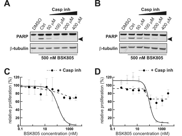
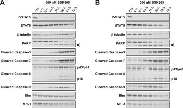
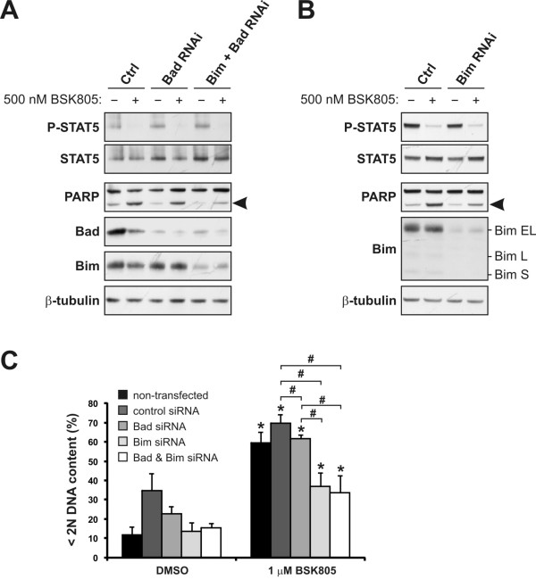
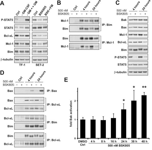
Methods: Pharmacological inhibition of JAK2/STAT5 signaling in JAK2V617F mutant SET-2 and MB-02 cells was used to study effects on signaling, cell proliferation and apoptosis by Western blot analysis, WST-1 proliferation assays and flow cytometry. Cells were transfected with siRNA oligos to deplete candidate pro- and anti-apoptotic proteins. Co-immunoprecipitation assays were performed to assess the impact of JAK2 inhibition on complexes of pro- and anti-apoptotic proteins.

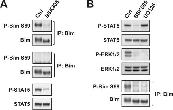
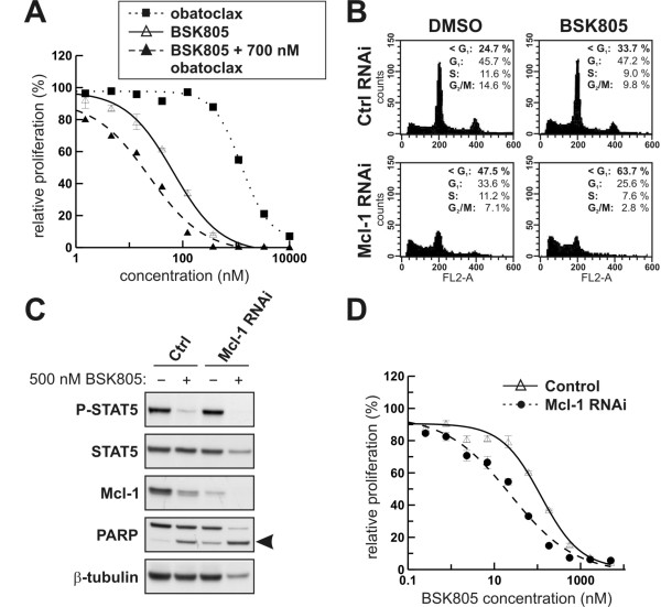
Results: Treatment of JAK2V617F mutant cell lines with a JAK2 inhibitor was found to trigger Bim activation. Furthermore, Bim depletion by RNAi suppressed JAK2 inhibitor-induced cell death. Bim activation following JAK2 inhibition led to enhanced sequestration of Mcl-1, besides Bcl-xL. Importantly, Mcl-1 depletion by RNAi was sufficient to compromise JAK2V617F mutant cell viability and sensitized the cells to JAK2 inhibition.

Conclusions: We conclude that Bim and Mcl-1 have key opposing roles in regulating JAK2V617F cell survival and propose that inactivation of aberrant JAK2 signaling leads to changes in Bim complexes that trigger cell death. Thus, further preclinical evaluation of combinations of JAK2 inhibitors with Bcl-2 family antagonists that also tackle Mcl-1, besides Bcl-xL, is warranted to assess the therapeutic potential for the treatment of chronic myeloproliferative neoplasms.


Figures

Figure 1
Figure 1
Caspase inhibition rescues JAK2V617F mutant cells from the apoptotic and anti-proliferative effects of the JAK2 inhibitor NVP-BSK805. SET-2 (A) and MB-02 (B) cells were pretreated for 1 hour with increasing concentrations of a pan-caspase inhibitor, followed by treatment for 24 hours with 500 nM NVP-BSK805. PARP cleavage (cleaved PARP is depicted by arrowheads) was assessed by Western blot analysis. β-tubulin was probed for as a loading control. Controls consisted of drug vehicle (DMSO) treated cells (baseline) and NVP-BSK805 treatment in the absence of the pan-caspase inhibitor. Proliferation assays with increasing concentrations of NVP-BSK805 in SET-2 (C) and MB-02 (D) cells were carried out in the absence (empty triangles/solid line) or presence (filled circles/stippled line) of 200 μM pan-caspase inhibitor.
Figure 2
Figure 2
Activation of intrinsic and extrinsic caspase cascades following JAK2 inhibition in JAK2V617F mutant cells lines. SET-2 (A) and MB-02 (B) cells were treated with 500 nM of the JAK2 inhibitor NVP-BSK805 and extracted at the indicated time points for Western blot analysis. Control (Ctrl) cells were treated with the drug vehicle DMSO for 72 hours. Induction of apoptosis markers becomes evident starting at the 16 hours time point in both cell lines. Arrowhead denotes cleaved PARP, running below the band for full-length PARP. The caspase-8 blot shows the p43/p41 cleavage intermediates and active caspase-8 fragment p18.
Figure 3
Figure 3
Bad and Bim depletion in JAK2V617F mutant SET-2 cells suppress NVP-BSK805-induced apoptosis. A: SET-2 cells were transfected with control siRNA (Ctrl), Bad siRNA or siRNAs to deplete both Bad and Bim simultaneously. After 72 hours, cells were treated with 500 nM NVP-BSK805 or the drug vehicle DMSO for 30 hours and extracted for Western blot analysis. Arrowhead depicts cleaved PARP running below the full-length PARP protein. β-tubulin was probed for as a loading control. B: SET-2 cells were transfected with control siRNA (Ctrl) or Bim siRNA and analyzed as described above. The Bim Western blot depicts an extended molecular weight range to show that Bim-EL is the predominant form of the protein expressed in SET-2 cells. C: SET-2 cells were transfected with control, Bad or Bim siRNAs, or siRNAs to deplete both Bad and Bim simultaneously, for 48 hours and then treated with 1 μM NVP-BSK805 or the drug vehicle DMSO for 40 hours. Controls also consisted of non-transfected cells. Then, DNA content was measured by FACS using propidium iodide (PI) staining and the percentage of cells with less than 2N DNA content was determined (data represent the mean ± SD of three independent experiments). *Significantly different from respective DMSO control using t-test (p < 0.05). #Significant difference between groups as assessed by t-test (p < 0.05). D: SET-2 cells were transfected with control, Bad or Bim siRNAs, or siRNAs to deplete both Bad and Bim simultaneously, for 24 hours and then treated with increasing concentrations of NVP-BSK805 in cell proliferation assays for 48 hours. The histogram depicts half-maximal growth-inhibitory (GI50) concentrations of NVP-BSK805 for each condition (data represent the mean ± SD of two independent experiments carried out in triplicate).
Figure 4
Figure 4
Bim depletion in JAK2V617F mutant MB-02 cells suppresses NVP-BSK805-induced apoptosis. A: MB-02 cells were transfected with control siRNA (Ctrl), Bad siRNA or siRNAs to deplete both Bad and Bim simultaneously. After 72 hours, cells were treated with 500 nM NVP-BSK805 or the drug vehicle DMSO for 16 hours and extracted for Western blot analysis. Arrowhead depicts cleaved PARP running below the full-length PARP protein. β-tubulin was probed for as a loading control. B: MB-02 cells were transfected with control siRNA (Ctrl) or Bim siRNA and analyzed as described above. The Bim Western blot depicts an extended molecular weight range to show that Bim-EL is the predominant form of the protein expressed in MB-02 cells. C: MB-02 cells were transfected with control, Bad or Bim siRNAs, or siRNAs to deplete both Bad and Bim simultaneously, for 48 hours and then treated with 1 μM NVP-BSK805 or the drug vehicle DMSO for 40 hours. Controls also consisted of non-transfected cells. Then, DNA content was measured by FACS using propidium iodide (PI) staining and the percentage of cells with less than 2N DNA content was determined (data represent the mean ± SD of three independent experiments). *Significantly different from respective DMSO control using t-test (p < 0.05). #Significant difference between groups as assessed by t-test (p < 0.05).
Figure 5
Figure 5
Analysis of the regulation of anti-apoptotic and pro-apoptotic proteins in JAK2V617F mutant SET-2 cells. A: JAK2 wild type TF-1 cells (left panels) were starved overnight in medium containing 0.1% FCS without GM-CSF. Cells were then stimulated with 2 ng/ml GM-CSF (+GM) for 8 hours and extracted. Control TF-1 cell extracts (Ctrl) were prepared from cells growing in full medium. SET-2 cells (right panels) were treated with DMSO (Ctrl) or 500 nM NVP-BSK805 for 16 hours. Cells were either extracted or washed and released into fresh medium (FM) without NVP-BSK805 for 8 hours and then extracted. B: SET-2 cells were treated with DMSO or 500 nM NVP-BSK805 for 4 or 24 hours, extracted, and lysates were subjected to co-immunoprecipitation studies. To control for potential unspecific binding of the proteins of interest to beads, extracts (same total protein input as used for the immunoprecipitations) were also incubated with beads without the respective antibodies (Ctrl). C: Levels of soluble Bak, Bax, Mcl-1, Bcl-xL and Bim in SET-2 whole cell extracts following treatment of cells for 4 and 24 hours with DMSO or NVP-BSK805. D: SET-2 cell lysates from cells treated as described above in panel B were subjected to immunoprecipitation of Bax (using an antibody recognizing an amino-terminal epitope) and co-immunoprecipitation studies. Potential unspecific binding of the proteins of interest to beads was controlled (Ctrl) as described above. E: SET-2 cells were incubated with DMSO for 48 hours or with 500 nM NVP-BSK805 for the indicated times. Bak activation was assessed using a Bak active conformation-specific antibody by flow cytometry analysis. Results are depicted as the means of three independent experiments ± SD. *Significantly different from DMSO control using t-test (p < 0.05). **Significantly different from DMSO control using t-test (p < 0.001).
Figure 6
Figure 6
Analysis of Bim phosphorylation in JAK2V617F mutant SET-2 cells. A: SET-2 cells were treated with DMSO or 500 nM NVP-BSK805 for 24 hours. Bim was immunoprecipitated and levels of Bim-EL Ser69 as well as Ser59 phosphorylation were analyzed by Western blotting. Levels of P-STAT5 and total STAT5 were probed for as controls for the drug treatment. B: SET-2 cells were treated with DMSO, 500 nM NVP-BSK805 or 10 μM UO126 for 4 hours. Levels of P-STAT5, STAT5, P-ERK1/2 and ERK1/2 were assessed by Western blotting. Bim was immunoprecipitated and levels of Bim-EL Ser69 phosphorylation were detected by Western blotting.
Figure 7
Figure 7
Mcl-1 depletion in JAK2V617F mutant SET-2 cells increases apoptosis and sensitizes the cells to NVP-BSK805-induced cell death. A: Cell proliferation assays were carried out in SET-2 cells treated with increasing concentrations of NVP-BSK805 (empty triangles/solid line), obatoclax (filled squares/dotted line) or NVP-BSK805 in combination with a fixed obatoclax concentration of 700 nM (filled triangles/stippled line) for 72 hours. B: SET-2 cells were transfected with control (Ctrl) or Mcl-1 siRNAs. After 48 hours cells were treated with DMSO or 500 nM NVP-BSK805 for 24 hours. Then, DNA content was measured by FACS using propidium iodide (PI) staining. The x- and y-axes represent fluorescent channel 2-area (FL2-A) fluorescence intensity for PI staining and cell count, respectively. The percentage of cells in the respective cell cycle phases is indicated for each treatment condition. C: SET-2 cells were transfected with control (Ctrl) or Mcl-1 siRNAs. After 48 hours, cells were treated with 500 nM NVP-BSK805 or the drug vehicle DMSO for 24 hours and extracted for Western blot analysis. Arrowhead depicts cleaved PAPR running below the full-length PARP protein. β-tubulin was probed for as a loading control. D: SET-2 cells were transfected with control (Ctrl) or Mcl-1 siRNAs. After 24 hours cells were incubated for 48 hours with increasing concentrations of NVP-BSK805 to assess cell proliferation relative to the respective DMSO treated controls.

Similar articles

Cited by

References

    1. Nelson ME, Steensma DP. JAK2 V617F in myeloid disorders: What do we know now, and where are we headed? Leuk Lymphoma. 2006;47:177–194. doi: 10.1080/10428190500301348. - DOI - PubMed
    1. Saharinen P, Vihinen M, Silvennoinen O. Autoinhibition of Jak2 Tyrosine Kinase Is Dependent on Specific Regions in Its Pseudokinase Domain. Mol Biol Cell. 2003;14:1448–1459. doi: 10.1091/mbc.E02-06-0342. - DOI - PMC - PubMed
    1. Levine RL, Wadleigh M, Cools J, Ebert BL, Wernig G, Huntly BJP, Boggon TJ, Wlodarska I, Clark JJ, Moore S. et al.Activating mutation in the tyrosine kinase JAK2 in polycythemia vera, essential thrombocythemia, and myeloid metaplasia with myelofibrosis. Cancer Cell. 2005;7:387–397. doi: 10.1016/j.ccr.2005.03.023. - DOI - PubMed
    1. Vannucchi AM. How do JAK2-inhibitors work in myelofibrosis: An alternative hypothesis. Leukemia Res. 2009;33:1581–1583. doi: 10.1016/j.leukres.2009.06.001. - DOI - PubMed
    1. Mesa R, Gale RP. Hypothesis: How do JAK2-inhibitors work in myelofibrosis. Leukemia Res. 2009;33:1156–1157. doi: 10.1016/j.leukres.2009.04.011. - DOI - PubMed

MeSH terms