. 2011 May;39(9):3695-709.
doi: 10.1093/nar/gkq1336. Epub 2011 Jan 17.

Identification of residues in the N-terminal PAS domains important for dimerization of Arnt and AhR

Affiliations

Identification of residues in the N-terminal PAS domains important for dimerization of Arnt and AhR

Nan Hao et al. Nucleic Acids Res. 2011 May.

Abstract

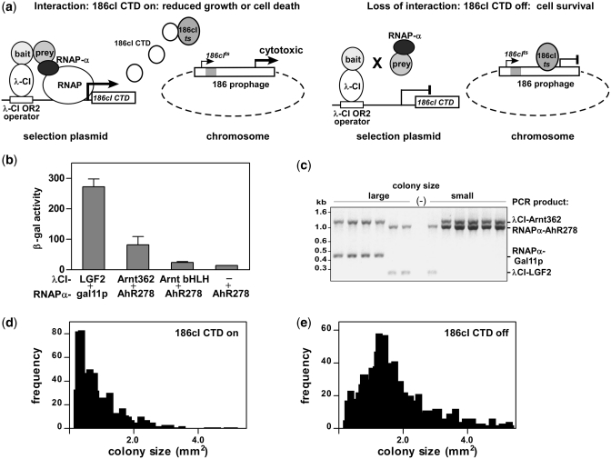
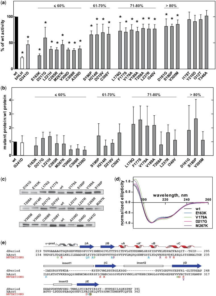
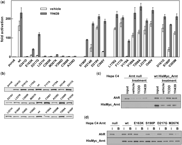
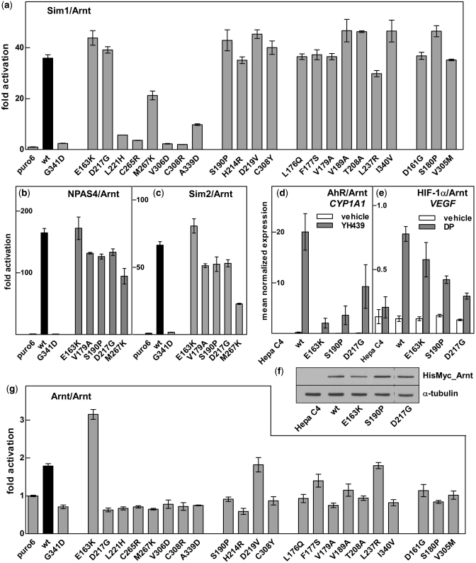
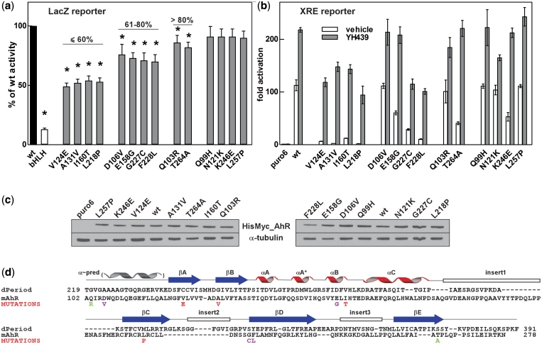
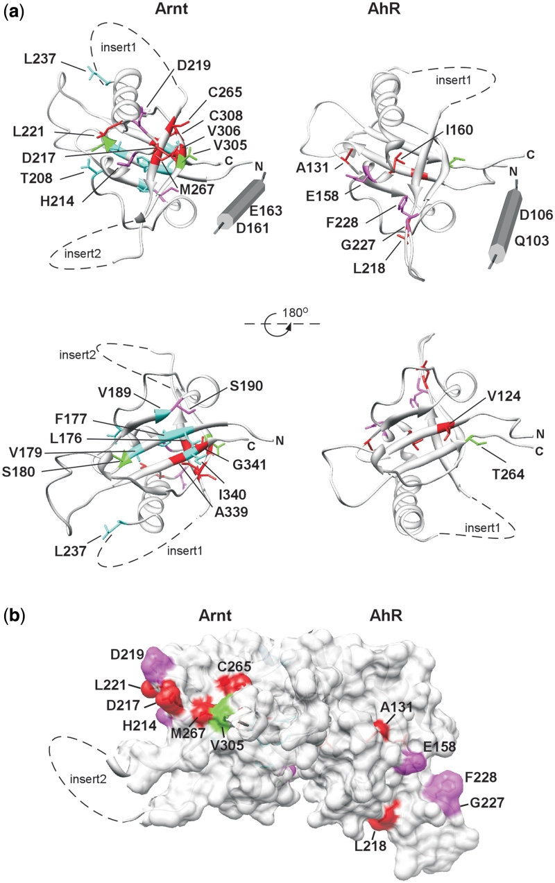
The basic helix-loop-helix (bHLH).PAS dimeric transcription factors have crucial roles in development, stress response, oxygen homeostasis and neurogenesis. Their target gene specificity depends in part on partner protein choices, where dimerization with common partner Aryl hydrocarbon receptor nuclear translocator (Arnt) is an essential step towards forming active, DNA binding complexes. Using a new bacterial two-hybrid system that selects for loss of protein interactions, we have identified 22 amino acids in the N-terminal PAS domain of Arnt that are involved in heterodimerization with aryl hydrocarbon receptor (AhR). Of these, Arnt E163 and Arnt S190 were selective for the AhR/Arnt interaction, since mutations at these positions had little effect on Arnt dimerization with other bHLH.PAS partners, while substitution of Arnt D217 affected the interaction with both AhR and hypoxia inducible factor-1α but not with single minded 1 and 2 or neuronal PAS4. Arnt uses the same face of the N-terminal PAS domain for homo- and heterodimerization and mutational analysis of AhR demonstrated that the equivalent region is used by AhR when dimerizing with Arnt. These interfaces differ from the PAS β-scaffold surfaces used for dimerization between the C-terminal PAS domains of hypoxia inducible factor-2α and Arnt, commonly used for PAS domain interactions.


Figures

Figure 1.
Figure 1.
Characterizing the reverse bacterial two hybrid system (RevB2H). (a) Mechanism: interaction between bait fused to bacteriophage λ-CI repressor (λCI-bait) and prey fused to RNA polymerase α-subunit (RNAPα-prey) (28) drives expression of the C-terminal domain of bacteriophage 186cI repressor [186cI-CTD (27)] from the plac λ-CI OR2-62 promoter (28) on the lacOR2-62_186cI-CTD selection plasmid. 186cI-CTD sequesters chromosomally encoded 186cI repressor (186cIts), from its binding site in the int- 186 prophage. This activates aberrant lytic gene transcription from the prophage, which results in significantly inhibited growth or cell death, depending on the strength of the bait–prey interaction. Mutations in the bait or prey fusion proteins that disrupt interaction reduce 186cI-CTD expression, allowing 186cIts to maintain repression of prophage lytic transcription, permitting normal cell growth. (b) Strength of the indicated λCI-bait and RNAPα-prey fusion proteins interaction measured as β-gal activity from the lacOR2-62_lacZ reporter gene in KS1. Data are mean ± SD, n = 3. (c) Selectivity for interaction: four plasmids encoding two pairs of interacting bait and prey proteins [λCI_Arnt362 and RNAPα_AhR278, λCI_LGF2 and RNAPα_gal11p (35)] were co-transformed into KS1033 carrying the lacOR2-62_186cI-CTD selection plasmid, with induction of fusion protein expression. Plasmid incompatibility permits establishment of only one of the two available λCI and RNAPα plasmids. Genotyping revealed the presence of non-interacting and interacting pairs in large and small colonies, respectively. (d) and (e) Effect of 186cI-CTD production on colony size: induction of protein expression in KS1033 harbouring the lacOR2-62_186cI-CTD selection and λCI_Arnt362 expression plasmids either with (d) or without (e) transformation of the RNAP_AhR278_lacZα mutagenic library.
Figure 2.
Figure 2.
Single amino acid mutations in Arnt PAS.A domain with disrupted AhR/Arnt heterodimerization isolated using RevB2H. (a) The strength of interaction between RNAPα_AhR278 and the potential λCI_Arnt362 loss-of-interaction mutants selected as large blue colonies from RevB2H was measured in a standard forward bacterial two hybrid system with the lacZ reporter gene (28). Data are mean with 95% confidence intervals, shown as a percentage of β-gal activity obtained with wt λCI_Arnt362 protein, n = 9. bHLH: λCI_Arnt bHLH domain only; *P < 0.05 (one tailed unpaired Student’s t-test). (b) Expression of mutant λCI_Arnt362 relative to wt; mean ± SEM. from three independent bacterial extracts. Percent symbol denotes grouping by β-gal activity relative to wt from (a). (c) Shows a representative western blot. (d) CD spectra of wt H6_Arnt362 and selected loss-of-interaction mutants, normalized to allow comparison with wt (30). CD spectra of all mutants are shown in Supplementary Figure S2. (e) The dimerization deficient mutations identified from RevB2H mapped onto the sequence of Arnt PAS.A, aligned with dPer PAS.A. dPer PAS.A is representative of PAS.A domains, which have one to three insert loops within the PAS domain fold (25). The positions of secondary-structure elements, including the predicted N-terminal α-helix are indicated above. The mutations are coloured according to the strength of disruption observed in the β-gal assay; red: <60%, magenta: 61–70%, blue: 71–80% and green: >80% of wt activity, yellow: Hepa c4 mutation G341D (37).
Figure 3.
Figure 3.
Arnt PAS.A mutations identified from RevB2H disrupt full-length AhR/Arnt heterodimerization in mammalian cells. (a) Activation of the pML-6c-X XRE-driven reporter gene in 293T cells by HisMyc-tagged AhR and full-length wt or mutant HisMyc-tagged Arnt in response to vehicle or the ligand YH439, shown as fold activation over basal levels (puro6) from endogenous protein. Data are mean ± SD of transfections performed in triplicate and representative of two independent experiments. (b) Expression of HisMyc_Arnt wt and mutants in the transfected cells used for the reporter gene assay in (a), detected with anti-Arnt monoclonal antibody and Cy5 labelled anti-mouse secondary antibody. (c) Cytosolic extracts from parental Arnt-deficient mouse Hepa C4 cells (Arnt null) and the derived line constitutively expressing HisMyc-tagged wt Arnt, from an integrated lentiviral vector, were treated in vitro with vehicle, TCDD or YH439. Protein captured by Ni resin was analysed by western blot, using anti-Myc antibody for HisMyc_Arnt or anti-AhR antibody to detect co-immobilized AhR. (d) Mutations in Arnt reduced co-capture of AhR in Ni affinity pull-downs. Cytosolic extracts from the Arnt null and derived cell lines constitutively expressing wt or the indicated mutant HisMyc_Arnt from integrated lentiviral vectors were in vitro transformed with YH439. Captured HisMyc_Arnt and AhR were detected by western blot using anti-Myc and anti-AhR antibodies. i, 5% of input; B, fraction bound to Ni resin. Data are representative of three independent experiments.
Figure 4.
Figure 4.
Arnt mutations differentially alter dimerization with other partner proteins. Activation of the CME driven reporter (pML-6c-wt) in 293T cells by wt or mutant HisMyc_Arnt together with hSim1_Myc (a), NPAS4_Myc (b) or hSim2s_Myc (c) shown as fold activation over basal levels (puro6). Data are mean ± SD of transfections performed in triplicate and are representative of two independent experiments. Hepa C4 cell lines expressing wt, E163K, S190P or D217G HisMyc_Arnt were treated with vehicle, YH439 or 2,2′-dipyridyl (DP) and assayed for CYP1A1 (d) or VEGF (e) mRNA induction by qRT-PCR. Data are mean ± SEM, n = 3. (f) Whole cell extracts from the Hepa C4 Arnt deficient and derived cell lines constitutively expressing wt, E163K, S190P or D217G HisMyc_Arnt were separated by SDS–PAGE, followed by western blotting for Myc tagged Arnt and α-tubulin loading control. Dashed line indicates removal of an empty lane from the image. (g) Activation of the E-box driven 4(CACGTG)TKMPluc reporter gene in 293T cells by wt or mutant HisMyc_Arnt. Data are mean ± SD of transfections performed in triplicate and are representative of two independent experiments.
Figure 5.
Figure 5.
Mutations in AhR PAS.A that alter dimerization with Arnt. (a) The strength of interaction between λCI_Arnt362 and the potential RNAPα_AhR278 loss-of-interaction mutants selected as large blue colonies from RevB2H was measured in the forward bacterial two hybrid system with the lacZ reporter gene (28). Data are mean with 95% confidence intervals, shown as a percentage of β-gal activity obtained with wt RNAPα_AhR278. bHLH: RNAPα_AhR bHLH domain only; *P < 0.05 (one tailed unpaired Student’s t-test). (b) AhR PAS.A mutations identified from RevB2H disrupted full-length AhR/Arnt function. Activation of the XRE-driven reporter gene in 293T cells by wt or mutant HisMyc_AhR and HisMyc_Arnt in response to vehicle or the ligand YH439, shown as fold activation over basal levels (puro6) from endogenous protein. Data are mean ± SD of transfections performed in triplicate and representative of two independent experiments. (c) Expression of wt and mutant HisMyc_AhR in the transfected cells used for the reporter gene assay in (b), detected by western blotting for Myc-tagged AhR and α-tubulin loading control. (d) AhR mutations deficient in dimerization with Arnt mapped onto the PAS.A sequence, illustrated with dPer PAS.A. See Figure 2e legend for details. Colouring of mutations according to the strength of disruption observed in the β-gal assay (a): red: <60%, magenta: 61–80%, green: >80% of wt activity.
Figure 6.
Figure 6.
Mutations altering dimerization of Arnt and AhR PAS.A domains define a putative dimerization interface. (a) Residues found to be important for Arnt and AhR dimerization are illustrated on homology models of Arnt and AhR PAS.A domain. The mutations are coloured according to the severity of the defect in the lacZ reporter assay. For Arnt red: <60%, magenta: 61–70%, blue: 71–80% and green: >80% of wt activity (Figure 2a) and for AhR red: <60%, magenta: 61–80%, green: >80% of wt activity (Figure 5a). (b) Superimposition of PAS.A models of Arnt and AhR onto the structure 3F1P of HIF-2α/Arnt PAS.B (49) suggests dimerization occurs through different interfaces for PAS A and PAS B domains.

Similar articles

Cited by

References

    1. McIntosh BE, Hogenesch JB, Bradfield CA. Mammalian Per-Arnt-Sim proteins in environmental adaptation. Annu. Rev. Physiol. 2010;72:625–645. - PubMed
    1. Zhong H, De Marzo AM, Laughner E, Lim M, Hilton DA, Zagzag D, Buechler P, Isaacs WB, Semenza GL, Simons JW. Overexpression of hypoxia-inducible factor 1alpha in common human cancers and their metastases. Cancer Res. 1999;59:5830–5835. - PubMed
    1. DeYoung MP, Tress M, Narayanan R. Identification of Down's syndrome critical locus gene SIM2-s as a drug therapy target for solid tumors. Proc. Natl Acad. Sci. USA. 2003;100:4760–4765. - PMC - PubMed
    1. Kewley RJ, Whitelaw ML, Chapman-Smith A. The mammalian basic helix-loop-helix/PAS family of transcriptional regulators. Int. J. Biochem. Cell Biol. 2004;36:189–204. - PubMed
    1. Arpiainen S, Lamsa V, Pelkonen O, Yim SH, Gonzalez FJ, Hakkola J. Aryl hydrocarbon receptor nuclear translocator and upstream stimulatory factor regulate Cytochrome P450 2a5 transcription through a common E-box site. J. Mol. Biol. 2007;369:640–652. - PubMed

Publication types

MeSH terms

Substances