. 2011 May;84(5):900-9.
doi: 10.1095/biolreprod.110.090035. Epub 2011 Jan 12.

Poly(ADP-ribose) polymerases PARP1 and PARP2 modulate topoisomerase II beta (TOP2B) function during chromatin condensation in mouse spermiogenesis

Affiliations

Poly(ADP-ribose) polymerases PARP1 and PARP2 modulate topoisomerase II beta (TOP2B) function during chromatin condensation in mouse spermiogenesis

Mirella L Meyer-Ficca et al. Biol Reprod. 2011 May.

Abstract

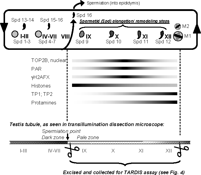
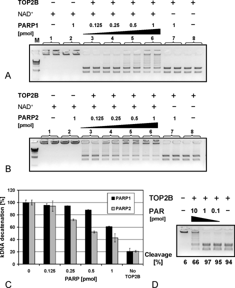
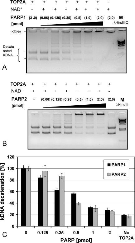
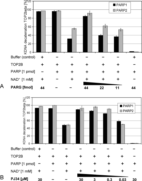
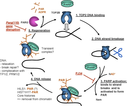
To achieve the specialized nuclear structure in sperm necessary for fertilization, dramatic chromatin reorganization steps in developing spermatids are required where histones are largely replaced first by transition proteins and then by protamines. This entails the transient formation of DNA strand breaks to allow for, first, DNA relaxation and then chromatin compaction. However, the nature and origin of these breaks are not well understood. We previously reported that these DNA strand breaks trigger the activation of poly(ADP-ribose) (PAR) polymerases PARP1 and PARP2 and that interference with PARP activation causes poor chromatin integrity with abnormal retention of histones in mature sperm and impaired embryonic survival. Here we show that the activity of topoisomerase II beta (TOP2B), an enzyme involved in DNA strand break formation in elongating spermatids, is strongly inhibited by the activity of PARP1 and PARP2 in vitro, and this is in turn counteracted by the PAR-degrading activity of PAR glycohydrolase. Moreover, genetic and pharmacological PARP inhibition both lead to increased TOP2B activity in murine spermatids in vivo as measured by covalent binding of TOP2B to the DNA. In summary, the available data suggest a functional relationship between the DNA strand break-generating activity of TOP2B and the DNA strand break-dependent activation of PARP enzymes that in turn inhibit TOP2B. Because PARP activity also facilitates histone H1 linker removal and local chromatin decondensation, cycles of PAR formation and degradation may be necessary to coordinate TOP2B-dependent DNA relaxation with histone-to-protamine exchange necessary for spermatid chromatin remodeling.


Figures

FIG. 1.
FIG. 1.
Schematic overview of spermiogenic development and isolation of relevant steps 9–12 spermatids. After meiosis, haploid spermatids (Spd) undergo an intricate differentiation process, called spermiogenesis, that comprises fundamental remodeling of the nucleus. In mice, spermatids are categorized into 16 developmental steps (Spd 1–16) that are found throughout the 12 stages of spermatogenesis (I–XII). Shapes of spermatid nuclei vary as they progress through development, which is shown only very schematically. Round step 1 spermatids (Spd 1) appear after stage XII, the stage where the meiotic divisions (M1 and M2) take place, that is, in stage I. In stage I, condensing spermatids (Spd 13) are also present. Spd 16 are moved into the epididymis during tubule stage VIII (spermiation). Therefore, tubule of stages IX–XII only contain Spd 9–12 that are in varying degrees of elongation. These developmental stages are marked by the formation of DSBs [12, 13] causing H2AFX phosphorylation (γH2AFX) [14, 15], the formation of PAR [14], and the replacement of histones by transition proteins TP1 and TP2 and protamines (reviewed in [67]). While TOP2B expression is detectable in all spermatid steps, TOP2B association with the nucleus is mostly restricted to tubule stages X and XI (see also Supplemental Fig. S2). This schematic shows that elongating spermatids (Spd 9–12) are only present in stages IX–XII where they represent the only haploid cell fraction. This is relevant to this study because these tubule sections were specifically isolated and utilized for in vivo TOP2B DNA binding studies (TARDIS assays). Because condensing spermatids diffract light more efficiently than other cells, transmission illumination was utilized to specifically excise tubule stages that appear darker or clearer using a dissecting scope. Stages IX–XII were readily identified as a pale zone, and such sections were collected and prepared for TARDIS assays (see Fig. 3).
FIG. 2.
FIG. 2.
TOP2B is inhibited by PARP1 and PARP2 activity in an NAD+-dependent manner in vitro. TOP2B activity was measured by quantifying the conversion rate of high molecular kDNA, which in its native catenated form does not enter the agarose gel, into two low molecular weight bands. A) TOP2B activity is inhibited by PARP1 in the presence, but not in the absence, of NAD+ in an enzyme concentration-dependent manner. B) Like PARP1, PARP2 inhibits TOP2B activity in a comparable manner. C) Quantification of conversion rates from two independent experiments performed in duplicate. The small amount of decatenation in the absence of topoisomerase (right column) represents trace background amounts of kDNA degradation already present in the kDNA substrate. Error bars indicate the standard deviations. D) TOP2B activity is inhibited by purified PAR, albeit only at relatively high levels (1 pmol/μl), indicating that TOP2B may be able to interact with PAR and that covalent posttranslational modification with PAR is not necessary for TOP2B inhibition in vitro.
FIG. 3.
FIG. 3.
TOP2A is inhibited by PARP1 and PARP2 activity. A) PARP1 inhibited TOP2A activity in the presence, but not in the absence, of NAD+ in an enzyme concentration-dependent manner. PARP1 itself exhibited no decatenating activity (lane 8). B) Similarly, PARP2 inhibited TOP2A activity in the presence, but not absence, of NAD+ in an enzyme concentration-dependent manner. C) TOP2A-dependent kDNA conversion rates from two independent experiments performed in duplicate are shown. Error bars indicate the standard deviations.
FIG. 4.
FIG. 4.
PARG activity and inhibition of PARPs by PJ34 restore TOP2B-mediated kDNA decatenating activity in a dose-dependent manner in vitro. A) Addition of PARG to reactions where TOP2B is inhibited by the addition of 1 pmol PARP1 or PARP2 in the presence of 1 mM NAD+ reduced the inhibition in a PARG enzyme concentration-dependent fashion. PARG itself had no decatenating activity and no inhibitory effect on TOP2B activity. B) Addition of PJ34 to inhibit PARP1 and PARP2 activity prevented the inhibition of TOP2B in a dose-dependent manner but by itself did not inhibit or activate TOP2B. Similarly, PARP enzymes did not cause kDNA decatenation or DNA strand breakage. Error bars indicate the standard deviations.
FIG. 5.
FIG. 5.
PARP inhibition increases testicular TOP2B activity in spermatids in vivo. Tubule sections enriched for stages that contain condensing spermatids (see Fig. 1) were analyzed for TOP2B activity using the TARDIS assay method. Live cells from stages IX–XII testis tubules isolated from wild-type mice (i.e., Wt129SV) or mice with genetically altered PAR pathways were embedded in low melting point agarose and lysed in the presence of protease inhibitors; proteins that were not covalently bound to the DNA were extracted using the high salt buffer. A) TOP2B bound to the DNA (red pseudocolored Hoechst 33258 dye) of decondensed spermatid nuclei was stained by indirect immunofluorescence detection (FITC, green) and photographed for digital quantification of signal intensities. Salt extraction-resistant TOP2B is detected as diffuse punctate staining and additional distinct foci restricted to the nucleus (arrows); developing spermatid flagella and midpieces are also stained, indicating the possible presence of TOP2B in these extranuclear cell compartments. Secondary antibody controls were completely negative (data not shown). Immunostaining of epididymal sperm using TOP2B antibodies confirmed the presence of TOP2B in the sperm midpiece (data not shown). Bar = 15 μm. B) TARDIS assay quantification of TOP2B immunofluorescence signals of spermatids isolated from Wt129SVE, Parp1−/−, or Parg(110)−/− mice that had been previously treated with ETO (80 mg/kg) or PJ34 (10 mg/kg) or both for 2 h. For each data point, 3–10 males were used and 500–1200 nuclei were measured per mouse. Each of the separate experiments was designed to include saline- and PJ34-treated wild-type controls. Statistical analyses were done using Student t-test. Data sets highly significant from untreated wild-type controls (P < 0.0001) are indicated by two asterisks (**). Treatment groups within the Parg(110)−/− genotype were not statistically significant from each other (n.s.) but highly significantly different from the wild-type controls. Error bars indicate the standard error of the mean. C) Immunoblot analysis of testicular SDS extracts confirming comparable TOP2B expression levels in 129SVE, Parp1−/−, and Parg(110)−/− mice. D) Testicular PAR steady-state levels are reduced after PJ34 injection of 129SVE mice (left two lanes) compared to saline-injected control mice, as used in the TARDIS assays. Whole testis SDS lysates were used for immunoblotting with PAR-specific antibodies. The loading control was β-actin (ACTB).
FIG. 6.
FIG. 6.
Proposed model of TOP2B and PARP1/2 activation coordinating spermatid DNA relaxation with nucleoprotein exchange. TOP2B, and potentially TOP2A, binds to target DNA (1) and generates a DNA strand break (2). Such DNA strand breaks trigger PARP activation and consequently PAR synthesis (3). Modification with PAR leads to catalytic inactivation and release of the topoisomerase-PARP complex from the DNA, along with PAR acceptor proteins, for example, HILS1, HIST1H1T, and core histones (4). Hypothetically, decatenated DNA is now accessible for binding to other available DNA-binding proteins, for example, transition proteins (TP1/2) and protamines (PRM1/2). The PARsylated topoisomerase-PARP complex is released from the DNA, and degradation of PAR by PARG is essential to restore enzyme activities (5). After removing histones and relieving DNA supercoiling, regenerated PARP and TOP2B enzymes can then move to another site along the chromatin and perform the same actions (1). Pharmacological PARP inhibition with PJ34 inhibits PARP activation (red block at step 3), while PAR accumulation observed in the Parg gene-disrupted mouse model indicates partially compromised enzyme regeneration (red block at step 5). ADPR, adenosine diphosphate ribose; Nam, nicotinamide.

Comment in

Similar articles

Cited by

References

    1. Zhao M, Shirley CR, Hayashi S, Marcon L, Mohapatra B, Suganuma R, Behringer RR, Boissonneault G, Yanagimachi R, Meistrich ML. Transition nuclear proteins are required for normal chromatin condensation and functional sperm development. Genesis 2004; 38: 200 213 - PubMed
    1. Li Y, Lalancette C, Miller D, Krawetz SA. Characterization of nucleohistone and nucleoprotamine components in the mature human sperm nucleus. Asian J Androl 2008; 10: 535 541 - PMC - PubMed
    1. Arpanahi A, Brinkworth M, Iles D, Krawetz SA, Paradowska A, Platts AE, Saida M, Steger K, Tedder P, Miller D. Endonuclease-sensitive regions of human spermatozoal chromatin are highly enriched in promoter and CTCF binding sequences. Genome Res 2009; 19: 1338 1349 - PMC - PubMed
    1. Hammoud SS, Nix DA, Zhang H, Purwar J, Carrell DT, Cairns BR. Distinctive chromatin in human sperm packages genes for embryo development. Nature 2009; 460: 473 478 - PMC - PubMed
    1. Brykczynska U, Hisano M, Erkek S, Ramos L, Oakeley EJ, Roloff TC, Beisel C, Schubeler D, Stadler MB, Peters AH. Repressive and active histone methylation mark distinct promoters in human and mouse spermatozoa. Nat Struct Mol Biol 2010; 17: 679 687 - PubMed

Publication types

MeSH terms

Substances