. 2010 Dec 28;5(12):e15587.
doi: 10.1371/journal.pone.0015587.

Role of sphingomyelin synthase in controlling the antimicrobial activity of neutrophils against Cryptococcus neoformans

Affiliations

Role of sphingomyelin synthase in controlling the antimicrobial activity of neutrophils against Cryptococcus neoformans

Asfia Qureshi et al. PLoS One. .

Abstract

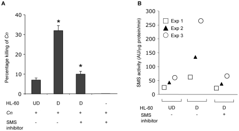
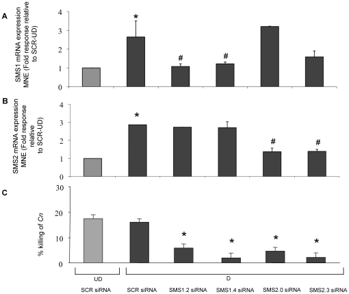
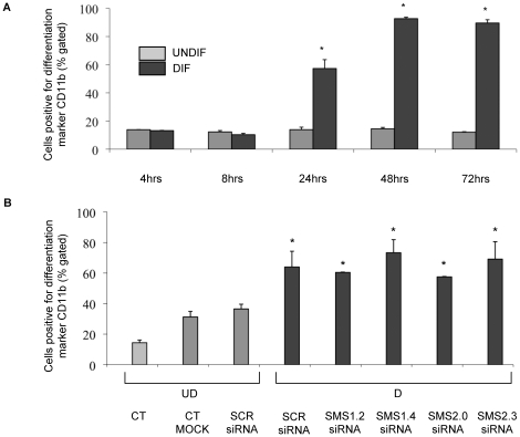
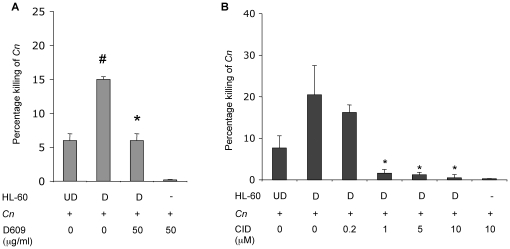
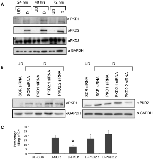
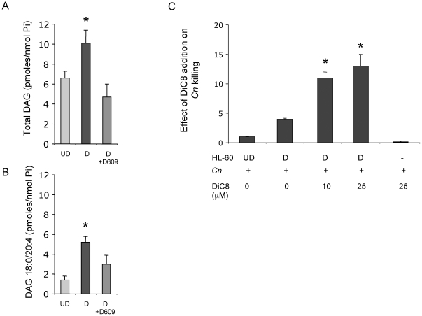
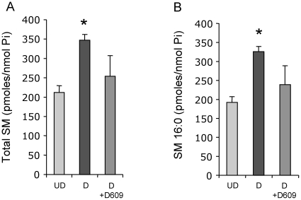
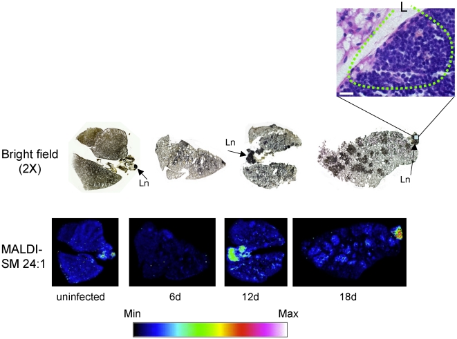
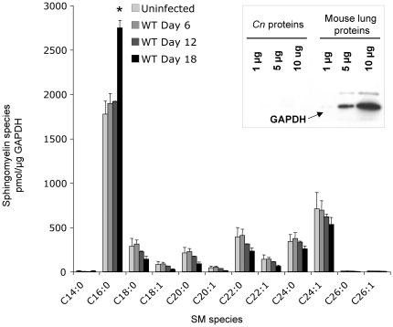
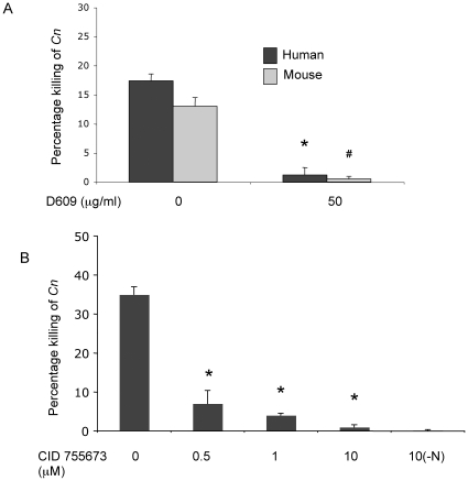
The key host cellular pathway(s) necessary to control the infection caused by inhalation of the environmental fungal pathogen Cryptococcus neoformans are still largely unknown. Here we have identified that the sphingolipid pathway in neutrophils is required for them to exert their killing activity on the fungus. In particular, using both pharmacological and genetic approaches, we show that inhibition of sphingomyelin synthase (SMS) activity profoundly impairs the killing ability of neutrophils by preventing the extracellular release of an antifungal factor(s). We next found that inhibition of protein kinase D (PKD), which controls vesicular sorting and secretion and is regulated by diacylglycerol (DAG) produced by SMS, totally blocks the extracellular killing activity of neutrophils against C. neoformans. The expression of SMS genes, SMS activity and the levels of the lipids regulated by SMS (namely sphingomyelin (SM) and DAG) are up-regulated during neutrophil differentiation. Finally, tissue imaging of lungs infected with C. neoformans using matrix-assisted laser desorption-ionization mass spectrometry (MALDI-MS), revealed that specific SM species are associated with neutrophil infiltration at the site of the infection. This study establishes a key role for SMS in the regulation of the killing activity of neutrophils against C. neoformans through a DAG-PKD dependent mechanism, and provides, for the first time, new insights into the protective role of host sphingolipids against a fungal infection.


Conflict of interest statement

Competing Interests: The authors have declared that no competing interests exist.

Figures

Figure 1
Figure 1
(A). Killing activity of HL-60 cells against C. neoformans (Cn) increases with differentiation and it is abrogated by inhibition of sphingomyelin synthase activity. Percentage killing of C. neoformans wild-type H99 by HL-60 cells undifferentiated (UD) and differentiated (D) with retinoic acid and DMSO. *, P<0.05. Treatment with sphingomyelin synthase inhibitor D609 completely abolished the killing activity of differentiated (D) cells. D609 has no effect on Cn cells. (B) Differentiation of HL-60 cells increases SMS activity. Sphingomyelin synthase (SMS) activity increases in HL-60 differentiated (D) compared to undifferentiated (UD) cells. Treatment with SMS inhibitor D609 decreases SMS activity of HL-60 D. Results show are from 3 independent experiments.
Figure 2
Figure 2. Effect of modulation of either SMS1 or SMS2 during differentiation of HL-60 cells.
Two million HL-60 cells were transfected with 4.5 µg of SCR, SMS1 siRNA (1.2 and 1.4), and SMS2 siRNA (2.0, 2.3) by nucleofection. Differentiation and down-regulation were induced for 48 hours. (A and B) Total RNA was extracted, and RT-PCR was performed using specific primers for SMS1 or SMS2 and GAPDH. The RT-PCR results were analyzed using Q-gene software which expresses data as the means of normalized expression (Fold response relative to SCR-UD). SMS1 mRNA expression was down-regulated by both SMS1.2 and SMS1.4 siRNA (A) and SMS2 mRNA expression was down-regulated by both SMS2.0 and SMS 2.3 siRNA (B). Data are the results of at least 3 independent experiments; error bars represent SD and * P<0.05 compared to SCR siRNA undifferentiated cells; # P<0.05 compared with SCR siRNA differentiated cells. (C) Inhibition of either SMS1 or SMS2 mRNA by siRNA blocks the killing activity of differentiated HL-60 cells; results are representative of at least 3 independent experiments. UD: undifferentiated cells; D: differentiated cells; SCR: scrambled control siRNA. MNE: mean of normalized expression. * P<0.05 compared with SCR siRNA differentiated cells.
Figure 3
Figure 3. Analysis of CD11b differentiation marker by flow cytometry.
(A) HL-60 cells were plated at 1×10⁵ cells/mL and differentiation induced. Undifferentiated cells received vehicle solution for the retinoic acid. Differentiated and undifferentiated cells were collected at the indicated time points and processed for flow cytometric analysis of CD11b positive cells. * P<0.05 compared with undifferentiated time-matched samples. (B) Effect of modulation of either SMS1 or SMS2 on differentiation of HL-60 cells by analysis of CD11b differentiation marker using flow cytometry. Two million HL-60 cells were transfected with 4.5 µg of SCR siRNA, SMS1 siRNA (1.2 and 1.4), and SMS2 siRNA (2.0, 2.3) by nucleofection. Downregulation and differentiation proceeded for 48 hours. Cells were then collected and processed for flow cytometric analysis of CD11b. * P<0.05 compared with SCR siRNA undifferentiated cells. UD: undifferentiated cells; D: differentiated cells; CT MOCK: control for transfection reagent; SCR: control scrambled siRNA.
Figure 4
Figure 4. Effect of conditioned media on the killing activity of HL-60 cells.
(A) Percentage killing of C. neoformans wild-type H99 (Cn) by conditioned media collected from HL-60 cells undifferentiated (UD), differentiated (D), from HL60 D treated with D609 or by D609 itself. (B) Effect of PKD inhibitor on the killing activity of differentiated HL-60 cells against C. neoformans (Cn) or against C. neoformans alone. Percentage killing of Cn wild-type H99 by conditioned media collected from HL-60 cells undifferentiated (UD), differentiated (D) and from HL60 D treated with the PKD1 inhibitor benzoxoloazepinolone (CID755673). # P<0.05 compared with UD cells. * P<0.05, compared to untreated D cells.
Figure 5
Figure 5. PKD isoforms during HL-60 cell differentiation.
(A) HL-60 cells were plated at 1×10⁵ cells/ml and differentiation was induced. Differentiated and undifferentiated cells were collected at 24, 48 and 72 hrs and cellular lysates analyzed by Western blot for PKD1, PKD2, PKD3 and GAPDH. Blots are representative of 3 experiments. (B) Effect of modulation of either PKD1 or PKD2 by siRNA on PKD1 and PKD2 protein levels and GAPDH as loading control. (C) Effect of PKDs siRNA on the killing activity of differentiated HL-60 cells against C. neoformans. Results are representative of at least 3 independent experiments. * P<0.05, compared to D-SCR cells. UD, undifferentiated cells; D, differentiated cells; SCR: control scrambled siRNA.
Figure 6
Figure 6. Mass spectrometry analysis of diacylglycerol in HL-60.
(A) Total levels of DAG in HL-60 undifferentiated (UD), differentiated (D) and HL-60 D treated with D609, as measured by LC-MS and normalized by nanomole of lipid inorganic phosphate (Pi); * P<0.05, compared to UD cells. (B) Specific lipid species for DAG (18:0/20:4) in HL-60 undifferentiated (UD), differentiated (D) and HL-60 D treated with D609, as measured by LC-MS and normalized by Pi. * P<0.05, compared to UD cells. (C) Effect of DiC8 addition on killing activity of differentiated HL-60 cells against C. neoformans (Cn) or C. neoformans alone, represented as a fold response relative to HL-60 undifferentiated cells (UD). * P<0.05, compared to untreated D cells.
Figure 7
Figure 7. Mass spectrometry analysis of sphingomyelin in HL-60.
(A) Total levels of SM in HL-60 undifferentiated (UD), differentiated (D) and HL-60 D treated with D609, as measured by LC-MS and normalized by nanomole of lipid inorganic phosphate (Pi). * P<0.05, compared to UD cells. (B) Specific lipid species for SM (16:0) in HL-60 undifferentiated (UD), differentiated (D) and HL-60 D treated with D609, as measured by LC-MS and normalized by Pi. * P<0.05, compared to UD cells.
Figure 8
Figure 8. Matrix-assisted laser desorption-ionization mass spectrometry (MALDI-MS) of SM C16:0 in lung infected with C. neoformans.
Lungs of CBA/J uninfected and infected mice with C. neoformans wild-type H99 strain were processed for MALDI tissue imaging and bright field photograph (2X) of a lung section stained with mucicarmine. Sphingomyelin (SM) 16:0 species ([M+Na]+ m/z 725) is concentrated in areas highly infiltrated with neutrophils, especially at 18 days of infection, as showed by hematoxylin and eosin staining at right. Min and Max, minimum and maximum intensity, respectively. N, neutrophils in dotted circle; black bar, 20 µm.
Figure 9
Figure 9. Matrix-assisted laser desorption-ionization mass spectrometry (MALDI-MS) of SM C24:1 in lung infected with C. neoformans.
Lungs of CBA/J uninfected and infected mice with C. neoformans wild-type strain were assayed for MALDI tissue imaging and bright field photograph (2X) of a lung section stained with mucicarmine. Sphingomyelin (SM) 24∶1 ([M+Na]+ m/z 835) species is not concentrated in the lung but in hilar and apical lymph nodes, as showed by hematoxylin and eosin staining. (Ln, lymph node). Min and Max, minimum and maximum intensity, respectively. L, lymphocytes in dotted circle. White bar, 20 µm.
Figure 10
Figure 10. Liquid Chromatography Mass spectrometry (LC/MS) analysis of mouse lungs infected with wild-type C neoformans (Cn).
Quantitative measurements by mass spectrometry (HPLC-MS/MS) of different sphingomyelin (SM) species in uninfected and Wild-type (WT)-infected lungs excised from CBA/J mice. Data were normalized to the level of mouse GAPDH protein in lungs using mammalian anti -glyceraldehyde 3-phosphate dehydrogenase (GAPDH) antibody from Ambion (Cat # AM4300), which shows specificity against the mammalian and not against the Cn GAPDH protein (inset). *, P<0.05, Day 18 versus Day 12.
Figure 11
Figure 11. Effect of inhibitors on C. neoformans killing in the presence of neutrophils.
(A) Effect of SMS inhibition on C. neoformans killing in the presence of neutrophils: 50 µg/ml D609 inhibits killing of C. neoformans in human and mouse neutrophils. *, P<0.05, treated versus human untreated cells; #, P<0.05, treated versus mouse untreated cells. (B) Effect of PKD1 inhibition (CID 755673) on killing of C. neoformans in the presence and in absence (-N) of human neutrophils. *, P<0.05, treated versus untreated cells.

Similar articles

Cited by

References

    1. Sanfelice F. Contributo alla morfologia e biologia dei blastomiceti che si sviluppano nei succhi di alcuni frutti. Ann Igien. 1894;4:463–495.
    1. Busse O. Uber parasitare zelleinschlusse und ihre zuchtung. Zentralbl Bakteriol. 1894;16:175–180.
    1. Buschke A. Uber eine durch Coccidien Hervergerufene Krankheit des menschen. Dtsch Med Wochenschr. 1895;21:14.
    1. Casadevall A, Perfect JR. Epidemiology. In: Casadevall A, Perfect JR, editors. Cryptococcus neoformans. Washington, DC: ASM; 1998. pp. 351–380.
    1. Fraser JA, Giles SS, Wenink EC, Geunes-Boyer SG, Wright JR, et al. Same-sex mating and the origin of the Vancouver Island Cryptococcus gattii outbreak. Nature. 2005;437:1360–1364. - PubMed

Publication types