. 2010 Dec 31:11:101.
doi: 10.1186/1471-2121-11-101.

The chromosomal association/dissociation of the chromatin insulator protein Cp190 of Drosophila melanogaster is mediated by the BTB/POZ domain and two acidic regions

Affiliations

The chromosomal association/dissociation of the chromatin insulator protein Cp190 of Drosophila melanogaster is mediated by the BTB/POZ domain and two acidic regions

Daniel Oliver et al. BMC Cell Biol. .

Abstract

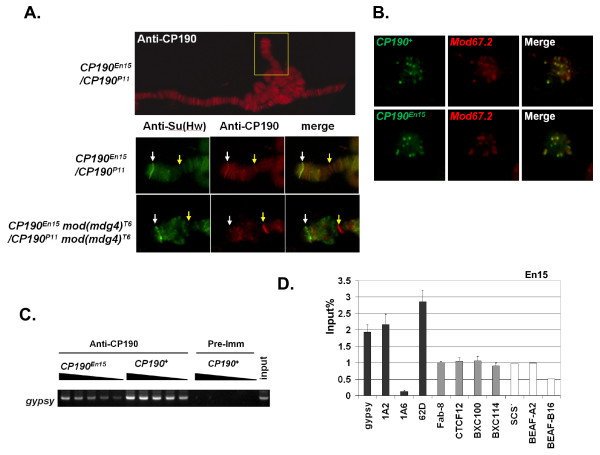
Background: Chromatin insulators or boundary elements are a class of functional elements in the eukaryotic genome. They regulate gene transcription by interfering with promoter-enhancer communication. The Cp190 protein of Drosophila melanogaster is essential to the function of at least three-types of chromatin insulator complexes organized by Su(Hw), CTCF and BEAF32.

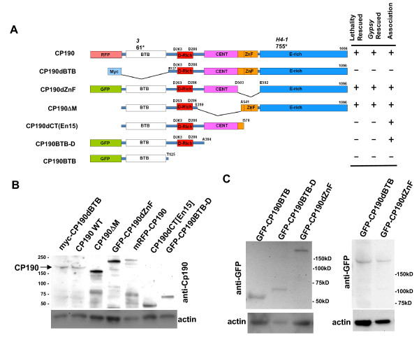
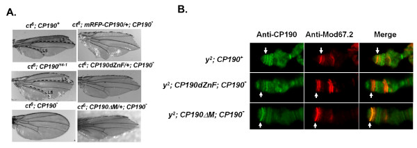
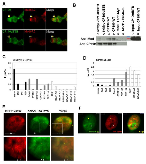
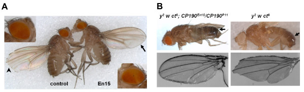
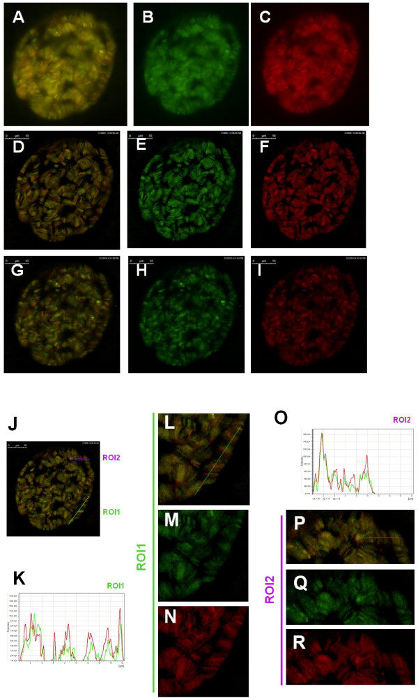
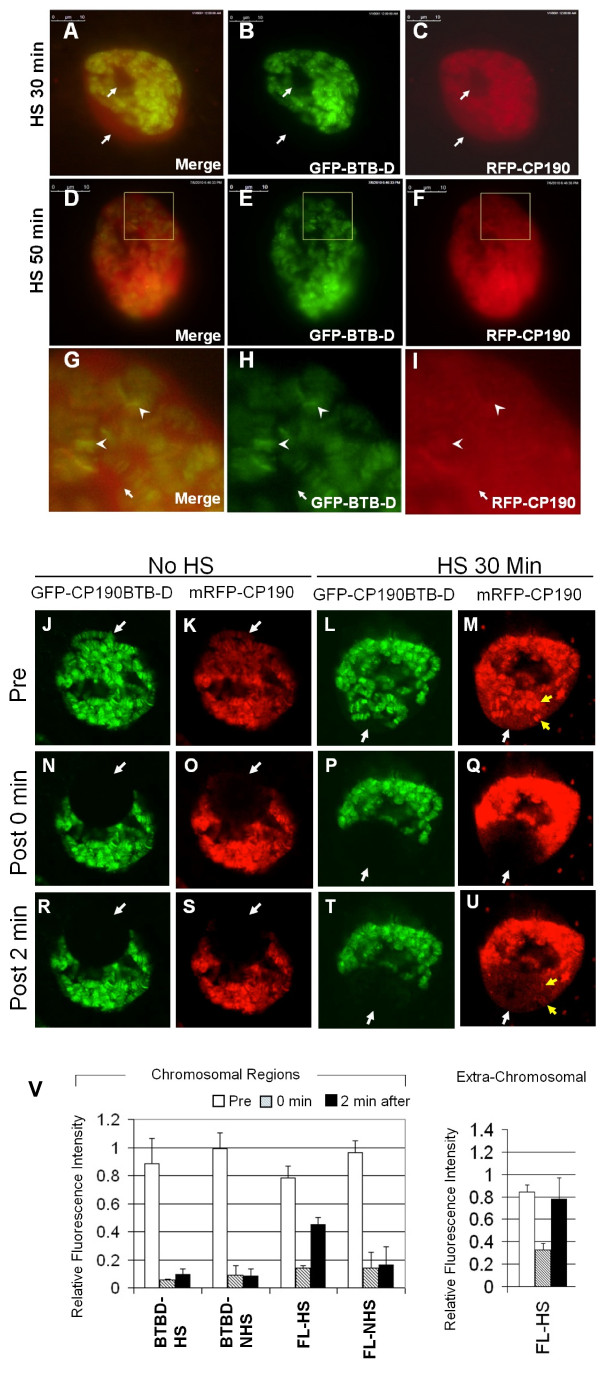
Results: We mapped functional regions of Cp190 in vivo and identified three domains that are essential for the insulator function and for the viability of flies: the BTB/POZ domain, an aspartic acid-rich (D-rich) region and a C-terminal glutamic acid-rich (E-rich) region. Other domains including the centrosomal targeting domain and the zinc fingers are dispensable. The N-terminal CP190BTB-D fragment containing the BTB/POZ domain and the D-rich region is sufficient to mediate association with all three types of insulator complexes. The fragment however is not sufficient for insulator activity or viability. The Cp190 and CP190BTB-D are regulated differently in cells treated with heat-shock. The Cp190 dissociated from chromosomes during heat-shock, indicating that dissociation of Cp190 with chromosomes can be regulated. In contrast, the CP190BTB-D fragment didn't dissociate from chromosomes in the same heat-shocked condition, suggesting that the deleted C-terminal regions have a role in regulating the dissociation of Cp190 with chromosomes.

Conclusions: The N-terminal fragment of Cp190 containing the BTB/POZ domain and the D-rich region mediates association of Cp190 with all three types of insulator complexes and that the E-rich region of Cp190 is required for dissociation of Cp190 from chromosomes during heat-shock. The heat-shock-induced dissociation is strong evidence indicating that dissociation of the essential insulator protein Cp190 from chromosomes is regulated. Our results provide a mechanism through which activities of an insulator can be modulated by internal and external cues.


Figures

Figure 1
Figure 1
Protein structure and expression levels of CP190 deletion mutants. (A) Schematic diagram of CP190 deletion mutants and their genetic complementation phenotypes. The full-length Cp190 is tagged with mRFP (mRFP-CP190). Each CP190 mutant contains a deletion of one of the functional domains. CP190dBTB lacks the BTB domain and is tagged with myc (myc-CP190dBTB) or with GFP (GFP-CP190dBTB, not shown); CP190dZnF lacks all three zinc fingers and is tagged with GFP (GFP-CP190dZnF); CP190ΔM lacks the centrosomal targeting domain (CENT); CP190BTB-D lacks CENT, zinc fingers and the E-rich domain and is tagged with GFP (GFP-CP190BTB-D); CP190BTB has only the BTB domain and is tagged with GFP (GFP-CP190BTB). The CP190dCT(En15) is the predicted protein from the EMS-induced CP190En15 mutant, which lacks two zinc fingers and the E-rich region. The amino acids at the junction of each deletion are indicated. (B-C) Expression of the mutated Cp190 proteins revealed by the anti-CP190 immunoblot (B) or by anti-GFP immunoblots (C). All transgenic lines were crossed into the homozygous CP1903 background. Proteins extracted from about two 3rd instar larvae containing the indicated transgene were loaded per each lane. Similar results were also obtained from pupae 24-48 hours after pupation. Underneath the blots were stripped filters re-probed with the anti-actin antibody as a loading control.
Figure 2
Figure 2
The GFP-CP190dZnF and the Cp190ΔM proteins are fully functional in the gypsy insulator. (A) The gypsy insulator-dependent wing phenotype of the ct6 mutation in the indicated genetic backgrounds. Wings of the ct6 flies have a cut shape and lack of margin bristles (top left). The two doted lines mark the veins L3 and L5 between which the margin bristles are not developed at all. The ct6 ; CP190H4-1 wing has some margin bristles between L3 and L5, and is rounder in shape (middle left). The wing shape of the CP190 deficient CP1903/CP190P1 flies is restored to a ct+-like shape with fully developed margin bristles (bottom left), indicated. A copy of the P[Ubi63e::mRFP-CP190] (top right), P[Ubi63e::GFP-CP190dZnF] (middle right) or the P[CP190ΔM] (bottom right) transgene rescues the defective gypsy insulator function in the CP190 deficient CP1903/CP190P1 background. The rescued flies have the cut wing shape similar to the ct6; CP190+ flies. (B) Localization of the mutated Cp190 proteins to the gypsy insulator insertion at the y locus on the y2 polytene chromosome. Shown are the tips of X chromosomes. The y locus that contains a copy of the gypsy insulator is indicated by white arrows. Distribution of the GFP-CP190dZnF protein (middle panel) and the Cp190ΔM protein (bottom panel) on the polytene chromosomes of the indicated flies was revealed by anti-Cp190 (left column). The anti-Mod(mdg4)67.2 (middle column) shows the distribution of the Su(Hw) insulator complex. The right column shows the merged images of the left and the middle columns.
Figure 3
Figure 3
The BTB domain is necessary but not sufficient for association with the Su(Hw) complex. (A) Distribution of myc-CP190dBTB and Cp190 on polytene chromosomes. The polytene chromosomes of the y2 (upper panel) and of the y2; P[Ubi63e::myc-CP190dBTB]; CP190P1/CP1903 (lower panel) flies were stained with anti-CP190 (left column) and anti-Mod(mdg4)67.2 (middle column). Shown are the tips of X chromosomes with the y locus (white arrows) and a band nears y (yellow arrows). (B) Co-immunoprecipitation of Cp190 and Mod(mdg4)67.2. Immunonblots of anti-Cp190 (top panel) and anti-Mod(mdg4)67.2 (bottom panel). Proteins were immunoprecipitated with anti-Myc from y2 ct6; P[Ubi63e::myc-CP190dBTB]/+; CP1903/TM6B pupae (lanes 1 and 2), with anti-Cp190 from y2 ct6 pupae (lanes 3 and 4), and with anti-Myc (lane 5) or pre-immune (lane 6) from y2 ct6 pupae. Input controls from myc-CP190dBTB (lane 7) and from y2 ct6 (lane 8) pupae. (C-D) Anti-Cp190 ChIP of known Su(Hw), CTCF, and BEAF32 loci assayed by Real-Time PCR (percentage of input DNA, n≥3), from y2 ct6 flies (C) and from ct6; P[Ubi63e::myc-CP190dBTB; CP1903/CP190P1 flies (D). The 1A6 region is the negative control [12]. All results were normalized to Fab-8. (E) Distribution of the mRFP-CP190 (red, left column), GFP-CP190dBTB (green, middle column) in a living salivary gland cell nucleus from a 3rd instar larva. An extra-chromosomal space containing GFP-CP190dBTB signals but not mRFP-CP190 signals (yellow arrows). The closer views (bottom row) are crops indicated by the white squares in the upper row. The white arrows point to two bands containing both mRFP-CP190 and GFP-CP190dBTB. (F) Distribution of GFP-CP190BTB-nls (green, left) and mRFP-CP190 (red) in the cell nucleus of a living salivary gland from a 3rd instar larva. The white arrows point an extra-chromosomal space containing GFP-CP190BTB-nls but not mRFP-CP190. The yellow arrows point to two mRFP-CP190 bands on polytene chromosomes that did not have detectable GFP-CP190BTB-nls signals.
Figure 4
Figure 4
The C-terminal E-rich domain is essential to Cp190's insulator function. (A) Genetic assays to test functionality of the gypsy insulator in CP190E15. Shown are morphological phenotypes of the y2 w ombP1-D11 ct6; mod(mdg4)T6 e female (left, control) and the y2 w ombP1-D11 ct6; CP190En15 mod(mdg4)T6 e/mod(mdg4)T6 e female (right, En15). The En15 fly has a darker abdomen cuticle color (enhanced y2 phenotype) compared to the control fly. The arrowhead points to the partially suppressed ct6 wing shape phenotype which lacks some wing margin bristle cells. The arrow points to the fully suppressed ct6 wing shape (enhanced ct6 phenotype). The ombP1-D11 pigmentation pattern of the eye of the control female fly is shown on the upper left and the ombP1-D11 pigmentation pattern of the En15 female which has expanded white region in the equatorial part of the eye (enhanced ombP1-D11 phenotype) is shown on the lower right. (B) The body cuticle pigmentation (upper panel) and the wing shape (lower panel) of the CP190En15/CP190P11 mutant (left column) and CP190+ flies (right column). Arrows point to different pigmentation of the abdomens.
Figure 5
Figure 5
The Cp190 fragment lacking the C-terminal E-rich domain localizes to Cp190-containing nuclear complexes. (A) Polytene chromosomes from y2; CP190En15/CP190P11 (top and middle panels) and from y2; CP190En15 mod(mdg4)T6/CP190P11 mod(mdg4)T6 (bottom panel) 3rd instar larvae were stained with anti-Su(Hw) (green, left column) and anti-Cp190 (red, middle column) antibodies. Merged images are shown on the right. The middle panel is the closer view, rotated 90 degrees counter clockwise, of the area indicated by the yellow square in the top image. White arrows point to the y locus at the tip of the X chromosome. Yellow arrows point to the Cp190-positive band at the constriction of the cytological location 3C. (B) Diploid cells of brain tissues from CP190+ flies (top) or from the CP190En15/CP190P11 (bottom) flies were stained with anti-Cp190 (green, left column) and anti-Mod(mdg4) (red, middle column) antibodies. Merged images are shown on the right. (C) The gypsy fragment amplified by PCR from anti-Cp190 precipitated chromatin from y2; CP190En15/CP190P11 pupae (left lanes, labeled CP190En15) and from y2; CP190+ pupae (middle lanes). The gypsy fragment amplified from the pre-immune serum precipitated y2 pupae (right lanes, labeled CP190+ and Pre-Imm) was the negative control and the gypsy fragments amplified from non-precipitated total y2 pupal genomic DNA (labeled as input on top) was the positive control. The black triangles indicate two-fold serial dilutions of the amount of the chromatin sample used in each PCR reaction. More volumes of the indicated template were in the PCR reactions on the left as indicated by the thicker part of each triangle. (D) The anti-Cp190 ChIP of y2 ct6; CP190En15 flies analyzed by Real-Time PCR. The same loci tested in Figure 3 ChIP assays were analyzed and were shown as percentage of input DNA (n ≥ 3). The results of all regions were normalized to Fab-8.
Figure 6
Figure 6
The N-terminal Cp190 fragment containing the BTB domain and D-rich region colocalizes with the full-length Cp190 protein. (A-I) The distribution of the GFP-CP190BTB-D (green, B, E, H, M, Q) and the mRFP-CP190 (red, C, F, I, N, R) proteins in the cell nucleus of a living salivary gland. (D-F) and (G-I) are two of the optical sections from the same cell shown in (A-C) and were analyzed by deconvolusion algorism. The same deconvolusion processed optical section as (D-F) is marked with the ROI1 (green) and ROI2 (purple) (J). (K-R) The intensity profile chart of the ROI1 (K) and the closer views of ROI1 (L-N). The ROI1 is indicated as a green line in (J) and in (L). The Intensity profile chart of the ROI2(O) and the closer views of ROI2 (P-R). The ROI2 is indicated as a purple line in (P) and in (J).
Figure 7
Figure 7
The full sized Cp190 dissociated from polytene chromosomes during heat-shock while the CP190BTB-D fragment remained bound. (A-I) Salivary glands dissected from 3rd instar larvae expressing both mRFP-CP190 and GFP-CP190BTB-D proteins were treated with heat-shock for 30 minutes (A-C), for 50 minutes (D-I). (J-V) Salivary glands without heat-shock (J, K, N, O, R, S) or heat-shock for 30 mins (L, M, P, Q, T, U) were analyzed by the Fluorescence Recovery After Photobleach (FRAP) technique. The distribution of GFP-CP190BTB-D (green, B, E, and H) and the distribution of mRFP-CP190 (red, C, F, I) indicate that mRFP-CP190 dissociated from the chromosomes and was present in extra-chromosomal spaces (arrows). The squares in D, E, and F mark the region that is enlarged in G, H, and I. The arrow heads in G, H, and I points to two randomly sampled bands of GFP-CP190BTB-D on polytene chromosomes. (J-U) Salivary glands without the pretreatment of heat-shock (J, K, N, O, R, S) or heat-shocked for 30 minutes (L, M, P, Q, T, U) were analyzed by the FRAP technique. Images were taken before photobleaching (J, K, L, M), right after photobleaching (N, O, P, Q) and 2 minutes after photobleaching (R, S, T, U). White arrows point to areas that were photobeached. Yellow arrows point to bands reappeared after photobeaching. (V) Quantitative analysis of the fluorescence of GFP-CP190BTB-D (BTBD) and mRFP-CP190 (FL) before photobleaching (white bars), 0 minute (grey bars) and 2 minutes (black bars) after photobleaching from multiple nuclei (n > = 3) in heat-shocked treated (HS) or non-heat-shocked treated (NHS) salivary glands. The Relative Fluorescence Intensity (RFI) of chromosomal regions (left chart) and the RFI of the mRFP-CP190 in HS nuclei in extra-chromosomal regions (right chart).

Similar articles

Cited by

References

    1. Gyurkovics H, Gausz J, Kummer J, Karch F. A new homeotic mutation in the Drosophila bithorax complex removes a boundary separating two domains of regulation. Embo J. 1990;9(8):2579–2585. - PMC - PubMed
    1. Geyer PK, Corces VG. DNA position-specific repression of transcription by a Drosophila zinc finger protein. Genes Dev. 1992;6(10):1865–1873. doi: 10.1101/gad.6.10.1865. - DOI - PubMed
    1. Jack J, Dorsett D, Delotto Y, Liu S. Expression of the cut locus in the Drosophila wing margin is required for cell type specification and is regulated by a distant enhancer. Development. 1991;113(3):735–747. - PubMed
    1. Spana C, Harrison DA, Corces VG. The Drosophila melanogaster suppressor of Hairy-wing protein binds to specific sequences of the gypsy retrotransposon. Genes Dev. 1988;2(11):1414–1423. doi: 10.1101/gad.2.11.1414. - DOI - PubMed
    1. Dorsett D. Potentiation of a polyadenylylation site by a downstream protein-DNA interaction. Proc Natl Acad Sci USA. 1990;87(11):4373–4377. doi: 10.1073/pnas.87.11.4373. - DOI - PMC - PubMed

Publication types

MeSH terms

LinkOut - more resources