. 2011 Jan;121(1):296-307.
doi: 10.1172/JCI42056. Epub 2010 Dec 22.

The TNFR family members OX40 and CD27 link viral virulence to protective T cell vaccines in mice

Affiliations

The TNFR family members OX40 and CD27 link viral virulence to protective T cell vaccines in mice

Shahram Salek-Ardakani et al. J Clin Invest. 2011 Jan.

Abstract

Induction of CD8+ T cell immunity is a key characteristic of an effective vaccine. For safety reasons, human vaccination strategies largely use attenuated nonreplicating or weakly replicating poxvirus-based vectors, but these often elicit poor CD8+ T cell immunity and might not result in optimal protection. Recent studies have suggested that virulence is directly linked to immunogenicity, but the molecular mechanisms underlying optimal CD8+ T cell responses remain to be defined. Here, using natural and recombinant vaccinia virus (VACV) strains, we have shown in mice that VACV strains of differing virulence induce distinct levels of T cell memory because of the differential use of TNF receptor (TNFR) family costimulatory receptors. With strongly replicating (i.e., virulent) VACV, the TNFR family costimulatory receptors OX40 (also known as CD134) and CD27 were engaged and promoted the generation of high numbers of memory CD8+ T cells, which protected against a lethal virus challenge in the absence of other mechanisms, including antibody and help from CD4+ T cells. In contrast, weakly replicating (i.e., low-virulence) VACV strains were poor at eliciting protective CD8+ T cell memory, as only the Ig family costimulatory receptor CD28 was engaged, and not OX40 or CD27. Our results suggest that the virulence of a virus dictates costimulatory receptor usage to determine the level of protective CD8+ T cell immunity.


Figures

Figure 1
Figure 1. Altered virulence of VACV strains.
C57BL/6 WT mice were infected i.p. with different strains of VACV (2 × 10⁵ PFU). (A) On the indicated days after infection, ovaries and spleens were removed from individual mice, and VACV titers were determined as described in Methods. (B) WT mice were infected i.n. with 104 PFU WR and 105 or 106 PFU WR-B18R, Lister, and NYCBOH as indicated. Animals were weighed daily. Mean percent of initial body weight is shown. Results are mean (n = 4 per group) from 1 of 3 experiments. *P < 0.05 vs. WR.
Figure 2
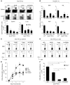
Figure 2. VACV virulence correlates with magnitude of CD8+ T cell memory.
WT mice were infected i.p. (A and CF) or i.n. (B) with the indicated VACV strains. Splenocytes and lung cells were harvested on day 40 (A), day 60 (B), day 540 (C and D), or days 4, 5, 6, 7, and 8 (E and F) after infection and stained with anti-CD8, -CD44, and -B8R or for intracellular IFN-γ after restimulation with the designated VACV peptides. (A) Representative plots of gated CD8+ T cells staining for CD44 and B8R in spleen and lung. Numbers indicate percent CD44+B8R+ cells after gating on CD8+ T cells. Total numbers of CD8+CD44+B8R+ T cells per organ were determined as described in Methods. *P < 0.05 vs. WR. (B) Number of CD8+IFN-γ+ T cells per lung specific for peptides B8R, A3L, A8R, and B2R. (C and D) Percent CD8+IFN-γ+ T cells per spleen (C) or lung (D) specific for B8R and A8R. (E and F) Total number of CD8+CD44+B8R+ (E) and CD8+IFN-γ+ (F) T cells per spleen at the indicated time points after infection with VACV variants. Results are mean ± SEM (n = 4 per group) from 1 of 2 experiments. *P < 0.05 vs. all other strains or as otherwise denoted.
Figure 3
Figure 3. OX40 does not drive CD8+ T cell memory with attenuated viruses or when viral replication is low.
(AG) WT or Ox40–/– mice were infected i.p. with various VACV strains (2 × 10⁵ or 2 × 10⁶ PFU) as indicated. (H) At 1 day after infection with WR, mice were injected i.p. with 200 μg poly:IC. At day 40 (A and B) or day 7 (C, D, F, and G), splenocytes were stained with CD8 plus CD44, CD62L, and B8R or intracellular IFN-γ, and the number of VACV-reactive CD8+ T cells was calculated. (F) Representative plots of gated CD8 cells staining for CD62L and B8R in spleen. Numbers indicate percent CD62L+B8R+ and CD62LB8R+ cells after gating on CD8+ T cells. Results are mean ± SEM (n = 6 per group). *P < 0.05 vs. WT. Similar results were obtained in 3 separate experiments. (E and H) On day 4 after infection, spleens from the indicated groups were removed from individual mice, and VACV titers were determined as described in Methods. *P < 0.05.
Figure 4
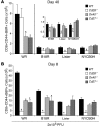
Figure 4. Virulence of VACV and stage of CD8+ T cell response determine the selective use of CD28, OX40, and CD27.
WT, Cd28–/–, Ox40–/–, or Cd27–/– mice were infected i.p. with VACV variants (2 × 10⁵ PFU) as indicated. At day 40 (A) or day 7 (B), splenocytes were stained with CD8 plus CD44 and B8R, and the number of VACV-reactive CD8+ T cells was calculated. Results are mean ± SEM (n = 6 per group). *P < 0.05 vs. WT. Similar results were obtained in 2 separate experiments. Inset in A shows the same data for WR-B18R, Lister, and NYCBOH with the scale enlarged.
Figure 5
Figure 5. CD27 signaling is critical for CD28-independent CD8+ T cell responses to WR.
(A and B) WT or Cd28–/– mice were infected i.p. with WR (2 × 10⁵ PFU). At the indicated days after infection, splenocytes were stained with CD8 plus CD44 and B8R (A), or intracellular IFN-γ or TNF (B), and the number or percentage of VACV-reactive CD8+ T cells was calculated. (C and D) WT, Cd28–/–, or Cd27–/– mice were infected i.p. with WR and injected i.p. on days 5, 6, 7, and 8 (C) with 150 μg nondepleting anti-CD70 blocking Ab in PBS. At day 13 after infection, splenocytes were harvested and stained for CD8, CD44, and B8R. (D) Representative plots of B8R staining, gating on CD8+ T cells, as well as total number of CD8+CD44+B8R+ T cells per spleen. Numbers within dot plots denote percentage of cells in the respective quadrants/gates. Results are mean ± SEM (n = 4 per group). *P < 0.05. Similar results were obtained in 2 separate experiments.
Figure 6
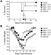
Figure 6. Vaccination of MHCII–/– mice with WR, but not NYCBOH or Lister, protects against lethal respiratory virus challenge.
MHCII–/– mice were immunized i.p. with VACV variants (2 × 10⁵ PFU). Naive MHCII–/– mice were used as control. 10 weeks after vaccination, mice were infected i.n. with a lethal dose of WR (4.5 × 10⁶ PFU; i.e., 400 × LD50). Some MHCII–/– groups were depleted of CD8+ (αCD8) T cells before i.n. challenge with VACV. Animals were weighed daily and euthanized if weight loss was greater than 30% body weight. Mean percent survival (A) and percent of initial body weight (B) are shown. Mean weight data in some cases were not plotted beyond the point at which mice died and beyond day 7 reflected only mice that survived infection. (C) On day 7 after challenge, tissues from individual mice that survived the infection were collected, and virus titers were determined by plaque assay as described in Methods. Results are mean (n = 4 per group) from 1 experiment. (D and E) Percent and total number of CD8+CD44+B8R+ cells and B8R-reactive, IFN-γ–producing CD8+CD62L cells in the spleen (D) and lungs (E) of MHCII–/– mice 90 days after immunization with WR or Lister. Results are mean ± SEM (n = 4 per group) from 1 experiment. *P < 0.05 vs. WR.
Figure 7
Figure 7. The frequency of CD8+ T cells in the lung prior to challenge directly correlates with the degree of protection against lethal VACV infection.
Naive CD8+CD44lo and VACV-reactive memory CD8+CD44hiB8R+ T cells were isolated from WR-infected mice, and varying numbers were instilled into the lungs of naive mice via the trachea. Some groups received 400 μl VACV-immune serum i.p. 1 day after transfer, mice were infected i.n. with a lethal dose of WR (1 × 10⁶ PFU; i.e., 100 × LD50). Animals were weighed daily and euthanized if weight loss was greater than 30% body weight. Mean percent survival (A) and percent of initial body weight (B) are shown. Mean weight data in some cases were not plotted beyond the point at which mice died and beyond day 7 reflected only mice that survived infection. Results are mean (n = 4 per group) from 1 experiment.
Figure 8
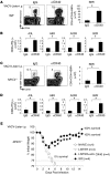
Figure 8. Enhanced CD8+ T cell responses to attenuated VACVs following agonist anti-OX40 treatment.
WT (A and B) and MHCII–/– (CE) mice were infected with 2 × 10⁵ PFU Lister i.p. 1 day later, mice were treated with 150 μg control rat IgG or anti-OX40 (αOX40). 8 days after infection, VACV-specific CD8+ T cells were assessed by tetramer (A and C) or by intracellular IFN-γ staining after stimulation with the indicated VACV peptides (B and D). Data are either representative plots of tetramer staining in gated CD8+ T cells, with percent positive indicated, or total number (mean ± SEM) of CD8+IFN-γ+ T cells per spleen from 4 individual mice. *P < 0.05. Similar results were obtained in 2 separate experiments. (E) MHCII–/– mice were immunized i.p. with WR or Lister (2 × 10⁵ PFU). 1 day later, cohorts of Lister-immunized mice were treated with 150 μg anti-OX40. Naive MHCII–/– mice were used as control. 10 weeks after vaccination, mice were infected i.n. with a lethal dose of WR (4.5 × 10⁶ PFU; i.e., 400 × LD50). Animals were weighed daily and euthanized if weight loss was greater than 25% body weight. Mean percent of initial body weight is shown, with percent survival indicated.
Figure 9
Figure 9. WR and Lister scarification generates superior protective CD8+ T cell immunity against i.n. viral challenge that is mediated by CD28, OX40, and CD27.
WT mice were infected by dermal scarification with WR and Lister (2 × 10⁵ PFU). (A) On the indicated days after infection, VACV titers at the site of infection were determined as described in Methods. *P < 0.05. (BD) 8 days after infection, splenocytes were harvested and stained with anti-CD8, -CD44, and -B8R or for intracellular IFN-γ after restimulation with B8R peptide. (B and C) Representative plots of gated CD8 cells staining for CD44 and B8R in spleen. Numbers indicate percent CD44+B8R+ cells after gating on CD8+ T cells. Total number of CD8+CD44+B8R+ T cells per organ was determined as described in Methods. (D) Percent CD8+IFN-γ+ T cells per spleen specific for B8R peptide. Results are mean ± SEM (n = 4 per group) from 1 of 3 experiments. *P < 0.05. (E) MHCII–/– mice were immunized by dermal scarification with WR and Lister (2 × 10⁵ PFU). Naive MHCII–/– mice were used as control. 10 weeks after vaccination, mice were infected i.n. with a lethal dose of WR (4.5 × 10⁶ PFU; i.e., 400 × LD50). Animals were weighed daily and euthanized if weight loss was greater than 30% body weight. Mean weight data in some cases were not plotted beyond the point at which mice died and beyond day 7 reflected only mice that survived infection.

Comment in

Similar articles

Cited by

References

    1. Lee MS, et al. Molecular attenuation of vaccinia virus: mutant generation and animal characterization. J Virol. 1992;66(5):2617–2630. - PMC - PubMed
    1. Midgley CM, Putz MM, Weber JN, Smith GL. Vaccinia virus strain NYVAC induces substantially lower and qualitatively different human antibody responses compared with strains Lister and Dryvax. J Gen Virol. 2008;89(pt 12):2992–2997. doi: 10.1099/vir.0.2008/004440-0. - DOI - PMC - PubMed
    1. Frey SE, et al. Dose-related effects of smallpox vaccine. N Engl J Med. 2002;346(17):1275–1280. doi: 10.1056/NEJMoa013431. - DOI - PubMed
    1. Wyatt LS, Earl PL, Eller LA, Moss B. Highly attenuated smallpox vaccine protects mice with and without immune deficiencies against pathogenic vaccinia virus challenge. Proc Natl Acad Sci U S A. 2004;101(13):4590–4595. doi: 10.1073/pnas.0401165101. - DOI - PMC - PubMed
    1. Stittelaar KJ, et al. Safety of modified vaccinia virus Ankara (MVA) in immune-suppressed macaques. Vaccine. 2001;19(27):3700–3709. doi: 10.1016/S0264-410X(01)00075-5. - DOI - PubMed

Publication types

MeSH terms