. 2011 Mar;85(5):2079-88.
doi: 10.1128/JVI.02279-10. Epub 2010 Dec 22.

EBNA3C attenuates the function of p53 through interaction with inhibitor of growth family proteins 4 and 5

Affiliations

EBNA3C attenuates the function of p53 through interaction with inhibitor of growth family proteins 4 and 5

Abhik Saha et al. J Virol. 2011 Mar.

Abstract

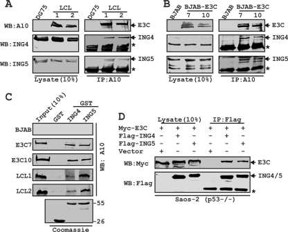
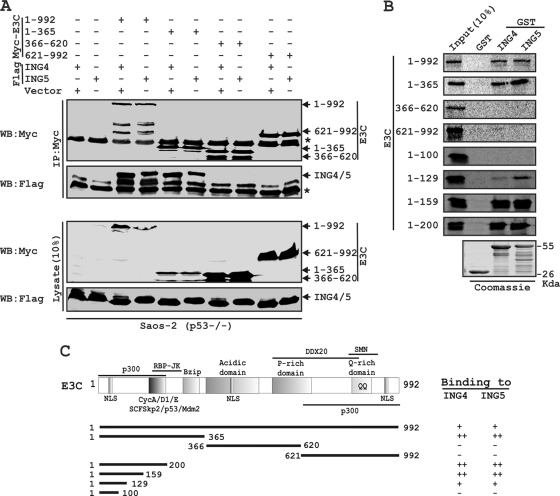
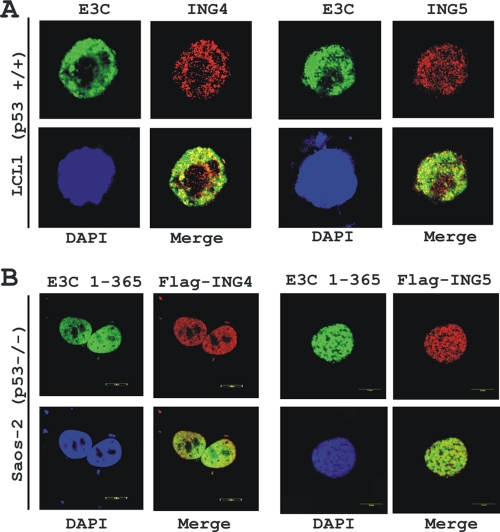
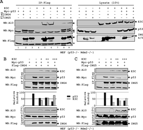
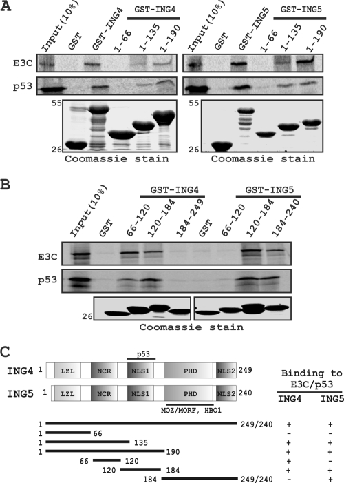
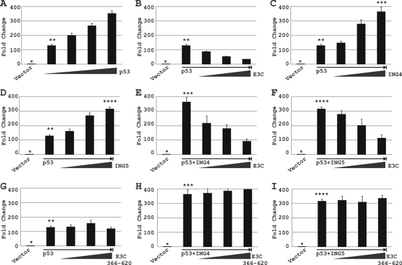
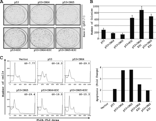
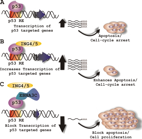
Epstein-Barr virus (EBV)-encoded EBNA3C is one of the latent proteins essential for the efficient transformation of human primary B lymphocytes into continuously proliferating lymphoblastoid cell lines (LCLs) in vitro through manipulation of a number of major cellular pathways. Although it does not have direct DNA-binding activity, EBNA3C plays a central role in the transcriptional modulation of a wide range of both viral and cellular genes during latent infection. Recently, we showed that EBNA3C can directly bind to the tumor suppressor protein p53 and repress its functions, in part by blocking its transcriptional activity as well as facilitating its degradation through stabilization of its negative regulator, Mdm2. In this study, we further showed that EBNA3C can negatively regulate p53-mediated functions by interacting with its regulatory proteins, the inhibitor of growth family proteins ING4 and ING5, shown to be frequently deregulated in different cancers. Functional mapping revealed that both ING4 and ING5 bound to N-terminal domain residues 129 to 200 of EBNA3C, which was previously demonstrated to associate with p53 and is also essential for LCL growth. In addition, we showed that a conserved domain of either ING4 or ING5 bound to both p53 and EBNA3C in a competitive manner, suggesting a potential role for EBNA3C whereby the ING4 or -5/p53 pathway is modulated in EBV-infected cells. Subsequently, we demonstrated that EBNA3C significantly suppresses both the ING4- and ING5-mediated regulation of p53 transcriptional activity in a dose-dependent manner. A colony formation assay as well as an apoptosis assay showed that EBNA3C nullified the negative regulatory effects on cell proliferation induced by coupled expression of p53 in the presence of either ING4 or ING5 in Saos-2 (p53(-/-)) cells. This report demonstrates a possible role for the candidate tumor suppressor ING genes in the biology of EBV-associated cancers.


Figures

FIG. 1.
FIG. 1.
EBNA3C forms a p53-independent complex with both ING4 and ING5. BJAB cells and two BJAB cell stable clones expressing EBNA3C (E3C7 and 10) (A) and DG75 cells and two LCL clones (LCL1 and 2) (B) were subjected to IP with EBNA3C specific antibody (A10). Samples were resolved by SDS-PAGE and detected by Western blotting (WB) for the indicated proteins by stripping and reprobing the same membrane. (C) Either purified GST or GST-ING4 or -ING5 beads were incubated with cell lysates, and EBNA3C was detected by WB with A10. Coomassie staining of GST-labeled proteins is shown in the bottom panel. Numbers at the right of the bottom blot are molecular masses (in kilodaltons). (D) Saos-2 (p53−/−) cells were cotransfected with myc-EBNA3C in the presence of either the vector control, flag-tagged ING4, or flag-tagged ING5, as indicated. At 36 h posttransfection, cells were harvested, lysed, and immunoprecipitated with flag antibody. Samples were subjected to WB with the indicated antibodies. The asterisks indicate the immunoglobulin bands.
FIG. 2.
FIG. 2.
A critical N-terminal domain of EBNA3C interacts with both ING4 and ING5. (A) Saos-2 (p53−/−) cells were cotransfected with either flag-ING4 or -ING5 in the presence of different myc-EBNA3C constructs, as indicated. Cells were harvested, lysed, and immunoprecipitated with anti-myc antibody. Samples were resolved by a 7-to-15% gradient SDS-PAGE and detected by Western blotting (WB) for the indicated proteins by stripping and reprobing the same membrane. (B) The wild-type plasmid or plasmids of EBNA3C expressing different truncated mutant proteins were in vitro translated in the presence of [S35]Met. After preclearing of all S35-radiolabeled translated proteins with GST beads for 1 h at 4°C, samples were subjected to GST pulldown assay by incubating them with either a GST control, GST-ING4, or GST-ING5 as indicated. Reactions were resolved by appropriate SDS-PAGE, exposed to a phosphorimager plate, and scanned using the Storm 850 imaging system. Coomassie staining of a parallel SDS-PAGE gel resolved purified GST proteins, seen in the bottom panel. (C) The schematic illustrates different structural and interaction domains of EBNA3C and summarizes the results of studies of the binding between different domains of EBNA3C and ING4 and ING5. ++, strong binding; +, moderate binding; −, no binding. NLS, nuclear localization signal; Bzip, basic leucine zipper domain; SMN, survival of motor neurons. The asterisk indicates the immunoglobulin bands.
FIG. 3.
FIG. 3.
EBNA3C colocalizes with both ING4 and ING5. (A) EBV-transformed LCL1 cells were plated and air dried onto slides. (B) Saos-2 (p53−/−) cells plated on coverslips were transfected with GFP-tagged EBNA3C (residues 1 to 365) in the presence of either flag-ING4 or -ING5 using Lipofectamine 2000. Endogenously (A) and ectopically (B) expressed ING4 and ING5 were detected using specific antibodies and M2 antibody, respectively, followed by specific anti-Alexa Fluor 594 20 antibody (red). (A) Endogenous EBNA3C in EBV-positive LCLs was detected using an EBNA3C-reactive mouse monoclonal antibody (A10 ascites) followed by anti-mouse Alexa Fluor 488 (green). Ectopically expressed GFP-EBNA3C (residues 1 to 365) was detected by GFP fluorescence. The nuclei were subsequently stained with DAPI (4′,6-diamino-2-phenylindole), and the images were captured using an Olympus confocal microscope. All panels are representative pictures from approximately 50 cells of five different fields of three independent experiments.
FIG. 4.
FIG. 4.
EBNA3C competes with p53 for binding with both ING4 and ING5. (A to C) MEF (p53−/− Mdm2−/−) cells were cotransfected with different combinations of untagged EBNA3C, myc-p53, and either flag-ING4 or -ING5 as indicated. (B and C) EBNA3C-expressing constructs were transfected in increasing amounts (0, 1, 5, and 15 μg). Transfected cells were harvested, lysed, and subjected to IP with anti-flag antibody. Samples were resolved by 7-to-15% gradient SDS-PAGE and detected by Western blotting (WB) using the indicated antibodies by stripping and reprobing the same membrane. The asterisks indicate the immunoglobulin bands.
FIG. 5.
FIG. 5.
Both EBNA3C and p53 bind to the same region of either ING4 or -5. (A and B) Both S35-radiolabeled, in vitro-translated EBNA3C and p53 were subjected to binding reactions with either a GST control or different GST-fused, truncated proteins of both ING4 and ING5. Precipitated proteins were resolved by 10% SDS-PAGE, dried, exposed to a phosphorimager plate, and scanned. (A and B) Coomassie stains of SDS-PAGE-resolved purified GST-fused proteins are shown in the bottom panels. Numbers at the left indicate molecular masses in kilodaltons. (C) The schematic illustrates different structural and interaction domains of both ING4 and ING5 and summarizes the results of studies of the binding of ING4 and ING5 with p53 and EBNA3C. +, binding; −, no binding. LZL, leucine zipper-like motif; NCR, novel conserved region; NLS, nuclear localization signal; PHD, plant homeodomain.
FIG. 6.
FIG. 6.
EBNA3C blocks ING4- and ING5-triggered p53 transcriptional activities. (A to I) Saos-2 (p53−/−) cells were cotransfected with 2 μg of a wild-type p21WAF1/CIP1 promoter construct and different combinations of plasmids expressing untagged EBNA3C, myc-p53, and either flag-tagged ING4 or flag-tagged ING5 as described in Materials and Methods. At 36 h posttransfection, cells were harvested and lysed in reporter lysis buffer. Total amounts of proteins were normalized by the Bradford assay. The bars plot the means of the results of two independent experiments. Error bars represent standard deviations (SD). Different numbers of asterisks represent identical bars in panels A to E. A single asterisk represents the p21WAF1/CIP1 promoter plus the vector control in panels A to I; two asterisks represent the p21WAF1/CIP1 promoter plus 1 μg of a myc-tagged p53-expressing vector in panels A to D and G; three asterisks represent the p21WAF1/CIP1 promoter plus 20 μg of a flag-tagged ING4-expressing vector in panels C, E, and H; and four asterisks represent the p21WAF1/CIP1 promoter plus 20 μg of a flag-tagged ING5-expressing vector in panels D, F, and I.
FIG. 7.
FIG. 7.
EBNA3C reverses the negative regulation of cell proliferation mediated by p53 coupled with either ING4 or -5. (A to B) Saos-2 (p53−/−) cells were electroporated with expression plasmids for myc-p53 with either a vector control, flag-ING4, or flag-ING5 or in combinations with EBNA3C, as indicated. Transfected cells were cultured in the selection medium (DMEM supplemented with 1 mg/ml G418). After a 2-week selection, cells were fixed on the plates with 4% formaldehyde and stained with 0.1% crystal violet. The area of the colonies (pixels) in each dish was calculated by Image J software. (B) The bar diagram represents the averages of data from two independent experiments. (C) Saos-2 cells were transfected with different combinations of expression constructs, as indicated, using the Ca3(PO4)2 precipitation method. Cells were collected at 36 h posttransfection after a 12-h serum starvation and fixed. Levels of cells undergoing apoptosis (sub-G0 phase) in individual PI-stained samples were analyzed by flow cytometry, and the data were analyzed by FlowJo software. The bar diagram represents the fold differences in sub-G0 value from that of the vector control sample. All graphs are from at least three similar repeated experiments.
FIG. 8.
FIG. 8.
Schematic representation of EBNA3C's role in modulating both ING4- and ING5-triggered p53 activity. (A) In response to genotoxic stress, p53 activates its downstream target genes, which subsequently leads to cell cycle arrest and apoptosis. Both ING4 and ING5 trigger a p53-mediated effect on cell proliferation. However, in EBV-positive cells, EBNA3C potentially inhibits both ING4- and ING5-mediated p53 transcriptional activity via displacing the interaction among p53, ING4, and ING5.

Similar articles

Cited by

References

    1. Bajaj, B. G., et al. 2008. Epstein-Barr virus nuclear antigen 3C interacts with and enhances the stability of the c-Myc oncoprotein. J. Virol. 82:4082-4090. - PMC - PubMed
    1. Doyon, Y., et al. 2006. ING tumor suppressor proteins are critical regulators of chromatin acetylation required for genome expression and perpetuation. Mol. Cell 21:51-64. - PubMed
    1. Halder, S., et al. 2009. Early events associated with infection of Epstein-Barr virus infection of primary B-cells. PLoS One 4:e7214. - PMC - PubMed
    1. Johannsen, E., C. L. Miller, S. R. Grossman, and E. Kieff. 1996. EBNA-2 and EBNA-3C extensively and mutually exclusively associate with RBPJkappa in Epstein-Barr virus-transformed B lymphocytes. J. Virol. 70:4179-4183. - PMC - PubMed
    1. Knight, J. S., K. Lan, C. Subramanian, and E. S. Robertson. 2003. Epstein-Barr virus nuclear antigen 3C recruits histone deacetylase activity and associates with the corepressors mSin3A and NCoR in human B-cell lines. J. Virol. 77:4261-4272. - PMC - PubMed

Publication types

MeSH terms