

Randomized transcoronary delivery of CD34(+) cells with perfusion versus stop-flow method in patients with recent myocardial infarction: Early cardiac retention of ⁹⁹(m)Tc-labeled cells activity
- PMID: 21161463
- PMCID: PMC3032199
- DOI: 10.1007/s12350-010-9326-z
Randomized transcoronary delivery of CD34(+) cells with perfusion versus stop-flow method in patients with recent myocardial infarction: Early cardiac retention of ⁹⁹(m)Tc-labeled cells activity
Abstract
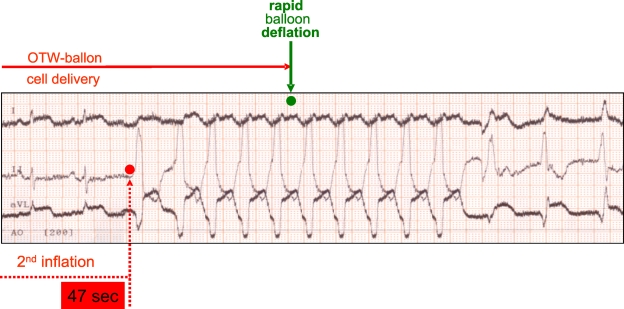
Background: For transcoronary progenitor cells' administration, injections under flow arrest (over-the-wire balloon technique, OTW) are used universally despite lack of evidence for being required for cell delivery or being effective in stimulating myocardial engraftment. Flow-mediated endothelial rolling is mandatory for subsequent cell adhesion and extravasation.
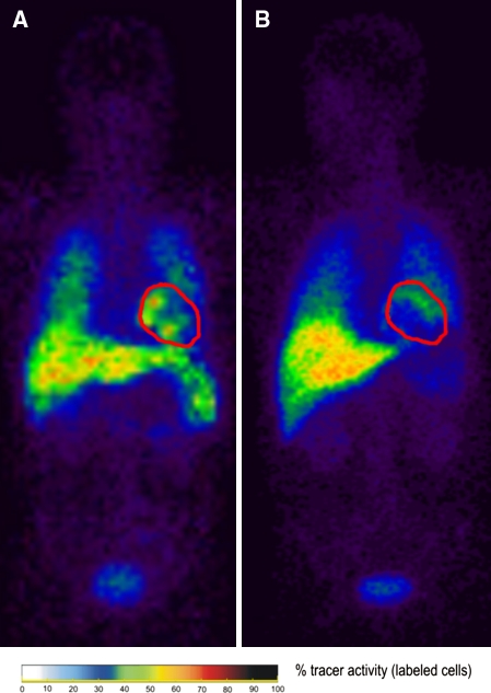
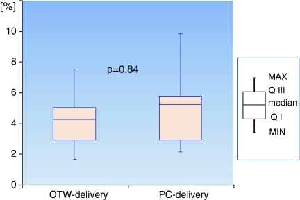
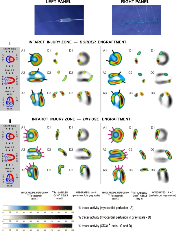
Methods: To optimize cell directing toward the coronary endothelium under maintained flow, the authors developed a cell-delivery side-holed perfusion catheter (PC). Thirty-four patients (36-69 years, 30 men) with primary stent-assisted angioplasty-treated anterior MI (peak TnI 151 [53-356]ng/dL, mean[range]) were randomly assigned to OTW or PC autologous ⁹⁹Tc-extametazime-labeled bone marrow CD34(+) cells (4.34 [0.92-7.54] × 10⁶) administration at 6-14 days after pPCI (LVEF 37.1 [24-44]%). Myocardial perfusion (⁹⁹(m)Tc-MIBI) and labeled cells' activity were evaluated (SPECT) at, respectively, 36-48 h prior to and 60 min after delivery.
Results: In contrast to OTW coronary occlusions, no intolerance or ventricular arrhythmia occurred with PC cells' administration (P < .001). One hour after delivery, 4.86 [1.7-7.6]% and 5.05 [2.2-9.9]% activity was detected in the myocardium (OTW and PC, respectively, P = .84). Labeled cell activity was clearly limited to the (viable) peri-infarct zone in 88% patients, indicating that the infarct core zone may be largely inaccessible to transcoronary-administered cells.
Conclusions: Irrespective of the transcoronary delivery method, only ≈ 5% of native (i.e., non-engineered) CD34(+) cells spontaneously home to the injured myocardium, and cell retention occurs preferentially in the viable peri-infarct zone. Although the efficacy of cell delivery is not increased with the perfusion method, by avoiding provoking ischemic episodes PC offers a rational alternative to the OTW delivery.
Figures
Similar articles
-
Infarct size determines myocardial uptake of CD34+ cells in the peri-infarct zone: results from a study of (99m)Tc-extametazime-labeled cell visualization integrated with cardiac magnetic resonance infarct imaging.Circ Cardiovasc Imaging. 2013 Mar 1;6(2):320-8. doi: 10.1161/CIRCIMAGING.112.979633. Epub 2012 Dec 27. Circ Cardiovasc Imaging. 2013. PMID: 23271789 Clinical Trial.
-
Transcoronary stem cell delivery using physiological endothelium-targeting perfusion technique: the rationale and a pilot study involving a comparison with conventional over-the-wire balloon coronary occlusions in patients after recent myocardial infarction.Kardiol Pol. 2006 May;64(5):489-98; discussion 499. Kardiol Pol. 2006. PMID: 16752333 Clinical Trial. English, Polish.
-
[Visualisation of early engraftment of transcoronary applied CD34+ cells in the infarct border zone].Kardiol Pol. 2008 Jan;66(1):73-7. Kardiol Pol. 2008. PMID: 18266190 Polish.
-
Single-photon emission computed tomography as a fundamental tool in evaluation of myocardial reparation and regeneration therapies.Postepy Kardiol Interwencyjnej. 2022 Dec;18(4):326-339. doi: 10.5114/aic.2023.124403. Epub 2023 Jan 23. Postepy Kardiol Interwencyjnej. 2022. PMID: 36967839 Free PMC article. Review.
-
SPECT and PET to optimize cardiac stem cell therapy.J Nucl Cardiol. 2012 Feb;19(1):118-25. doi: 10.1007/s12350-011-9485-6. J Nucl Cardiol. 2012. PMID: 22246968 Review. No abstract available.
Cited by
-
Effect of the stop-flow technique on cardiac retention of c-kit positive human cardiac stem cells after intracoronary infusion in a porcine model of chronic ischemic cardiomyopathy.Basic Res Cardiol. 2015 Sep;110(5):503. doi: 10.1007/s00395-015-0503-8. Epub 2015 Jul 7. Basic Res Cardiol. 2015. PMID: 26150250 Free PMC article.
-
Myocardial regeneration strategy using Wharton's jelly mesenchymal stem cells as an off-the-shelf 'unlimited' therapeutic agent: results from the Acute Myocardial Infarction First-in-Man Study.Postepy Kardiol Interwencyjnej. 2015;11(2):100-7. doi: 10.5114/pwki.2015.52282. Epub 2015 Jun 22. Postepy Kardiol Interwencyjnej. 2015. PMID: 26161101 Free PMC article.
-
Challenges for heart disease stem cell therapy.Vasc Health Risk Manag. 2012;8:99-113. doi: 10.2147/VHRM.S25665. Epub 2012 Feb 17. Vasc Health Risk Manag. 2012. PMID: 22399855 Free PMC article. Review.
-
Midterm outcomes of transmyocardial laser revascularization with intramyocardial injection of adipose derived stromal cells for severe refractory angina.Postepy Kardiol Interwencyjnej. 2018;14(2):176-182. doi: 10.5114/aic.2018.76409. Epub 2018 Jun 19. Postepy Kardiol Interwencyjnej. 2018. PMID: 30008770 Free PMC article.
-
Safety and efficacy of transcoronary transfer of human neonatal stem cells to ischemic myocardium using a novel cell-delivery system (CIRCULATE catheter) in swine model of acute myocardial infarction.Postepy Kardiol Interwencyjnej. 2022 Dec;18(4):431-438. doi: 10.5114/aic.2022.121697. Epub 2022 Nov 30. Postepy Kardiol Interwencyjnej. 2022. PMID: 36967844 Free PMC article.
References
-
- Lipinski MJ, Biondi-Zoccai GGL, Abbate A, Khianey R, Sheiban I, Bartunek J, et al. Impact of intracoronary cell therapy on left ventricular function in the setting of acute myocardial infarction. A meta-analysis of controlled clinical trials. J Am Coll Cardiol. 2007;50:1761–1767. doi: 10.1016/j.jacc.2007.07.041. - DOI - PubMed
-
- Tendera M, Wojakowski W, Ruzyllo W, Chojnowska L, Tracz W, Musiałek P, et al. Intracoronary infusion of bone marrow-derived selected CD34+CXCR4+ cells and non-selected mononuclear cells in patients with acute STEMI and reduced left ventricular ejection fraction. Results of REGENT Trial. Eur Heart J. 2009;30:1313–1321. doi: 10.1093/eurheartj/ehp073. - DOI - PubMed
-
- Musialek P, Tracz W, Skotnicki AB, Zmudka K, Pieniazek P, Walter Z, et al. Transcoronary stem cell delivery with the use of physiological endothelium-targeting perfusion technique: The rationale and a pilot study in patients after recent myocardial infarction. Pol Heart J. 2006;64:489–498. - PubMed
Publication types
MeSH terms
Substances
LinkOut - more resources
Full Text Sources
Other Literature Sources
Medical
