Analysis of subcellular localization of Myo7a, Pcdh15 and Sans in Ush1c knockout mice
- PMID: 21156003
- PMCID: PMC3052758
- DOI: 10.1111/j.1365-2613.2010.00751.x
Analysis of subcellular localization of Myo7a, Pcdh15 and Sans in Ush1c knockout mice
Abstract
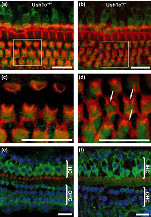
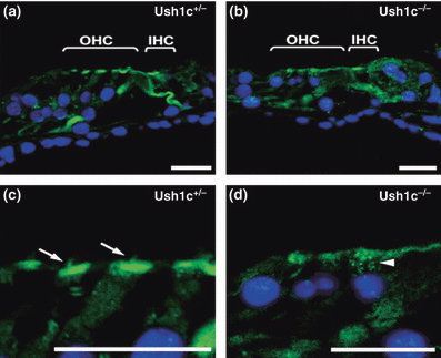
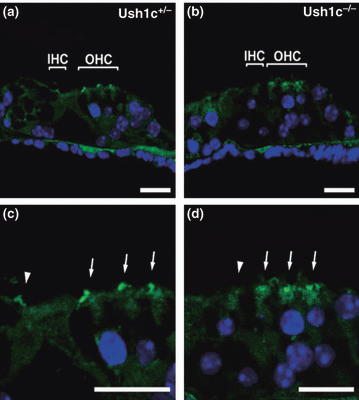
Usher syndrome (USH) is the most frequent cause of combined deaf-blindness in man. An important finding from mouse models and molecular studies is that the USH proteins are integrated into a protein network that regulates inner ear morphogenesis. To understand further the function of harmonin in the pathogenesis of USH1, we have generated a targeted null mutation Ush1c mouse model. Here, we examine the effects of null mutation of the Ush1c gene on subcellular localization of Myo7a, Pcdh15 and Sans in the inner ear. Morphology and proteins distributions were analysed in cochlear sections and whole mount preparations from Ush1c(-/-) and Ush1c(-/+) controls mice. We observed the same distribution of Myo7a throughout the cytoplasm in knockout and control mice. However, we detected Pcdh15 at the base of stereocilia and in the cuticular plate in cochlear hair cells from Ush1c(+/-) controls, whereas in the knockout Ush1c(-/-) mice, Pcdh15 staining was concentrated in the apical region of the outer hair cells and no defined staining was detected at the base of stereocilia nor in the cuticular plate. We showed localization of Sans in the stereocilia of controls mouse cochlear hair cells. However, in cochleae from Ush1c(-/-) mice, strong Sans signals were detected towards the base of stereocilia close to their insertion point into the cuticular plate. Our data indicate that the disassembly of the USH1 network caused by absence of harmonin may have led to the mis-localization of the Protocadherin 15 and Sans proteins in the cochlear hair cells of Ush1c(-/-) knockout mice.
© 2010 The Authors. International Journal of Experimental Pathology © 2010 International Journal of Experimental Pathology.
Figures


Similar articles
-
Photoreceptor expression of the Usher syndrome type 1 protein protocadherin 15 (USH1F) and its interaction with the scaffold protein harmonin (USH1C).Mol Vis. 2005 May 12;11:347-55. Mol Vis. 2005. PMID: 15928608
-
Molecular basis of human Usher syndrome: deciphering the meshes of the Usher protein network provides insights into the pathomechanisms of the Usher disease.Exp Eye Res. 2006 Jul;83(1):97-119. doi: 10.1016/j.exer.2005.11.010. Epub 2006 Mar 20. Exp Eye Res. 2006. PMID: 16545802 Review.
-
Interactions in the network of Usher syndrome type 1 proteins.Hum Mol Genet. 2005 Feb 1;14(3):347-56. doi: 10.1093/hmg/ddi031. Epub 2004 Dec 8. Hum Mol Genet. 2005. PMID: 15590703
-
Genetic analysis of Tunisian families with Usher syndrome type 1: toward improving early molecular diagnosis.Mol Vis. 2016 Jul 19;22:827-35. eCollection 2016. Mol Vis. 2016. PMID: 27440999 Free PMC article.
-
Usher I syndrome: unravelling the mechanisms that underlie the cohesion of the growing hair bundle in inner ear sensory cells.J Cell Sci. 2005 Oct 15;118(Pt 20):4593-603. doi: 10.1242/jcs.02636. J Cell Sci. 2005. PMID: 16219682 Review.
Cited by
-
Current understanding of usher syndrome type II.Front Biosci (Landmark Ed). 2012 Jan 1;17(3):1165-83. doi: 10.2741/3979. Front Biosci (Landmark Ed). 2012. PMID: 22201796 Free PMC article.
-
Integrin α8 and Pcdh15 act as a complex to regulate cilia biogenesis in sensory cells.J Cell Sci. 2017 Nov 1;130(21):3698-3712. doi: 10.1242/jcs.206201. Epub 2017 Sep 7. J Cell Sci. 2017. PMID: 28883094 Free PMC article.
-
Functional Analysis of the Transmembrane and Cytoplasmic Domains of Pcdh15a in Zebrafish Hair Cells.J Neurosci. 2017 Mar 22;37(12):3231-3245. doi: 10.1523/JNEUROSCI.2216-16.2017. Epub 2017 Feb 20. J Neurosci. 2017. PMID: 28219986 Free PMC article.
-
Digenic inheritance of deafness caused by 8J allele of myosin-VIIA and mutations in other Usher I genes.Hum Mol Genet. 2012 Jun 1;21(11):2588-98. doi: 10.1093/hmg/dds084. Epub 2012 Feb 29. Hum Mol Genet. 2012. PMID: 22381527 Free PMC article.
-
Plasma Membrane Targeting of Protocadherin 15 Is Regulated by the Golgi-Associated Chaperone Protein PIST.Neural Plast. 2016;2016:8580675. doi: 10.1155/2016/8580675. Epub 2016 Oct 27. Neural Plast. 2016. PMID: 27867666 Free PMC article.
References
-
- Adato A, Michel V, Kikkawa Y, et al. Interactions in the network of Usher syndrome type 1 proteins. Hum. Mol. Genet. 2005;14:347–356. - PubMed
-
- Ahmed ZM, Riazuddin S, Riazuddin S, Wilcox ER. The molecular genetics of Usher syndrome. Clin. Genet. 2003;63:431–444. - PubMed
-
- Alagramam KN, Yuan H, Kuehn MH, Murcia CL, Wayne S, Srisailpathy CR. Mutations in the novel protocadherin pcdh15 cause usher syndrome type 1f. Hum. Mol. Genet. 2001a;10:1709–1718. - PubMed
-
- Alagramam KN, Murcia CL, Kwon HY, Pawlowski KS, Wright CG, Woychik RP. The mouse ames waltzer hearing-loss mutant is caused by mutation of pcdh15, a novel protocadherin gene. Nat. Genet. 2001b;27:99–102. - PubMed
Publication types
MeSH terms
Substances
Grants and funding
LinkOut - more resources
Full Text Sources

