. 2011 Jan 15;186(2):1001-10.
doi: 10.4049/jimmunol.1002240. Epub 2010 Dec 8.

Negative regulation of IRF7 activation by activating transcription factor 4 suggests a cross-regulation between the IFN responses and the cellular integrated stress responses

Affiliations

Negative regulation of IRF7 activation by activating transcription factor 4 suggests a cross-regulation between the IFN responses and the cellular integrated stress responses

Qiming Liang et al. J Immunol. .

Abstract

Cells react to viral infection by exhibiting IFN-based innate immune responses and integrated stress responses, but little is known about the interrelationships between the two. In this study, we report a linkage between these two host-protective cellular mechanisms. We found that IFN regulatory factor (IRF)7, the master regulator of type I IFN gene expression, interacts with activating transcription factor (ATF)4, a key component of the integrated stress responses whose translation is induced by viral infection and various stresses. We have demonstrated that IRF7 upregulates ATF4 activity and expression, whereas ATF4 in return inhibits IRF7 activation, suggesting a cross-regulation between the IFN response and the cellular integrated stress response that controls host innate immune defense against viral infection.


Conflict of interest statement

DISCLOSURES

The authors have no financial conflicts of interest.

Figures

FIGURE 1
FIGURE 1
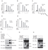
Association of ATF4 with IRF7. (A) ATF4 interacts with IRF7. Flag-tagged IRF7, IRF7 ID domain, IRF3, and luciferase were cotransfected with HA-tagged ATF4 expression vectors into HEK293T cells. Lysates of the transfected cells were immunoprecipitated with anti-Flag M2 affinity beads. The IP complexes and whole cell lysates were analyzed by western blot with antibodies as indicated. (B) ATF4 interacts with IRF7 mainly through the ZIP2 domain. Flag-tagged IRF7, GST-tagged ATF4 full-length and various truncation mutants were expressed in HEK293T cells. The GST-ATF4 fusion proteins were purified with glutathione beads. After washing and blocking, the bound beads were incubated with lysates of Flag-IRF7 expressing cells. After extensive washes, the bound proteins were eluted and analyzed by immunoblotting with anti-Flag and anti-GST antibodies. (C) A schematic presentation of full-length ATF4 and its mutants. BD, basic amino acid domain.
FIGURE 2
FIGURE 2
ATF4 inhibits IRF7 transactivation activity. (A) and (B) ATF4 inhibits IRF7-induced IFNα1 and IFNβ promoter activities in a dose-dependent manner. HEK293T cells were transfected with 100 ng of the IFNα1 (A) and IFNβ (B) luciferase reporter and increasing amounts (50 ng, 100 ng, 250 ng, 500 ng) of ATF4-expressing plasmids as indicated. Eight h after transfection, cells were infected with Sendai virus (SV) or left untreated as controls. Dual luciferase assay were performed at 24h after transfection. The relative luciferase activity was expressed as arbitrary units by normalizing firefly luciferase activity to renilla luciferase activity. Data represent the average of three independent experiments and error bars represent standard deviation. (C) Deletion of the Zip2 domain impairs the inhibition of IRF7 by ATF4. HEK293T cells were transfected the IFNα1 luciferase reporter and plasmids expressing wild type ATF4 or its mutants. Dual luciferase assays were performed similarly to those described above. (D) and (E) ATF4 inhibits IRF7 transactivation activities induced poly(I:C). HEK293T cells were transfected with the 100 ng of IFNα1 (D) or 100 ng of IFNβ (E) luciferase reporter, 1 µg/ml poly(I:C) and increasing amounts of ATF4-expressing plasmids (50 ng, 100 ng, 250 ng, 500 ng) as indicated. Dual luciferase assay were performed at 24 h after transfection. (F) and (G) ATF4 inhibits IRF7 phosphorylation by IKKε and TBK1. HEK293T cells were transfected with Flag-IRF7, HA-ATF4 plus HA-IKKε (F) or TBK1 (G) at the mass ratio of 10:20:1. Forty-eight h after transfection, cell lysates were analyzed by immunoblotting with anti-pIRF7 (Ser477/Ser479) phosphorylation-specific antibody and other antibodies as indicated. (H) Phosphorylation of ATF4 by IKKε/TBK1 but not IKKα/IKKβ. HEK293T cells were transfected with HA-ATF4 plus IKKα, IKKβ, IKKε, or TBK1 expression plasmids at the mass ratio of 10:1. Forty-eight h after transfection, cell lysates were analyzed by immunoblotting with antibodies as indicated.
FIGURE 3
FIGURE 3
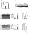
Knockout of ATF4 potentiates IRF7 activation and IFN production. (A) Knockout of ATF4 potentiates IRF7 transactivation activity. The wild-type and ATF4−/− MEF cells were transfected with mouse 200 ng of IFNα6 luciferase reporter plasmids. Cells were infected with SV, and dual luciferase assays were conducted as described in Fig. 1. (B) and (C) Knockout of ATF4 potentiates type I IFN induction. Wild-type and ATF4−/− MEFs were treated with Sendai virus (B) or poly(I:C) (C). Total RNAs were isolated at the times indicated, and the levels of IFNα, IFNβ, IRF7, IRF3, ISG56, and β-actin mRNA were determined by RT-PCR. Secreted IFNα in the medium collected at 8 h and 12 h were measured by ELISA. (D) and (E) Knockout of ATF4 suppresses VSV infection. Wild-type and ATF4−/− MEFs were infected with VSV at an MOI of 0.25. Cell lysates and culture medium were collected at the indicated times after infection. The lysates were analyzed by western blot for detection of VSV glycoprotein G; β-actin acted as a loading control (D). The titers in the culture medium at 24 h and 30 h after infection were determined by plaque assay (E). (F) Expression of ATF4 in MEFs potentiates VSV replication. The ATF4−/− MEFs were transduced with lentivirus vectors expressing ATF4 or GFP as a control. The transduced MEFs were infected with VSV and viral titers were determined 20 h after infection. Data in (A–C), (E), and (F) represent the average of at least three independent experiments and error bars represent standard deviation.
FIGURE 4
FIGURE 4
ATF4 inhibits transcription of IRF7. (A) Loss of ATF4 potentiates IRF7 promoter activity. The wild-type and ATF4−/− MEFs were transfected with 200 ng of IRF7 promoter reporter plasmid. Luciferase assays were performed 36 h after transfection. (B) ATF4 inhibits IRF7 promoter in a dose-dependent manner. HeLa cells were transfected with 200 ng of IRF7 promoter reporter and increasing amounts (50 ng, 100 ng, 250 ng) of ATF4-expressing plasmids. Luciferase assays were performed as described as above. (C) Schematic presentation of human IRF7 promoter. IRFE, ISRE, and CRE/ATF are putative regulatory elements in IRF7 promoter. (D) HeLa cells were transfected with 200 ng of IRF7 promoter reporter or the mutant constructs as depicted in (C). Luciferase assays were performed as described as above. Mutation of the ISRE (a known IRF7 binding site and positive regulatory element) but not the CRE/ATF (putative ATF4 binding site) abolishes the inhibition of IRF7 promoter by ATF4.
FIGURE 5
FIGURE 5
IRF7 regulates ATF4 expression and activity. (A) IRF7 but not IRF3 increases ATF4 transactivation activity. HEK293T cells were transfected with 100 ng of ATF4-responsive CHOP promoter reporter and increasing amounts (50 ng, 100 ng, 250 ng, and 500 ng) of IRF7 or IRF3 expression vectors as indicated. Dual luciferase assays were performed 24 h after transfection. (B)–(D) IRF7 can increase ATF4 transactivation activity when the IFN circuit is disrupted. The above experiment was repeated in the parental 2ftGH (B) and its derivatives, U3A (STAT1) (C) and U4A (JAK1) (D) cells. (E) IRF7 upregulates translation of ATF4. MEFs were transfected with 200 ng of the ATF4 5’UTR luciferase reporter and 200 ng of IRF7 or IRF3 expression plasmids as indicated. Stress inducers DL-homocysteine (DL-Hyc), tunicamycin (Tn), and thapsigargin (Tg) were added 20 h after transfection. A dual-luciferase assay was performed 24 h after transfection. Data represent the average of at least three independent experiments and error bars represent standard deviation.
FIGURE 6
FIGURE 6
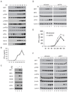
A cross regulation between IFN and the cellular integrated stress responses. (A) Expression kinetics of the major components of IFN and stress responses during Sendai virus infection. A549 cells were infected with Sendai virus in triplicate (160 HA units/ml). At the indicated times after infection, whole cell lysates or nuclear extract (for detection of ATF4) were prepared and analyzed by immunoblotting with indicated antibodies. Parallel samples were used to prepare RNA for RT-PCR analysis for detection of type I IFNs. (B) Activation of the integrated stress response impairs IRF7-induced expression of IFNs. A549 cells were infected with Sendai virus for 4 h and then treated with thapsigargin for another 4 h. Whole-cell lysates and nuclear extracts were prepared and subjected to immunoblotting with the indicated antibodies. (C) Knockdown of ATF4 by siRNA potentiates expression of IRF7 and IFNα. A549 cells stably transduced with siATF4 or siControl were infected with Sendai virus for time as indicated. Whole-cells lysates and nuclear extract were analyzed by immunoblotting with specified antibodies. IFNs in the culture medium were measure by ELISA. (F) Knockdown of IRF7 by siRNA reduces virus-induced cellular integrated stress responses. A549 cells stably transduced by siIRF7 or siControl were infected with Sendai virus. Whole-cell lysates and nuclear extract were analyzed by immunoblotting with the indicated antibodies.
Figure 7
Figure 7
Schematic diagram of cross regulation between IFN and integrated stress responses. Viral infection induces type I IFN expression and also activates multiple eIF2α kinases, mainly PKR and PERK. Phosphorylation of eIF2α and activation of IRF7 itself increases the translation of ATF4. The increased expression level of ATF4 protein induces stress-response genes to help cell recovery but inhibits the expression and transactivation of IRF7 to terminate IFN signaling. As a result, this negative feedback loop allows host cells to terminate the IFN responses effectively.

Similar articles

Cited by

References

    1. Honda K, Takaoka A, Taniguchi T. Type I interferon [corrected] gene induction by the interferon regulatory factor family of transcription factors. Immunity. 2006;25:349–360. - PubMed
    1. Honda K, Taniguchi T. IRFs: master regulators of signalling by Toll-like receptors and cytosolic pattern-recognition receptors. Nat Rev Immunol. 2006;6:644–658. - PubMed
    1. Paun A, Pitha PM. The IRF family, revisited. Biochimie. 2007;89:744–753. - PMC - PubMed
    1. Hiscott J. Triggering the innate antiviral response through IRF-3 activation. J Biol Chem. 2007;282:15325–15329. - PubMed
    1. Honda K, Yanai H, Takaoka A, Taniguchi T. Regulation of the type I IFN induction: a current view. Int Immunol. 2005;17:1367–1378. - PubMed

Publication types

MeSH terms