Prdm16 determines the thermogenic program of subcutaneous white adipose tissue in mice
- PMID: 21123942
- PMCID: PMC3007155
- DOI: 10.1172/JCI44271
Prdm16 determines the thermogenic program of subcutaneous white adipose tissue in mice
Abstract
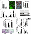
The white adipose organ is composed of both subcutaneous and several intra-abdominal depots. Excess abdominal adiposity is a major risk factor for metabolic disease in rodents and humans, while expansion of subcutaneous fat does not carry the same risks. Brown adipose produces heat as a defense against hypothermia and obesity, and the appearance of brown-like adipocytes within white adipose tissue depots is associated with improved metabolic phenotypes. Thus, understanding the differences in cell biology and function of these different adipose cell types and depots may be critical to the development of new therapies for metabolic disease. Here, we found that Prdm16, a brown adipose determination factor, is selectively expressed in subcutaneous white adipocytes relative to other white fat depots in mice. Transgenic expression of Prdm16 in fat tissue robustly induced the development of brown-like adipocytes in subcutaneous, but not epididymal, adipose depots. Prdm16 transgenic mice displayed increased energy expenditure, limited weight gain, and improved glucose tolerance in response to a high-fat diet. shRNA-mediated depletion of Prdm16 in isolated subcutaneous adipocytes caused a sharp decrease in the expression of thermogenic genes and a reduction in uncoupled cellular respiration. Finally, Prdm16 haploinsufficiency reduced the brown fat phenotype in white adipose tissue stimulated by β-adrenergic agonists. These results demonstrate that Prdm16 is a cell-autonomous determinant of a brown fat-like gene program and thermogenesis in subcutaneous adipose tissues.
Figures


Similar articles
-
Fasting induces a subcutaneous-to-visceral fat switch mediated by microRNA-149-3p and suppression of PRDM16.Nat Commun. 2016 May 31;7:11533. doi: 10.1038/ncomms11533. Nat Commun. 2016. PMID: 27240637 Free PMC article.
-
Wt1 haploinsufficiency induces browning of epididymal fat and alleviates metabolic dysfunction in mice on high-fat diet.Diabetologia. 2022 Mar;65(3):528-540. doi: 10.1007/s00125-021-05621-1. Epub 2021 Nov 30. Diabetologia. 2022. PMID: 34846543 Free PMC article.
-
Transcriptional control of brown fat determination by PRDM16.Cell Metab. 2007 Jul;6(1):38-54. doi: 10.1016/j.cmet.2007.06.001. Cell Metab. 2007. PMID: 17618855 Free PMC article.
-
Role of PRDM16 in the activation of brown fat programming. Relevance to the development of obesity.Histol Histopathol. 2013 Nov;28(11):1411-25. doi: 10.14670/HH-28.1411. Epub 2013 Jun 17. Histol Histopathol. 2013. PMID: 23771475 Review.
-
Brown adipose tissue: the molecular mechanism of its formation.Nutr Rev. 2009 Mar;67(3):167-71. doi: 10.1111/j.1753-4887.2009.00184.x. Nutr Rev. 2009. PMID: 19239631 Review.
Cited by
-
GQ-16, a TZD-Derived Partial PPARγ Agonist, Induces the Expression of Thermogenesis-Related Genes in Brown Fat and Visceral White Fat and Decreases Visceral Adiposity in Obese and Hyperglycemic Mice.PLoS One. 2016 May 3;11(5):e0154310. doi: 10.1371/journal.pone.0154310. eCollection 2016. PLoS One. 2016. PMID: 27138164 Free PMC article.
-
Overexpression of PRDM16 attenuates acute kidney injury progression: genetic and pharmacological approaches.MedComm (2020). 2024 Sep 21;5(10):e737. doi: 10.1002/mco2.737. eCollection 2024 Oct. MedComm (2020). 2024. PMID: 39309696 Free PMC article.
-
Brown adipose tissue as a regulator of energy expenditure and body fat in humans.Diabetes Metab J. 2013 Feb;37(1):22-9. doi: 10.4093/dmj.2013.37.1.22. Epub 2013 Feb 15. Diabetes Metab J. 2013. PMID: 23441053 Free PMC article.
-
Can Brown Fat Win the Battle Against White Fat?J Cell Physiol. 2015 Oct;230(10):2311-7. doi: 10.1002/jcp.24986. J Cell Physiol. 2015. PMID: 25760392 Free PMC article. Review.
-
Inhibition of in vitro and in vivo brown fat differentiation program by myostatin.Obesity (Silver Spring). 2013 Jun;21(6):1180-8. doi: 10.1002/oby.20117. Epub 2013 Jul 19. Obesity (Silver Spring). 2013. PMID: 23868854 Free PMC article.
References
-
- Wang Y, Rimm EB, Stampfer MJ, Willett WC, Hu FB. Comparison of abdominal adiposity and overall obesity in predicting risk of type 2 diabetes among men. Am J Clin Nutr. 2005;81(3):555–563. - PubMed
-
- Klein S, et al. Waist circumference and cardiometabolic risk: a consensus statement from shaping America’s health: Association for Weight Management and Obesity Prevention; NAASO, the Obesity Society; the American Society for Nutrition; and the American Diabetes Association. Diabetes Care. 2007;30(6):1647–1652. doi: 10.2337/dc07-9921. - DOI - PubMed
Publication types
MeSH terms
Substances
Grants and funding
LinkOut - more resources
Full Text Sources
Other Literature Sources
Molecular Biology Databases

