. 2011 Mar;23(3):555-65.
doi: 10.1016/j.cellsig.2010.11.007. Epub 2010 Nov 25.

Serine phosphorylation regulates disabled-1 early isoform turnover independently of Reelin

Affiliations

Serine phosphorylation regulates disabled-1 early isoform turnover independently of Reelin

Zhihua Gao et al. Cell Signal. 2011 Mar.

Abstract

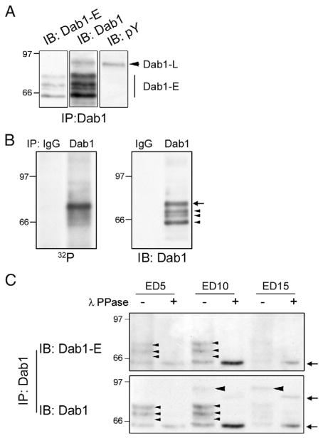
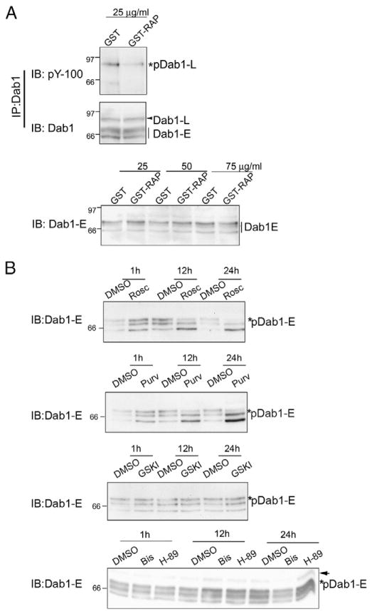
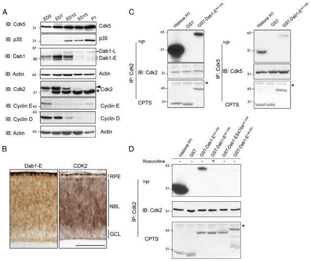
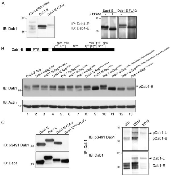
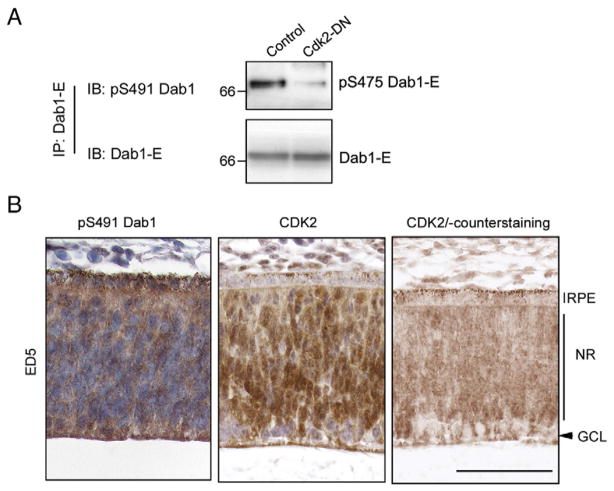
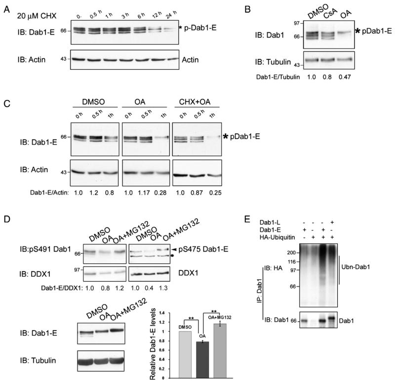
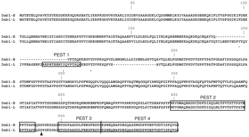
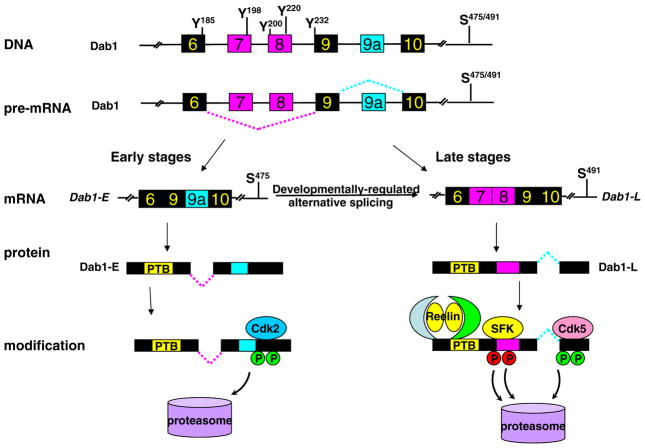
The Reelin-Disabled 1 (Dab1) signaling pathway plays an important role in neuronal cell migration during brain development. Dab1, an intracellular adapter protein which is tyrosine phosphorylated upon Reelin stimulation, has been directly implicated in the transmission and termination of Reelin-mediated signaling. Two main forms of Dab1 have been identified in the developing chick retina, an early isoform (Dab1-E) expressed in progenitor cells and a late isoform (Dab1-L, a.k.a. Dab1) expressed in differentiated cells. Dab1-E is missing two Src family kinase (SFK) phosphorylation sites that are critical for Reelin-Dab1 signaling and is not tyrosine phosphorylated. We have recently demonstrated a role for Dab1-E in the maintenance of retinal progenitor cells. Here, we report that Dab1-E is phosphorylated at serine/threonine residues independent of Reelin. Cdk2, highly expressed in retinal progenitor cells, mediates Dab1-E phosphorylation at serine 475 which in turn promotes ubiquitination-triggered proteasome degradation of Dab1-E. Inhibition of protein phosphatase 1 and/or protein phosphatase 2A leads to increased Dab1-E instability. We propose that Dab1 turnover is regulated by both Reelin-independent serine/threonine phosphorylation and Reelin-dependent tyrosine phosphorylation.


Figures

Fig. 1
Fig. 1
Dab1-E phosphorylation in the developing retina. Dab1 proteins were immunoprecipitated from ED10 retinal lysates using anti-Dab1 antibody and analyzed by western blotting. The blot was immunostained with anti-Dab1-E antibody (left panel), anti-Dab1 antibody (middle panel) and anti-phosphotyrosine antibody (right panel). The arrowhead indicates Dab1-L, whereas the vertical bar indicates the different forms of Dab1-E. (B) ED5 chick retinal cells were metabolically labeled with 32Pi. Endogenous Dab1 proteins were immunoprecipitated with anti-Dab1 antibody. The immunoprecipitates were resolved by SDS-PAGE and transferred to a PVDF membrane. Phosphorylated Dab1 proteins were visualized by autoradiography. The membrane was immunostained with anti-Dab1 antibody. The arrow indicates the major Dab1-E band labeled with 32Pi, whereas arrowheads indicate weaker bands labeled with 32Pi. (C) Endogeneous Dab1 was immunoprecipitated from lysates prepared from ED5, ED10 and ED15 chick retinas. The immunoprecipitates were treated with lambda phosphatase (λ PPase) (+) or left untreated (−). Blots were immunostained with anti-Dab1-E antibody (top panel) and anti-Dab1 antibody (bottom panel). Thin arrowheads indicate phosphorylated Dab1-E and thick arrowheads indicate phosphorylated Dab1-L, whereas arrows point to dephosphorylated Dab1-E and -L.
Fig. 2
Fig. 2
Cdk inhibition reduces Dab1-E phosphorylation. (A) Lysates were prepared from ED10 retinal cultures treated with 25 μg/ml of GST or GST-RAP for 24 h and subjected to immunoprecipitation with anti-Dab1 antibody. The immunoprecipitates were resolved by 8% SDS-PAGE and blots immunostained with anti-phosphotyrosine and anti-Dab1 antibodies (top panel). ED10 retinal cultures were treated with GST or GST-RAP for 24 h at the indicated concentrations. Western blot analysis was carried out using anti-Dab1-E antibody. (B) ED5 retinal cultures were treated with DMSO, 20 μM roscovitine (Rosc, Cdk inhibitor), 20 μM purvananol A (Purv, Cdk inhibitor), 20 μM GSKI (Gsk 3 inhibitor), 1 μM bisindolylmaleimide I (Bis, PKC inhibitor) or 1 μM H-89 (PKA inhibitor) for 1, 12 and 24 h. Cell lysates were resolved by 8% SDS-PAGE and immunoblotted using an anti-Dab1-E antibody. The arrow indicates a non-specific band labeled by the antibody.
Fig. 3
Fig. 3
Cdk phosphorylates Dab1-E in vitro. (A) Expression of Cdk and cyclins in the developing retina. Retinal lysates prepared from ED5, ED7, ED10, ED15 and P1 chick embryos were resolved by SDS-PAGE and blots immunostained with anti-Cdk5, anti-p35, anti-Dab1, anti-Cyclin D1, anti-Cyclin E, anti-Cdk2 and anti-actin antibodies. The two Cdk2 bands (indicated by arrowhead and asterisk) may represent posttranslationally-modified or alternatively-spliced Cdk2 isoforms. (B) Immunohistochemical analysis of Dab1-E and Cdk2 in ED7 chick retina. Tissue sections from formalin-fixed paraffin-embedded ED7 chick retina were immunostained with anti-Dab1-E or anti-Cdk2 antibody. Abbreviations: RPE, retinal pigmented epithelium; NBL, neuroblastic layer; GCL, ganglion cell layer. Scale bar, 50 μm. (C) Endogenous Cdk2 and Cdk5 proteins were immunoprecipitated from ED5 retinal tissue lysates and incubated with histone H1, GST and GST-Dab1-E441-535, in the presence of [γ-32P] ATP. The complexes were separated in a 10% SDS-polyacrylamide gel and transferred to a nitrocellulose membrane. Protein phosphorylation was visualized by autoradiography. Histone H1, GST and GST-Dab1-E fragments were visualized by CPTS staining. Cdk2 and Cdk5 identity was verified by immunostaining the membranes with anti-Cdk2 and anti-Cdk5 antibodies. (D) The Cdk2 complex was immunoprecipitated and incubated with putative substrates as indicated in (B) in the absence/presence of 20 μM roscovitine. Autoradiography, CPTS and western blot analysis are described in (B).
Fig. 4
Fig. 4
Dab1-E serine 475 phosphorylation in transfected cells and in the retina. (A) ED10 chick retinal lysates and lysates prepared from HeLa cells transfected with pcDNA3-Dab1-E and pCMV-Tag4A-Dab1-E (Dab1-E-FLAG) expression constructs were subjected to western blot analysis using anti-Dab1 antibody (left panel). Dab1-E immunoprecipitations were carried out using HeLa cells transfected with pcDNA3-Dab1-E or pCMV-Tag4A-Dab1-E expression constructs. The immunoprecipitates were treated with λ protein phosphatase (λ PPase +) or left untreated (−) before separating the proteins in an 8% SDS-polyacrylamide gel and immunoblotting with anti-Dab1-E antibody. (B) Schematic representation of putative Cdk phosphorylation sites in Dab1-E protein (top panel). Lysates prepared from HeLa cells transfected with pCMV-Tag4A-Dab1-E wild-type and mutants were subjected to western blot analysis using anti-Dab1 and anti-actin antibodies (bottom panel). The asterisk indicates phosphorylated Dab1-E. (C). Protein extracts prepared from HEK293T cells transfected with pcDNA3-Dab1-E, pcDNA3-Dab1-L, pCMV-Tag4A-Dab1-E wild-type and pCMV-Tag4A-Dab1-ES475A were analysed by western blotting using the anti-S491 Dab1 phosphorylation specific antibody (pS491Dab1, left panel). Dab1 immunoprecipitates prepared from ED7, ED10 and ED15 chick retina lysates were resolved by 8% SDS-PAGE (20×20 cm), transferred to a PVDF membrane and immunostained with anti-pS491 Dab1 antibody and Dab1 antibody.
Fig. 5
Fig. 5
Cdk2 phosphorylates Dab1-E in the retina. (A) Dab1-E was immunoprecipitated from ED5 retinal cultures transfected with pcDNA3 and pcDNA3-HA-Cdk2 dominant negative (Cdk2-DN) expression constructs. The immunoprecipitates were analysed by western blotting using anti-pS491 Dab1 (pS475 Dab1-E) and anti-Dab1-E antibodies. (B) Immunohistochemical analysis of pS491 Dab1 and Cdk2 in ED5 chick retina. Consecutive tissue sections from formalin-fixed paraffin-embedded ED5 chick retina were immunostained with anti-pS491 Dab1 or anti-Cdk2 antibody. Sections were either counterstained with hematoxylin (left and middle panels) or not counterstained (right panel) in order to better visualize the cellular distribution of Cdk2 protein in the retina. Abbreviations: RPE, retinal pigmented epithelium; NR, neural retina; GCL, ganglion cell layer. Scale bar, 50 μm.
Fig. 6
Fig. 6
Serine 475 phosphorylation destabilizes Dab1-E and promotes ubiquitination. (A) Lysates were prepared from ED5 retinal cultures treated with 20 μM cycloheximide (CHX) for 0 to 24 h (as indicated) and subjected to western blot analysis using anti-Dab1-E and anti-actin antibodies. (B) ED5 retinal cultures were treated with DMSO, 1 μM cyclosporin A (CsA) or 250 nM okadaic acid (OA) for 1 h. Cell lysates were resolved by 8% SDS-PAGE, transferred and immunostained with anti-Dab1-E and anti-tubulin antibodies. Protein band density was quantified by Image J. Values indicate the ratio between Dab1-E and tubulin levels, with the ratio arbitrarily set at 1 for DMSO-treated samples. (C) ED5 retinal cultures were treated with DMSO, 250 nM okadaic acid (OA) or 250 nM OA in the presence of 20 μM CHX for the indicated times. Western blot analysis and quantitation were carried out as in (B), with actin used as the loading control. (D) ED5 retinal cultures were treated with DMSO, 100 nM okadaic acid (OA) or 100 nM OA in the presence of 10 μM MG132 for 4 h. Western blot analysis and quantitation were carried out as in (B), with DDX1 and tubulin serving as loading controls, respectively. The histogram shows relative Dab1-E protein levels from three independent experiments. Significant differences between DMSO, OA and OA+MG132 treated cultures are indicated by double asterisks (P<0.01, ANOVA and t test). (E) HEK293T cells were transfected with pcDNA3-Dab1-E, pcDNA3-HA-Ubiquitin (HA-Ubiquitin), pcDNA3-HA-Ubiquitin and pcDNA3-Dab1-E, or pcDNA3-HA-Ubiquitin and pcDNA3-Dab1-L. Dab1 proteins were immunoprecipitated from cell lysates using anti-Dab1 antibody and immunoprecipitates analysed by western blotting using anti-HA and anti-Dab1 antibodies.
Fig. 7
Fig. 7
The protein sequences of both Dab1-E (AY242122) and Dab1-L (AY242123) were scanned for potential PEST motifs using a web-based algorithm, ePESTfind (http://emboss.bioinformatics.nl/cgi-bin/emboss/epestfind). Four potential PEST motifs with a PESTfind Score >5 are highlighted in black boxes. Tyrosines that are phosphorylated upon Reelin stimulation in Dab1-L are marked with asterisks, whereas the S475/S491 residue phosphorylated by Cdk in Dab1-E/Dab1-L is indicated with a triangle. Amino acid numbering is based on the Dab1-L sequence.
Fig. 8
Fig. 8
Model depicting the dual regulatory mechanism controlling Dab1-E and Dab1-L levels during development. The Dab1 gene structure (exons 6, 7, 8, 9, 9a and 10), pre-mRNA (dotted lines indicate alternative splicing), mRNA and protein are diagrammatically represented. At early developmental stages, Dab1-E is the main isoform expressed in the retina. As development progresses, alternative splicing results in a switch from Dab1-E to Dab1-L isoform. S/T phosphorylation of Dab1-E occurs independently of Reelin, whereas tyrosine phosphorylation of Dab1-L is dependent on Reelin stimulation. Dab1 levels are tightly regulated by Reelin-induced Dab1-L tyrosine phosphorylation and Cdk-mediated S/T serine phosphorylation through proteasome degradation.

Similar articles

Cited by

References

    1. Howell BW, Hawkes R, Soriano P, Cooper JA. Nature. 1997;389(6652):733. - PubMed
    1. Howell BW, Herrick TM, Cooper JA. Genes Dev. 1999;13(6):643. - PMC - PubMed
    1. Howell BW, Lanier LM, Frank R, Gertler FB, Cooper JA. Mol Cell Biol. 1999;19(7):5179. - PMC - PubMed
    1. Trommsdorff M, Borg JP, Margolis B, Herz J. J Biol Chem. 1998;273(50):33556. - PubMed
    1. Songyang Z, Shoelson SE, Chaudhuri M, Gish G, Pawson T, Haser WG, King F, Roberts T, Ratnofsky S, Lechleider RJ, et al. Cell. 1993;72(5):767. - PubMed

Publication types

MeSH terms