. 2011 Feb 5;410(1):129-41.
doi: 10.1016/j.virol.2010.10.037. Epub 2010 Nov 26.

Recombination of 5' subgenomic RNA3a with genomic RNA3 of Brome mosaic bromovirus in vitro and in vivo

Affiliations

Recombination of 5' subgenomic RNA3a with genomic RNA3 of Brome mosaic bromovirus in vitro and in vivo

Joanna Sztuba-Solińska et al. Virology. .

Abstract

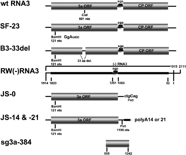
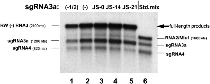
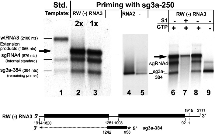
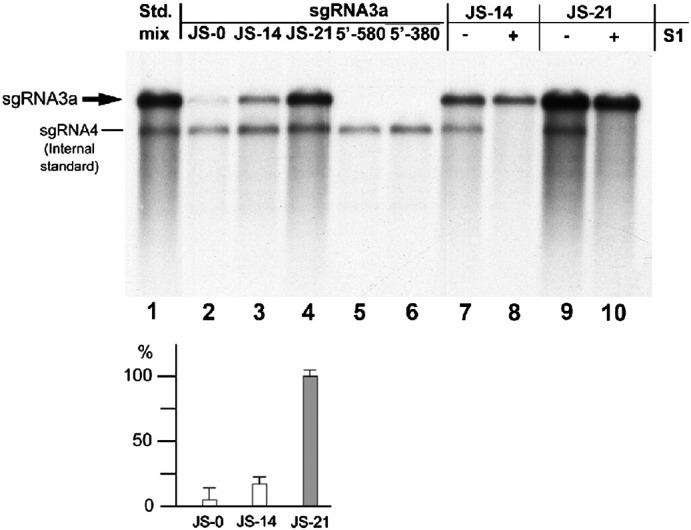
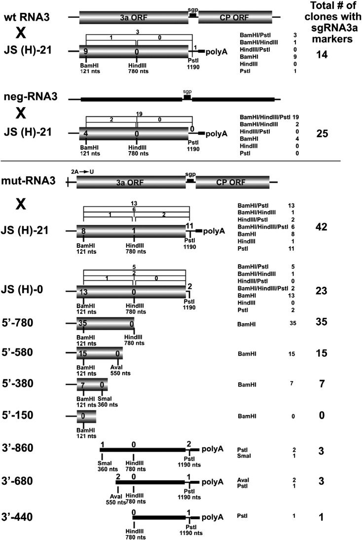
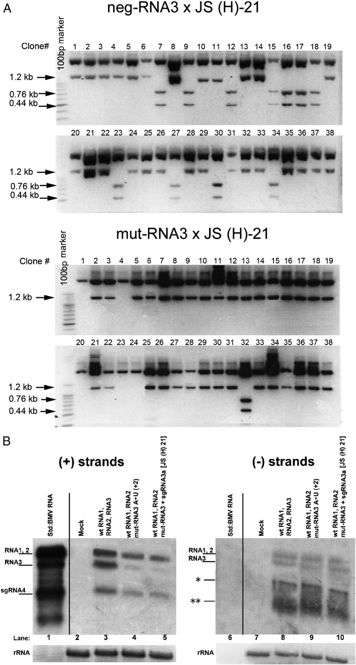
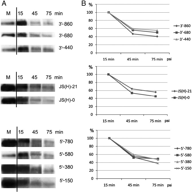
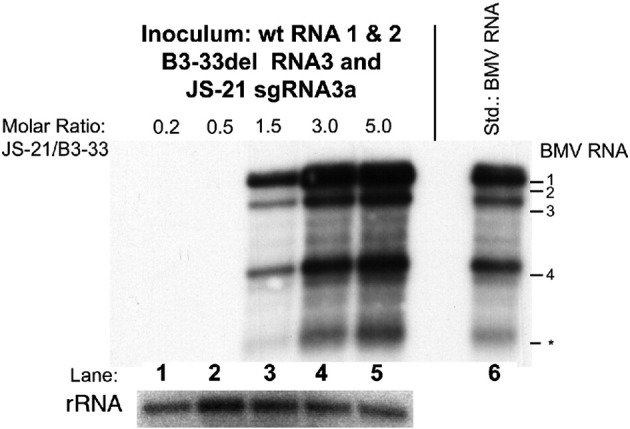
RNA-RNA recombination salvages viral RNAs and contributes to their genomic variability. A recombinationally-active subgenomic promoter (sgp) has been mapped in Brome mosaic bromovirus (BMV) RNA3 (Wierzchoslawski et al., 2004. J. Virol.78, 8552-8864) and mRNA-like 5' sgRNA3a was characterized (Wierzchoslawski et al., 2006. J. Virol. 80, 12357-12366). In this paper we describe sgRNA3a-mediated recombination in both in vitro and in vivo experiments. BMV replicase-directed co-copying of (-) RNA3 with wt sgRNA3a generated RNA3 recombinants in vitro, but it failed to when 3'-truncated sgRNA3a was substituted, demonstrating a role for the 3' polyA tail. Barley protoplast co-transfections revealed that (i) wt sgRNA3a recombines at the 3' and the internal sites; (ii) 3'-truncated sgRNA3as recombine more upstream; and (iii) 5'-truncated sgRNA3 recombine at a low rate. In planta co-inoculations confirmed the RNA3-sgRNA3a crossovers. In summary, the non-replicating sgRNA3a recombines with replicating RNA3, most likely via primer extension and/or internal template switching.


Figures

Fig. 1
Fig. 1
BMV RNA3 variants used in the in vitro and whole-plant experiments. Wt RNA3 is shown on the top with the ClaI restriction site indicated. An intermediate SF-23 construct carries a sequence corresponding to the BamHI marker site, whereas B3-33del carries a 33-aa deletion in the 3a ORF. JS-14 and JS-21 are sgRNA3a constructs carrying 3′ polyA tails of 14 and 21 A residues, respectively, plus RNA sequences corresponding to BamHI and PstI marker sites, whereas JS-0 does not carry a 3′ polyA tail. sg3a-384 is a (+) strand RNA primer corresponding to a middle region of the RNA3 sequence. Thin lines mark non-coding regions, whereas shaded rectangles represent ORFs for the 3a movement and coat (CP) proteins. The positions of marker sites are indicated by small vertical bars. To construct RW (−) RNA3, a portion of (−) RNA3 between nts 1 and 1914 (the nucleotide positions correspond to those in wt genomic (+) RNA3) was fused immediately downstream of the (+) RNA3 promoter (nts 1915–2111). The sequences complementary to the 3a and CP ORFs are shown as thick lines with the corresponding nucleotide positions below (for more details, see Wierzchoslawski and Bujarski, 2006).
Fig. 2
Fig. 2
Characterization of BMV RdRp in vitro copying products of mixtures of RW(−) RNA3 and sgRNA3a templates. Template RNA mixtures were copied as described in Materials and methods, and the radioactive RdRp products were separated in a 1% denaturing agarose gel. The sgRNA3a variants are specified above (lanes 3 to 5). Lanes 1 and 2 contain copying products of RW(−) RNA3 alone [symbolized by (−)] with half the amount loaded in lane 1. Lane 6 contains a mixture of three radioactive in vitro transcripts as migration standards (the RNA2 marker was synthesized from plasmid pB2TP5 after linearization with MluI).
Fig. 3
Fig. 3
Analysis of the products of in vitro RdRp extension reactions of radioactive sg3a-384 RNA on the RW (−) RNA3 template. The products were separated in denaturing 1.2% agarose gels. Lane 1, mixture of in vitro-transcribed radioactive wt BMV RNA3, sgRNA4, and sg3a-384 RNA as size standards; lanes 2 and 3, primer extension reaction with a single (lane 3) or double (lane 2) amount of the radioactive sg3a-384 RNA. In addition, lanes 2 and 3 contain 32P-labeled sgRNA4 as an internal loading standard that was added after the reaction. The reaction in lane 2 contains a double amount of the sg3a-384 primer. Lanes 4 and 5, control extension reactions of sg3a-384 primer on the BMV RNA2 template (lane 4) or without an RNA2 template (lane 5). Lanes 6 and 7, digestion of sg3a-384 extension products without (lane 6) or with (lane 7) S1 nuclease. Both, sgRNA4 (added post-reaction) and ss sg3a-384 RNA disappeared after S1 nuclease treatment (lane 7). Lanes 8 and 9, analysis of TNT activity of BMV RdRp, with the extension reactions completed with (lane 8) or without (lane 9) GTP. Both the RW(−) RNA3 template and the corresponding sg3a-384 RNA primer (* symbolizes the radioactivity) are shown schematically below (see Fig. 1 for more details).
Fig. 4
Fig. 4
In vitro copying of sgRNA3a templates with BMV RdRp. Equimolar amounts of sgRNA3a constructs JS-0 (lane 2), JS-14 (lane 3), JS-21 (lane 4), 5′-580 (lane 5) and 5′-380 (lane 6) were copied in standard BMV replicase reactions as the only templates. The radioactive products were separated in a 1.0% formamide/formaldehyde agarose gel and blotted to nylon membrane followed by autoradiography. Bands representing the copying products are marked by thick arrows. Lane 1, 32P-labeled mixture of JS-21 and sgRNA4 transcripts as size standards. In addition, lanes 2 to 10 contain the sgRNA4 loading marker, added post-copying reaction. The densitometric quantification results are shown below. The open bars represent the relative density of bands corresponding to the in vitro copying products of JS-0 to JS-21 (which was taken as 100%) after normalizing to the internal sgRNA4 standard. Lanes 7 to 10, analysis of the ds nature of the copying products of JS-14 and JS-21 RNAs by S1 nuclease digestion (lanes 8 and 10, respectively) versus undigested products (lanes 7 and 9).
Fig. 5
Fig. 5
Recombination of BMV RNA3 templates with sgRNA3a constructs in barley protoplasts. wt RNA3 × JS(H)-21 indicates an experiment with wt genomic RNA3 (wt RNA3) and full-length sgRNA3a (JS(H)-21). neg-RNA3 × JS(H)-21 indicates an experiment with negative-strand RNA3 (neg-RNA3) and full-length sgRNA3a (JS(H)-21). mut-RNA3 × JS(H)-21 indicates an experiment with an RNA3 derivative bearing a point mutation (A → U) at position 2 near the 5′ end. The remaining sgRNA3a-derivatives that were tested for recombination with mut-RNA3 are shown below, including JS(H)-0: an sgRNA3a derivative without a polyA tail; 5′-780, 5′-580, 5′-380, 5′-150: the 5′-nested deletion sgRNA3a fragments; and 3′-860, 3′-680, 3′-440: the 3′-nested deletion sgRNA3a fragments. The positions of marker restriction sites are indicated below each construct. The column on the right shows the general recombination frequency (per-cent) based on an analysis of one-hundred insert-bearing cDNA clones. The frequency of recombinants bearing single restriction markers is shown inside the shaded rectangles, whereas that of those carrying double or triple markers is represented above the constructs as brackets. The 3′nested RNAs do not carry the 3a ORF initiation codon and they are therefore represented by thick black lines. The other elements are as in Fig. 1. The numbers summarize the results from two independent transfection experiments with each sgRNA3a derivative.
Fig. 6
Fig. 6
Recombination of BMV RNA3 with sgRNA3a in barley protoplasts. A. Restriction enzyme digestion of recombinant RNA3 cDNA clones obtained from barley protoplasts that were transfected with a mixture of wt BMV RNAs 1 and 2 and (top) Neg-RNA3 plus JS(H)-21 sgRNA3a or (bottom) mut-RNA3 and JS(H)-21 sgRNA3a. The RNA3 region representing the sgRNA3a sequence was amplified from total RNA extracts by RT-PCR, and the cDNA products were cloned into the pGEM-T Easy system. The insert sequences were released by HindIII/EcoRI digestion and separated by electrophoresis in a 1.5% agarose gel. An intact 1.2-kb fragment reflected the lack of the HindIII marker site at nt position 780, whereas the double 0.44 kb and 0.76 kb bands indicate the presence of the HindIII marker. B. Northern blot analysis showing the accumulation in protoplasts of either (+) or (−) strands (left and right panels, respectively) of BMV RNAs. Total protoplast RNA was separated in a 1.2% denaturing agarose gel, blotted, and probed with a 3′-specific RNA probe detecting either (+) or (−) strands (see Materials and methods). Lanes 1 and 6, virion BMV RNA used as size standards; lanes 2 and 7, negative controls from mock inoculated protoplasts; lanes 3 and 8, protoplasts transfected with equimolar amounts of BMV RNAs 1 and 2 and wt RNA3; lanes 4 and 9, protoplasts transfected with BMV RNAs 1 and 2 and (A → U) RNA3; lanes 5 and 10, protoplasts transfected with BMV RNAs 1 and 2, (A → U) RNA3, and sgRNA3a. Ribosomal RNA (rRNA) bands (after staining with ethidium bromide) are shown below. The position corresponding to the (−) RNA4 band is marked with a single asterisk on the right panel, whereas the migration position of degradation products (likely because minus strands are not encapsidated and thus less protected) is marked with two asterisks.
Fig. 7
Fig. 7
The stability of the sgRNA3a constructs used for transfection experiments in barley protoplasts. A. Autoradiogram representing the intensity of bands corresponding to individual radioactive sgRNA3as that were transfected into protoplasts and extracted after incubation for 15, 45, or 75 min. M stands for the size marker; the names of the constructs are indicated on the right hand side. B. The results of densitometric analysis of the bands shown in A, calculated as the percentage of the initial band intensity.
Fig. 8
Fig. 8
Recombination in planta between 3a-truncated BMV RNA3 and sgRNA3a. C. quinoa seedlings were co-inoculated with wt RNAs 1 and 2, B3-33del RNA3, and various amounts of JS-21 sgRNA3a (the molar ratios of JS-21 to B3-33del are indicated on the top, lanes 1–5). Total RNA was extracted from the inoculated leaves seven days later, separated by electrophoresis in a denaturing agarose gel, blotted onto nylon membrane, and probed with the BMV (+) strand 3′ probe (see Materials and methods). BMV RNAs were detected in plants infected with higher molar ratios of JS-21 to B3-33 del RNAs (lanes 3 to 5). Lane 6 shows the migration of marker BMV RNAs extracted from a viral preparation that was isolated from local lesion tissue after infection with wt transcript BMV RNAs. The lower panel shows the concentration of ribosomal RNA (rRNA) after staining with ethidium bromide. The positions of individual BMV RNAs are indicated on the right. The asterisk marks possible degradation product (in necrotic lesions of C. quinoa tissue).

Similar articles

Cited by

References

    1. Adair T.L., Kearney C.M. Recombination between a 3-kilobase Tobacco mosaic virus transgene and a homologous viral construct in the restoration of viral and nonviral genes. Arch. Virol. 2000;145:1867–1883. - PubMed
    1. Agol V.I. Molecular mechanisms of Poliovirus variation and evolution. Curr. Top. Microbiol. Immunol. 2006;299:211–259. - PubMed
    1. Alejska M., Kurzyńska-Kokorniak A., Broda M., Kierzek R., Figlerowicz M. How RNA viruses exchange their genetic material. Acta Biochim. Pol. 2001;48:391–407. - PubMed
    1. Allison R., Janda M., Ahlquist P. Sequence of Cowpea chlorotic mottle virus RNAs 2 and 3 and evidence of a recombination event during bromoviral evolution. Virology. 1989;172:321–330. - PubMed
    1. Allison R., Thompson C., Ahlquist P. Regeneration of a functional RNA virus genome by recombination between deletion mutants and requirement for cowpea chlorotic mottle virus 3a and coat genes for systemic infection. Proc. Natl Acad. Sci. USA. 1990;87:1820–1824. - PMC - PubMed

Publication types