

Malignant cells facilitate lung metastasis by bringing their own soil
- PMID: 21098274
- PMCID: PMC3003109
- DOI: 10.1073/pnas.1016234107
Malignant cells facilitate lung metastasis by bringing their own soil
Abstract
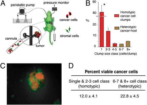
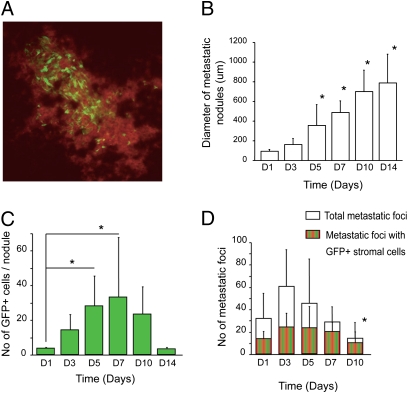
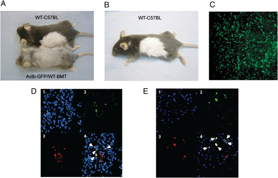
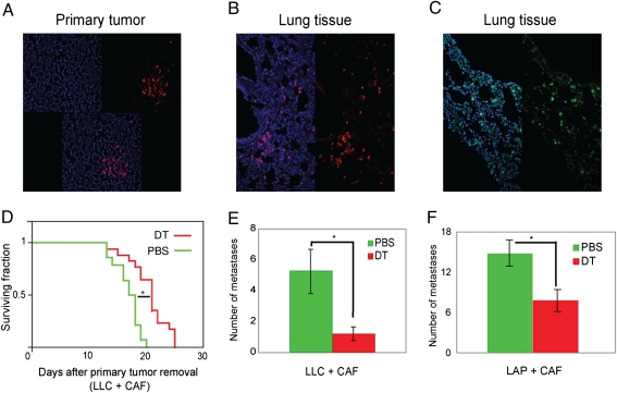
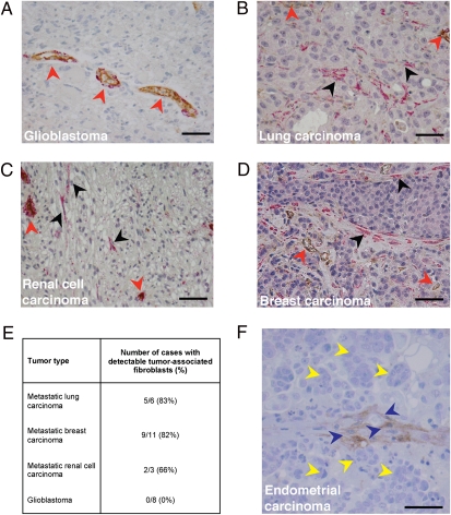
Metastatic cancer cells (seeds) preferentially grow in the secondary sites with a permissive microenvironment (soil). We show that the metastatic cells can bring their own soil--stromal components including activated fibroblasts--from the primary site to the lungs. By analyzing the efferent blood from tumors, we found that viability of circulating metastatic cancer cells is higher if they are incorporated in heterotypic tumor-stroma cell fragments. Moreover, we show that these cotraveling stromal cells provide an early growth advantage to the accompanying metastatic cancer cells in the lungs. Consistent with this hypothesis, we demonstrate that partial depletion of the carcinoma-associated fibroblasts, which spontaneously spread to the lung tissue along with metastatic cancer cells, significantly decreases the number of metastases and extends survival after primary tumor resection. Finally, we show that the brain metastases from lung carcinoma and other carcinomas in patients contain carcinoma-associated fibroblasts, in contrast to primary brain tumors or normal brain tissue. Demonstration of the direct involvement of primary tumor stroma in metastasis has important conceptual and clinical implications for the colonization step in tumor progression.
Conflict of interest statement
The authors declare no conflict of interest.
Figures
Similar articles
-
Mesenchymal stem cells within tumour stroma promote breast cancer metastasis.Nature. 2007 Oct 4;449(7162):557-63. doi: 10.1038/nature06188. Nature. 2007. PMID: 17914389
-
Studying primary tumor-associated fibroblast involvement in cancer metastasis in mice.Nat Protoc. 2012 Mar 22;7(4):756-62. doi: 10.1038/nprot.2012.031. Nat Protoc. 2012. PMID: 22441294 Free PMC article.
-
Syndecan-1 induction in lung microenvironment supports the establishment of breast tumor metastases.Breast Cancer Res. 2018 Jul 5;20(1):66. doi: 10.1186/s13058-018-0995-x. Breast Cancer Res. 2018. PMID: 29976229 Free PMC article.
-
The metastatic niche and stromal progression.Cancer Metastasis Rev. 2012 Dec;31(3-4):429-40. doi: 10.1007/s10555-012-9373-9. Cancer Metastasis Rev. 2012. PMID: 22699312 Free PMC article. Review.
-
The role of tumor stroma in cancer progression and prognosis: emphasis on carcinoma-associated fibroblasts and non-small cell lung cancer.J Thorac Oncol. 2011 Jan;6(1):209-17. doi: 10.1097/JTO.0b013e3181f8a1bd. J Thorac Oncol. 2011. PMID: 21107292 Review.
Cited by
-
Collective cancer cell invasion requires RNA accumulation at the invasive front.Proc Natl Acad Sci U S A. 2020 Nov 3;117(44):27423-27434. doi: 10.1073/pnas.2010872117. Epub 2020 Oct 15. Proc Natl Acad Sci U S A. 2020. PMID: 33060293 Free PMC article.
-
Multifaceted tumor stromal fibroblasts.Cancer Microenviron. 2012 Dec;5(3):187-93. doi: 10.1007/s12307-012-0109-8. Epub 2012 May 25. Cancer Microenviron. 2012. PMID: 22627670 Free PMC article.
-
Metastasis-Initiating Cells and Ecosystems.Cancer Discov. 2021 Apr;11(4):971-994. doi: 10.1158/2159-8290.CD-21-0010. Cancer Discov. 2021. PMID: 33811127 Free PMC article. Review.
-
[Tumor microenvironment--the critical element of tumor metastasis].Zhongguo Fei Ai Za Zhi. 2015 Jan;18(1):48-54. doi: 10.3779/j.issn.1009-3419.2015.01.08. Zhongguo Fei Ai Za Zhi. 2015. PMID: 25603873 Free PMC article. Review. Chinese.
-
Myxoid stroma is associated with postoperative relapse in patients with stage II colon cancer.BMC Cancer. 2020 Sep 3;20(1):842. doi: 10.1186/s12885-020-07335-w. BMC Cancer. 2020. PMID: 32883261 Free PMC article.
References
Publication types
MeSH terms
Substances
Grants and funding
- T32 CA073479-11/CA/NCI NIH HHS/United States
- P01 CA080124-09/CA/NCI NIH HHS/United States
- R01 CA126642-03/CA/NCI NIH HHS/United States
- R01 CA085140-07/CA/NCI NIH HHS/United States
- R01 CA115767-04/CA/NCI NIH HHS/United States
- R01 CA085140/CA/NCI NIH HHS/United States
- P01 CA080124-10/CA/NCI NIH HHS/United States
- R01 CA085140-06/CA/NCI NIH HHS/United States
- R01 CA115767-01A1/CA/NCI NIH HHS/United States
- T32 CA073479-12/CA/NCI NIH HHS/United States
- R24 CA085140/CA/NCI NIH HHS/United States
- R01 CA085140-09/CA/NCI NIH HHS/United States
- P01 CA080124-04/CA/NCI NIH HHS/United States
- T32 CA073479-08/CA/NCI NIH HHS/United States
- T32 CA073479/CA/NCI NIH HHS/United States
- R01 CA115767-02/CA/NCI NIH HHS/United States
- R01CA096915/CA/NCI NIH HHS/United States
- R01 CA126642/CA/NCI NIH HHS/United States
- R01 CA085140-10/CA/NCI NIH HHS/United States
- T32 CA073479-07/CA/NCI NIH HHS/United States
- R01 CA115767-03/CA/NCI NIH HHS/United States
- R01 CA115767-05/CA/NCI NIH HHS/United States
- T32 CA073479-13/CA/NCI NIH HHS/United States
- P01 CA080124-05/CA/NCI NIH HHS/United States
- R01CA085140/CA/NCI NIH HHS/United States
- R01 CA126642-02/CA/NCI NIH HHS/United States
- P01 CA080124/CA/NCI NIH HHS/United States
- R24 CA085140-04/CA/NCI NIH HHS/United States
- R01 CA126642-02S1/CA/NCI NIH HHS/United States
- R01 CA126642-01A1/CA/NCI NIH HHS/United States
- P01 CA080124-07/CA/NCI NIH HHS/United States
- T32 CA073479-09/CA/NCI NIH HHS/United States
- R01CA115767/CA/NCI NIH HHS/United States
- R01CA126642/CA/NCI NIH HHS/United States
- T32 CA073479-10/CA/NCI NIH HHS/United States
- P01 CA080124-06A2/CA/NCI NIH HHS/United States
- R01 CA115767/CA/NCI NIH HHS/United States
- P01 CA080124-05S1/CA/NCI NIH HHS/United States
- R01 CA096915/CA/NCI NIH HHS/United States
- R01 CA085140-08/CA/NCI NIH HHS/United States
- P01CA080124/CA/NCI NIH HHS/United States
- R24 CA085140-05/CA/NCI NIH HHS/United States
- P01 CA080124-08/CA/NCI NIH HHS/United States
LinkOut - more resources
Full Text Sources
Other Literature Sources
Medical
