. 2010 Nov 24;40(4):671-81.
doi: 10.1016/j.molcel.2010.11.002.

ATP-dependent steps in the binding of ubiquitin conjugates to the 26S proteasome that commit to degradation

Affiliations

ATP-dependent steps in the binding of ubiquitin conjugates to the 26S proteasome that commit to degradation

Andreas Peth et al. Mol Cell. .

Abstract

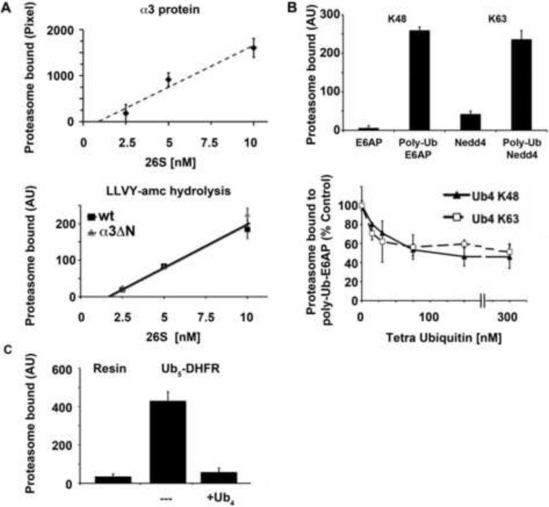
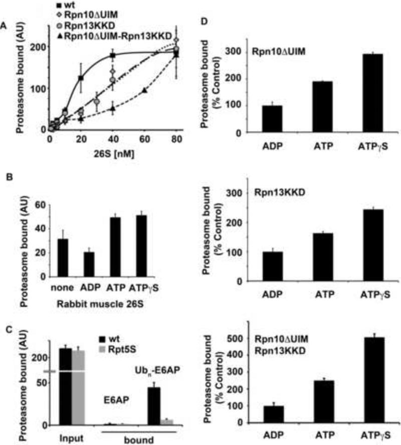
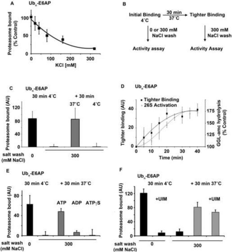
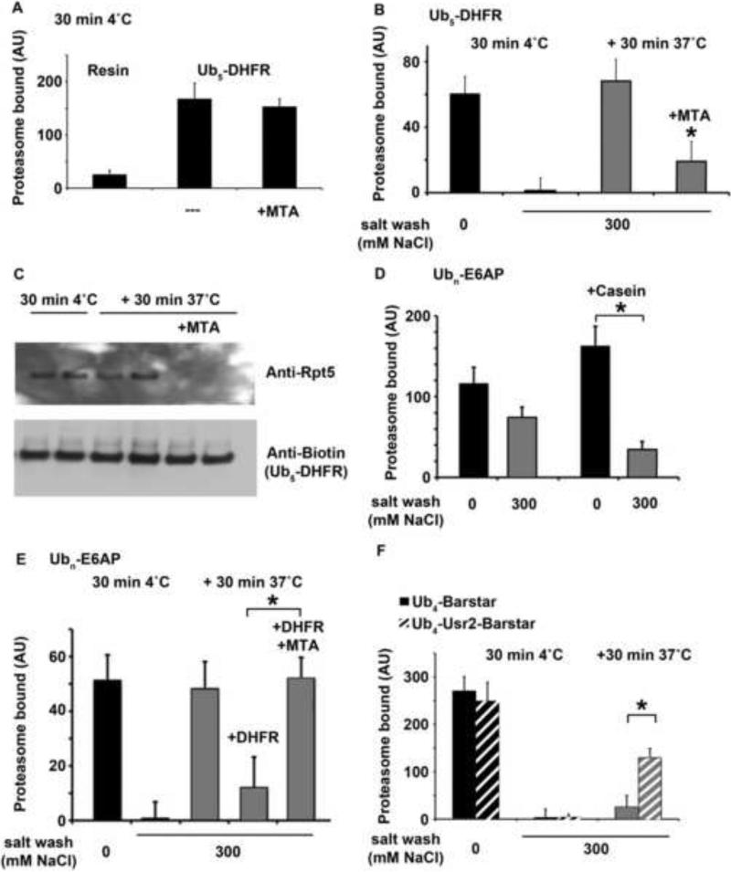
Eukaryotic cells target proteins for degradation by the 26S proteasome by attaching a ubiquitin chain. Using a rapid assay, we analyzed the initial binding of ubiquitinated proteins to purified 26S particles as an isolated process at 4°C. Subunits Rpn10 and Rpn13 contribute equally to the high-affinity binding of ubiquitin chains, but in their absence, ubiquitin conjugates bind to another site with 4-fold lower affinity. Conjugate binding is stimulated 2- to 4-fold by binding of ATP or the nonhydrolyzable analog, ATPγS (but not ADP), to the 19S ATPases. Following this initial, reversible association, ubiquitin conjugates at 37°C become more tightly bound through a step that requires ATP hydrolysis and a loosely folded domain on the protein, but appears independent of ubiquitin. Unfolded or loosely folded polypeptides can inhibit this tighter binding. This commitment step precedes substrate deubiquitination and allows for selection of ubiquitinated proteins capable of being unfolded and efficiently degraded.


Figures

Fig. 1
Fig. 1. K48 and K63 linked poly-Ub chains bind to the same site on 26S proteasomes
(A) The amount of 26S proteasomes bound to poly-Ub conjugates was determined by Western Blot analysis (upper panel) using an anti-α3 antibody or 26S activity (lower panel) measured by hydrolysis of LLVY-amc. Proteasome activity and protein amount correlated tightly over the range of 26S concentration (2.5-10 nM). The activity based readout was measured with wt and open gated 26S particles (α3ΔN). (B) Binding of 26S proteasomes to K48 (Poly-Ub-E6AP) and K63 linked chains (Poly-Ub-Nedd4) was measured. The unmodified E3 ligases were used as control (upper panel). Binding of 26S to Poly-Ub-E6AP in the presence of increasing concentrations of K48 or K63 linked Ub4 (lower panel). Binding of 26S proteasomes without the any addition was taken as 100%. (C) Binding of 26S to Ub5-DHFR was measured with and without an excess (300 nM) of K48 linked Ub4 during the binding reaction. All values are the means of at least three independent experiments ± SEM.
Fig. 2
Fig. 2. ATP and ATPγS stimulate proteasome binding to Poly-Ub conjugates
(A) 26S proteasomes were isolated from wt, rpn10ΔUIM, rpn13KKD and rpn10ΔUIM/rpn13KKD yeast strains. Increasing concentrations of 26S (1.25-80 nM) were added to 30 nM of Poly-Ub-E6AP and resin bound activity was measured. Proteasome activity bound to E6AP alone has been subtracted from these values. (B) Binding of muscle 26S proteasomes to Poly-Ub E6AP was measured in the presence of no added nucleotide, ADP, ATP, or ATPγS. (C) 26S were purified from wt yeast or a rpt5S (K228S) mutant which bears a mutation in Rpt5's ATP-binding site, and proteasome binding to poly-Ub conjugates was measured. (D) 26S were isolated from rpn10ΔUIM (upper panel), rpn13KKD (middle panel) and rpn10ΔUIM/rpn13KKD (lower panel) yeast strains and binding of the 26S in the presence of 1mM ADP, ATP or ATPγS was determined. Binding of the 26S in the presence of ADP was taken as 100%. All values are the means of at least three independent experiments ± SEM.
Fig. 3
Fig. 3. A temperature- and ATP-dependent binding step after the initial binding of Ub conjugates to the proteasome leads to a tighter, salt resistant binding
(A) Binding at 4°C is salt-sensitive. After the initial binding reaction, resin bound 26S were washed with increasing concentration of KCl (0-300 mM) at 4°C. Binding after washing without KCl was taken as 100%. (B) Scheme for the binding assay to distinguish initial and tighter binding of Ub conjugates to proteasomes (C) 26S purified from muscle were pre-bound to Poly-Ub-E6AP at 4°C. This initial binding (black bars) is completely reversed by high salt washes (300 mM NaCl). After the initial binding reaction, proteasomes were incubated for 30 min at 37°C or 4°C and washed with high salt. Resin bound proteasome activity after the second incubation was assayed, as described before and is represented by grey bars. (D) The transition to the tighter binding of 26S to poly-Ub-E6AP was measured over time and compared to the stimulation of proteasomes. After a 30 min incubation at 4°C, the unbound fraction of proteasomes was removed and the resin was incubated at 37°C. The salt wash-resistant proteasome activity was monitored at the indicated times (0 - 40 min). The time course leading to the maximal extent of tight binding was compared with that for the stimulation of gate-opening (as monitored by the enhanced peptidase activity) after conjugate binding under the same conditions. (E) Binding of 26S was performed as described in (C). After the initial binding, bound 26S were incubated at 37°C for 30 minutes in the presence of ATP, ADP or ATPγS. Resin bound proteasome activity after the second. Incubation was assayed as after washing with 300 mM NaCl and is represented in grey bars. (F) The S5a UIM domain inhibits only the initial, but not the tighter binding of poly-Ub conjugates to proteasomes. Proteasome binding to poly-Ub-E6AP was measured with and without the addition of 5 μM of the UIM domain of S5a during or after the initial binding. The UIM domain inhibits the initial binding but when added after the initial binding has already occurred no significant reduction in the transition to the tighter binding was observed. The initial, salt sensitive binding after 30 min at 4°C is shown in black bars, the tighter, salt resistant binding is shown in grey bars. All values are the means of at least three independent experiments ± SEM.
Fig. 4
Fig. 4. The transition to tighter binding is dependent on an unfolded or loosely folded domain of the protein and can be inhibited by unfolded proteins
(A) Initial binding of 26S proteasomes at 4°C to immobilized Ub5-DHFR was measured with and without the addition of 200 nM Methotrexate (MTA). (B) Binding of 26S particles to Ub5-DHFR with and without the addition of 200 nM Methotrexate (MTA) was measured as described in (Fig. 3C). The initial binding at 4°C is shown in black bars. After the initial binding, unbound 26S were removed, and the resin bound particles were incubated for an additional 30 min at 37°C or 4°C. After this second incubation, the resin was washed with buffer containing 300 mM NaCl (High salt). Bound 26S activity after the second incubation is shown in grey bars. (C) Binding of 26S particles to Ub5-DHFR was assayed at 4°C and then at 37°C as described in (B). The amount of bound proteasomes was detected using Western Blot analysis with an anti-Rpt5 antibody. The amount of full-length biotin labeled Ub5-DHFR was determined using Streptavidin coupled alkaline phosphatase. Thus, no evidence for deubiquitination was evident. (D) Binding of 26S to Poly-Ub-E6AP was measured with and without the addition of 200 nM of casein as described in Fig. 3C Initial binding at 4°C in shown in black bars, the binding after the second incubation at 37°C is shown in grey bars. Bound proteasomes were washed with 0 (no salt) or 300 mM NaCl (High salt). (E) Binding of 26S to Poly-Ub-E6AP was measured as in Fig. 3C with and without the addition of DHFR during the second incubation. After the initial binding reaction (in black bars), unbound 26S were removed. Then 500 nM of DHFR was added before the 30 min incubation at 37°C or DHFR that had been preincubated for 30 min at 4°C with Methotrexate (1μM) before addition. Proteasome activity measured after the second incubation is shown in grey bars. (F) Binding of 26S to immobilized Ub4-Barstar and Ub4-USR2-Barstar was measured as in Fig. 3C. Proteasome activity associated with the Ub conjugates was measured after the initial binding (black bars) at 4°C and after additional 30 min incubation at 37°C (tighter binding, grey bars). Nonspecific binding of 26S to the resin has been subtracted from the values. All values are the means of at least three independent experiments ± SEM. * P < 0.05

Similar articles

Cited by

References

    1. Beal R, Deveraux Q, Xia G, Rechsteiner M, Pickart C. Surface hydrophobic residues of multiubiquitin chains essential for proteolytic targeting. Proc Natl Acad Sci U S A. 1996;93:861–866. - PMC - PubMed
    1. Bech-Otschir D, Helfrich A, Enenkel C, Consiglieri G, Seeger M, Holzhutter HG, Dahlmann B, Kloetzel PM. Polyubiquitin substrates allosterically activate their own degradation by the 26S proteasome. Nat Struct Mol Biol. 2009;16:219–225. - PubMed
    1. Benaroudj N, Zwickl P, Seemuller E, Baumeister W, Goldberg AL. ATP hydrolysis by the proteasome regulatory complex PAN serves multiple functions in protein degradation. Mol Cell. 2003;11:69–78. - PubMed
    1. Besche H, Haas W, Gygi S, Goldberg A. Isolation of mammalian 26S proteasomes and p97/VCP complexes using the ubiquitin-like domain from HHR23B reveals novel proteasome-associated proteins. Biochemistry. 2009 - PMC - PubMed
    1. Beskow A, Grimberg KB, Bott LC, Salomons FA, Dantuma NP, Young P. A conserved unfoldase activity for the p97 AAA-ATPase in proteasomal degradation. J Mol Biol. 2009;394:732–746. - PubMed

Publication types

MeSH terms

LinkOut - more resources