

In vivo confocal microscopy of corneal nerves: analysis and clinical correlation
- PMID: 21090996
- PMCID: PMC3148212
- DOI: 10.3109/08820538.2010.518133
In vivo confocal microscopy of corneal nerves: analysis and clinical correlation
Abstract
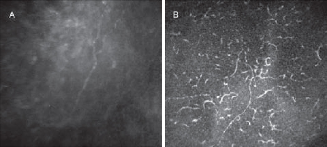
Corneal confocal microscopy is a growing technique for the study of the cornea at the cellular level, providing images comparable to ex vivo histochemical methods. In vivo confocal microscopy (IVCM) has an enormous potential, being a noninvasive procedure that images the living cornea, to study both its physiological and pathological states. Corneal nerves are of great interest to clinicians and scientists due to their important roles in regulating corneal sensation, epithelial integrity, proliferation, wound healing, and for their protective functions. IVCM enables the noninvasive examination of corneal nerves, allowing the study of nerve alterations in different ocular diseases, after corneal surgery, and in systemic diseases. To date, the correlation of sub-basal corneal nerves and their function has been studied in normal eyes, keratoconus, dry eye, contact lens wearers, and in neurotrophic keratopathy, among others. Further, the effect of corneal surgery on nerves has been studied, demonstrating the regenerative capacity of corneal nerves and the recovery of sensation. Moreover, IVCM has been applied in the diagnosis of peripheral diabetic neuropathy and the assessment of progression in this systemic disease. The purpose of this review is to describe the principles, applications, and clinical correlation of IVCM in the study of corneal nerves in different ocular and systemic diseases.
Conflict of interest statement
Figures

Similar articles
-
In Vivo Confocal Microscopy of Corneal Nerves in Health and Disease.Ocul Surf. 2017 Jan;15(1):15-47. doi: 10.1016/j.jtos.2016.09.004. Epub 2016 Oct 19. Ocul Surf. 2017. PMID: 27771327 Free PMC article. Review.
-
In vivo confocal microscopy of human corneal nerves in health, in ocular and systemic disease, and following corneal surgery: a review.Br J Ophthalmol. 2009 Jul;93(7):853-60. doi: 10.1136/bjo.2008.150615. Epub 2008 Nov 19. Br J Ophthalmol. 2009. PMID: 19019923 Review.
-
Pathologic epithelial and anterior corneal nerve morphology in early-stage congenital aniridic keratopathy.Ophthalmology. 2012 Sep;119(9):1803-10. doi: 10.1016/j.ophtha.2012.02.043. Epub 2012 Apr 17. Ophthalmology. 2012. PMID: 22512983
-
In Vivo Confocal Microscopy of Prominent Conjunctival and Corneal Nerves in Multiple Endocrine Neoplasia Type 2B.Cornea. 2019 Nov;38(11):1453-1455. doi: 10.1097/ICO.0000000000002028. Cornea. 2019. PMID: 31205161
-
Calcified corneal nerves.Cornea. 2015 Jun;34(6):707-9. doi: 10.1097/ICO.0000000000000430. Cornea. 2015. PMID: 25850704
Cited by
-
Translational Immunoimaging and Neuroimaging Demonstrate Corneal Neuroimmune Crosstalk.Cornea. 2016 Nov;35 Suppl 1(Suppl 1):S20-S24. doi: 10.1097/ICO.0000000000001014. Cornea. 2016. PMID: 27631352 Free PMC article. Review.
-
Understanding Neuropathic Corneal Pain--Gaps and Current Therapeutic Approaches.Semin Ophthalmol. 2016;31(1-2):59-70. doi: 10.3109/08820538.2015.1114853. Semin Ophthalmol. 2016. PMID: 26959131 Free PMC article. Review.
-
Characterization of the Corneal Subbasal Nerve Plexus in Limbal Stem Cell Deficiency.Cornea. 2017 Mar;36(3):347-352. doi: 10.1097/ICO.0000000000001092. Cornea. 2017. PMID: 27941384 Free PMC article.
-
Select noxious stimuli induce changes on corneal nerve morphology.J Comp Neurol. 2017 Jun 1;525(8):2019-2031. doi: 10.1002/cne.24191. Epub 2017 Mar 14. J Comp Neurol. 2017. PMID: 28213947 Free PMC article.
-
Multimodal Approach in Dry Eye Disease Combining In Vivo Confocal Microscopy and HLA-DR Expression.Transl Vis Sci Technol. 2024 Aug 1;13(8):39. doi: 10.1167/tvst.13.8.39. Transl Vis Sci Technol. 2024. PMID: 39177993 Free PMC article.
References
-
- Oliveira-Soto L, Efron N. Morphology of corneal nerves using confocal microscopy. Cornea. 2001;20:374–384. - PubMed
-
- Guthoff RF, Wienss H, Hahnel C, Wree A. Epithelial innervation of human cornea: a three-dimensional study using confocal laser scanning fluorescence microscopy. Cornea. 2005;24:608–613. - PubMed
-
- Beuerman RW, Schimmelpfennig B. Sensory dennervation of the rabbit cornea affects epithelial properties. Exp Neurol. 1980;69:196–201. - PubMed
-
- Stern ME, Beuerman RW, Fox RI, Gao J, Mircheff AK, Pflugfelder SC. The pathology of dry eye: the interaction between the ocular surface and lacrimal glands. Cornea. 1998;17:584–589. - PubMed
-
- Nishida T. Neurotrophic mediators and corneal wound healing. Ocul Surf. 2005;3:194–202. - PubMed
Publication types
MeSH terms
Grants and funding
LinkOut - more resources
Full Text Sources
Other Literature Sources
Medical