Inhibition of NF-kappa B activity in mammary epithelium increases tumor latency and decreases tumor burden
- PMID: 21076466
- PMCID: PMC3063854
- DOI: 10.1038/onc.2010.521
Inhibition of NF-kappa B activity in mammary epithelium increases tumor latency and decreases tumor burden
Abstract
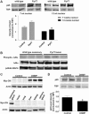
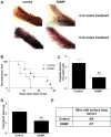
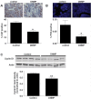
The transcription factor nuclear factor kappa B (NF-κB) is activated in human breast cancer tissues and cell lines. However, it is unclear whether NF-κB activation is a consequence of tumor formation or a contributor to tumor development. We developed a doxycycline (dox)-inducible mouse model, termed DNMP, to inhibit NF-κB activity specifically within the mammary epithelium during tumor development in the polyoma middle T oncogene (PyVT) mouse mammary tumor model. DNMP females and PyVT littermate controls were treated with dox from 4 to 12 weeks of age. We observed an increase in tumor latency and a decrease in final tumor burden in DNMP mice compared with PyVT controls. A similar effect with treatment from 8 to 12 weeks indicates that outcome is independent of effects on postnatal virgin ductal development. In both cases, DNMP mice were less likely to develop lung metastases than controls. Treatment from 8 to 9 weeks was sufficient to impact primary tumor formation. Inhibition of NF-κB increases apoptosis in hyperplastic stages of tumor development and decreases proliferation at least in part by reducing Cyclin D1 expression. To test the therapeutic potential of NF-κB inhibition, we generated palpable tumors by orthotopic injection of PyVT cells and then treated systemically with the NF-κB inhibitor thymoquinone (TQ). TQ treatment resulted in a reduction in tumor volume and weight as compared with vehicle-treated control. These data indicate that epithelial NF-κB is an active contributor to tumor progression and demonstrate that inhibition of NF-κB could have a significant therapeutic impact even at later stages of mammary tumor progression.
Figures


Similar articles
-
Activation of nuclear factor kappa B in mammary epithelium promotes milk loss during mammary development and infection.J Cell Physiol. 2010 Jan;222(1):73-81. doi: 10.1002/jcp.21922. J Cell Physiol. 2010. PMID: 19746431 Free PMC article.
-
Aberrant activation of NF-κB signaling in mammary epithelium leads to abnormal growth and ductal carcinoma in situ.BMC Cancer. 2015 Sep 30;15:647. doi: 10.1186/s12885-015-1652-8. BMC Cancer. 2015. PMID: 26424146 Free PMC article.
-
RelB/p52 NF-kappaB complexes rescue an early delay in mammary gland development in transgenic mice with targeted superrepressor IkappaB-alpha expression and promote carcinogenesis of the mammary gland.Mol Cell Biol. 2005 Nov;25(22):10136-47. doi: 10.1128/MCB.25.22.10136-10147.2005. Mol Cell Biol. 2005. PMID: 16260626 Free PMC article.
-
NF-kappaB in mammary gland development and breast cancer.J Mammary Gland Biol Neoplasia. 2003 Apr;8(2):215-23. doi: 10.1023/a:1025905008934. J Mammary Gland Biol Neoplasia. 2003. PMID: 14635796 Review.
-
NF-kappaB and apoptosis in mammary epithelial cells.J Mammary Gland Biol Neoplasia. 1999 Apr;4(2):165-75. doi: 10.1023/a:1018725207969. J Mammary Gland Biol Neoplasia. 1999. PMID: 10426395 Review.
Cited by
-
Adverse outcome pathways for ionizing radiation and breast cancer involve direct and indirect DNA damage, oxidative stress, inflammation, genomic instability, and interaction with hormonal regulation of the breast.Arch Toxicol. 2020 May;94(5):1511-1549. doi: 10.1007/s00204-020-02752-z. Epub 2020 May 13. Arch Toxicol. 2020. PMID: 32399610 Free PMC article. Review.
-
Immunity drives TET1 regulation in cancer through NF-κB.Sci Adv. 2018 Jun 20;4(6):eaap7309. doi: 10.1126/sciadv.aap7309. eCollection 2018 Jun. Sci Adv. 2018. PMID: 29938218 Free PMC article.
-
The crossroads of breast cancer progression: insights into the modulation of major signaling pathways.Onco Targets Ther. 2017 Nov 20;10:5491-5524. doi: 10.2147/OTT.S142154. eCollection 2017. Onco Targets Ther. 2017. PMID: 29200866 Free PMC article. Review.
-
Down-regulation of neogenin accelerated glioma progression through promoter Methylation and its overexpression in SHG-44 Induced Apoptosis.PLoS One. 2012;7(5):e38074. doi: 10.1371/journal.pone.0038074. Epub 2012 May 29. PLoS One. 2012. PMID: 22666451 Free PMC article.
-
A requirement for p120-catenin in the metastasis of invasive ductal breast cancer.J Cell Sci. 2021 Mar 17;134(6):jcs250639. doi: 10.1242/jcs.250639. J Cell Sci. 2021. PMID: 33097605 Free PMC article.
References
-
- Baeuerle PA, Baltimore D. I kappa B: a specific inhibitor of the NF-kappa B transcription factor. Science. 1988;242:540–6. - PubMed
-
- Baldwin AS., Jr. The NF-kappa B and I kappa B proteins: new discoveries and insights. Annu Rev Immunol. 1996;14:649–83. - PubMed
-
- Biswas DK, Iglehart JD. Linkage between EGFR family receptors and nuclear factor kappaB (NF-kappaB) signaling in breast cancer. J Cell Physiol. 2006;209:645–52. - PubMed
Publication types
MeSH terms
Substances
Grants and funding
LinkOut - more resources
Full Text Sources
Other Literature Sources
Molecular Biology Databases
Research Materials

