. 2010 Dec;12(12):1177-85.
doi: 10.1038/ncb2121. Epub 2010 Nov 14.

The N-end rule pathway is mediated by a complex of the RING-type Ubr1 and HECT-type Ufd4 ubiquitin ligases

Affiliations

The N-end rule pathway is mediated by a complex of the RING-type Ubr1 and HECT-type Ufd4 ubiquitin ligases

Cheol-Sang Hwang et al. Nat Cell Biol. 2010 Dec.

Abstract

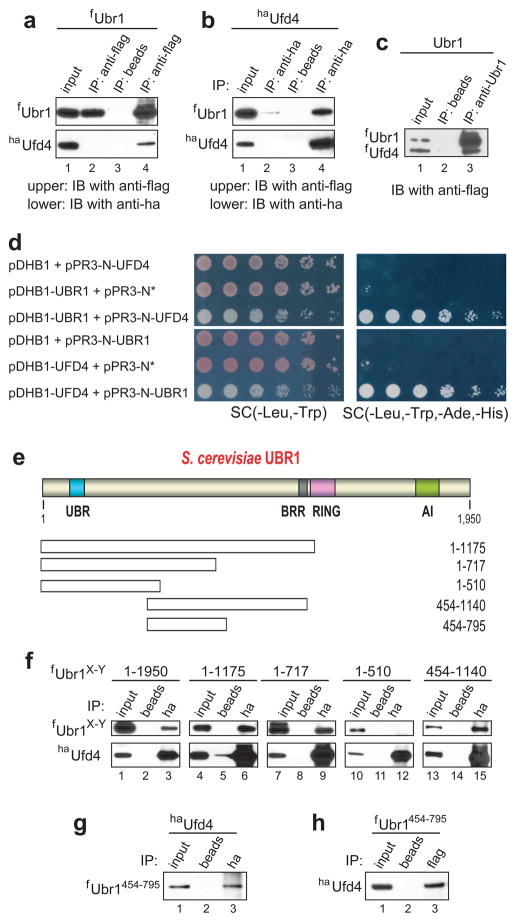
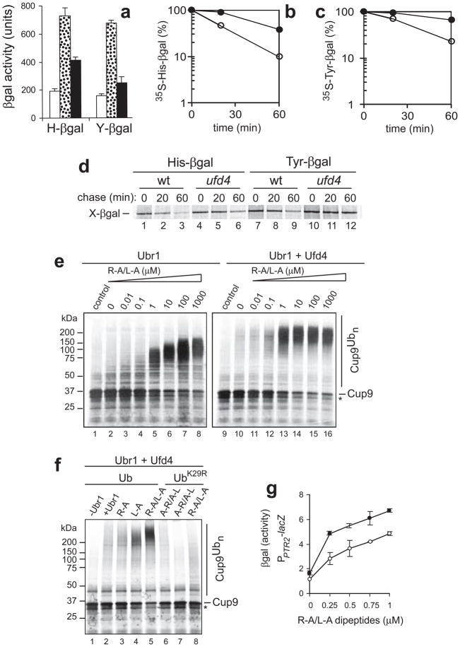
Substrates of the N-end rule pathway are recognized by the Ubr1 E3 ubiquitin ligase through their destabilizing amino-terminal residues. Our previous work showed that the Ubr1 E3 and the Ufd4 E3 together target an internal degradation signal (degron) of the Mgt1 DNA repair protein. Ufd4 is an E3 enzyme of the ubiquitin-fusion degradation (UFD) pathway that recognizes an N-terminal ubiquitin moiety. Here we show that the RING-type Ubr1 E3 and the HECT-type Ufd4 E3 interact, both physically and functionally. Although Ubr1 can recognize and polyubiquitylate an N-end rule substrate in the absence of Ufd4, the Ubr1-Ufd4 complex is more processive in that it produces a longer substrate-linked polyubiquitin chain. Conversely, Ubr1 can function as a polyubiquitylation-enhancing component of the Ubr1-Ufd4 complex in its targeting of UFD substrates. We also found that Ubr1 can recognize the N-terminal ubiquitin moiety. These and related advances unify two proteolytic systems that have been studied separately for two decades.


Conflict of interest statement

COMPETING INTERESTS

The authors declare that they have no competing financial interest.

Figures

Figure 1
Figure 1
The Arg/N-end rule and UFD pathways. (a) The S. cerevisiae Arg/N-end rule pathway. See Introduction for terminology. N-terminal residues are indicated by single-letter abbreviations for amino acids. Yellow ovals denote the rest of a protein substrate. (b) The S. cerevisiae UFD (Ub-fusion degradation) pathway,,. One class of UFD substrates are engineered protein fusions that have in common a ‘nonremovable’ N-terminal Ub moiety that acts as a degron. Mgt1 is a physiological substrate of both the Arg/N-end rule and UFD pathways. A degron of Mgt1 is close to its N-terminus but is distinct from an N-degron. Polyubiquitylation and degradation of Cup9 is mediated by the Ubr1-Ufd4 complex. (c) Both the Arg/N-end rule pathway and a subset of the UFD pathway are mediated by the Ubr1/Rad6-Ufd4/Ubc4 complex discovered in the present work. Also cited are physiological substrates of these pathways in S. cerevisiae. Mgt1 and Cup9 contain internal degrons,,. The separase-produced fragment of the Scc1 subunit of cohesin contains an Arg-based N-degron,.
Figure 2
Figure 2
Ubiquitylation of Mgt1 by Ubr1-Ufd4. The in vitro ubiquitylation assay is described in Methods. Reaction mixtures were incubated at 30° C for 15 min, followed by SDS-PAGE and autoradiography. 35S-Mgt1f3 and its polyubiquitylated derivatives are indicated on the right. Lane 1, Mgt1f3 in the complete reaction but without added Ub and with Ubr1 as the sole E3. Lane 2, same as lane 1 but with wild-type Ub. Lane 3, same as lane 1 but with UbK29R. Lane 4, same as lane 1 but with UbK48R. Lane 5, same as lane 1 but with UbK63R. Lane 6, S-Mgt1f3 in the complete reaction (containing both Ubr1 and Ufd4) but without added Ub. Lane 7, same as lane 6 but with wild-type Ub. Lane 8, same as lane 6 but with UbK29R. Lane 9, same as lane 6 but with UbK48R. Lane 10, same as lane 6 but with UbK63R. Lane 11, Mgt1f3 in the complete reaction but without added Ub and with Ufd4. Lane 12, same as lane 11 but with wild-type Ub. Lane 13, same as lane 11 but with UbK29R. Lane 14, same as lane 1 but with UbK48R. Lane 15, same as lane 11 but with UbK63R.
Figure 3
Figure 3
Physical interaction between Ubr1 and Ufd4. (a) Coimmunoprecipitation of fUbr1 and haUfd4 with anti-flag antibody. JD52 S. cerevisiae expressed N-terminally flag-tagged Ubr1 (fUbr1), either alone (lane 2) or together with N-terminally ha-tagged Ufd4 (haUfd4) (lanes 3 and 4). Cell extracts were immunoprecipitated with anti-flag (lanes 2 and 4) or with antibody-free beads (lane 3). The upper and lower panels show the results of immunoblotting with anti-flag (detection of fUbr1) and with anti-ha (detection of haUfd4), respectively. Lane 1, 1% input of extract from cells expressing both fUbr1 and haUfd4. Lane 2, extract from cells expressing only fUbr1 was incubated with anti-flag pre-bound to beads. Immunoblotting with anti-flag and anti-ha (upper and lower panels, respectively). Lane 3, extract from cells expressing both fUbr1 and haUfd4, but with beads lacking antibody. Lane 4, same as lane 3 but immunoprecipitation with anti-flag. (b) Same as in a but extracts were immunoprecipitated with anti-ha pre-bound to beads (lanes 2 and 4) or with beads lacking antibody (lane 3), followed by SDS-PAGE of immunoprecipitates and immunoblotting with anti-flag and anti-ha. (c) Direct interaction of fUbr1 and fUfd4. Lane 1, 10% inputs of fUbr1 and fUfd4 (purified as described in ref. ; see also Fig. S1a). fUbr1 (125 ng) and fUfd4 (125 ng) were mixed and incubated with beads lacking antibody (lane 2) or with previously characterized affinity-purified anti-Ubr1 antibody pre-bound to beads (lane 3), for 1 hr at 4° C in 0.25 ml of binding buffer, followed by SDS-PAGE and immunoblotting with anti-flag. (d) In vivo detection of Ubr1-Ufd4 interactions using split-Ub assay. S. cerevisiae coexpressing bait (pDHB1, pDHB1-UBR1, or pDHB1-UFD4) and prey (pPR3-N*, pPR3-N-UFD4, or pPR3-N-UBR1) plasmids were grown to A600 of ~1, serially diluted (5-fold), and plated on either ‘permissive’ SC(-Leu, -Trp) (left column) or SC(-Leu, -Trp, -Ade, -His) medium (right column). pDHB1 and pPR3-N are the initial (vector) plasmids. pPR3-N* contained a stop codon immediately after the ORF encoding the mutant N-terminal half of Ub (NUb) in pPR3-N. (e) The UBR box, the BRR region, the RING domain, and the AI (autoinhibitory) domain of the S. cerevisiae Ubr1 N-recognin,,. Fragments of Ubr1 employed to map its Ufd4-interacting region are below the diagram. (f) Extracts from JD52 S. cerevisiae that expressed haUfd4 and either full-length fUbr1 or its flag-tagged fragments were incubated with antibody-lacking beads (lanes 2, 5, 8, 11 and 14) or with anti-ha pre-bound to beads (lanes 3, 6, 9, 12 and 15). Bound proteins were eluted from washed beads, followed by SDS-PAGE and immunoblotting with anti-flag. Input lanes, samples of extracts that corresponded to 1% of initial extracts. (g) Coimmunoprecipitation of fUbr1454-795 and haUfd4 with anti-ha. Lane 1, 1% input of the initial extract. Lane 2, extracts from cells that expressed the fUbr1454-795 fragment and full-length haUfd4 were incubated with antibody-free beads (lanes 2). Bound proteins were eluted from washed beads and fractionated by SDS-PAGE, followed by immunoblotting with anti-flag. Lane 3, same as lane 2 but immunoprecipitation with anti-ha pre-bound to beads. (h) Lanes 1-3, same as lanes 1-3 in g, but immunoprecipitation with anti-flag (instead of anti-ha), followed by immunoblotting with anti-ha.
Figure 4
Figure 4
Enhancement of ubiquitylation and degradation of Arg/N-end rule substrates by Ufd4. (a) X-eK-DHFRha (X=Met, Arg, Leu), denoted as X-DHFRha, are C-terminally ha-tagged Arg/N-end rule reporters produced from Ub-X-DHFRha using in vitro deubiquitylation (Fig. S1b). X-DHFRha contained the mouse dihydrofolate reductase (DHFR) moiety and the ~40-residue N-terminal extension called eK [extension (e) containing lysine (K)]. Purified X-DHFRha (X=Met, Arg, Leu) (1.25 μM; 2 μl) were incubated in 20 μl of a ubiquitylation assay for 15 min at 30° C, followed by SDS-PAGE and immunoblotting with anti-ha. Lanes 1, 7, 13: X-DHFRha in the absence of indicated assay’s components. Lanes 2, 8, 14: same as lanes l, 7, 13 but with Rad6 E2. Lane 3, 9, 15, same as lanes 1, 7, 13 but with Ubr1 and Rad6. Lanes 4, 10, 16 same as lanes 1, 7, 13 but with Ubc4 E2. Lane 5, 11, 17 same as lane 1, 7, 13 but with Ufd4 and Ubc4. Lane 6, 12, 18 same as lane 1 but with Ubr1, Rad6, Ufd4 and Ubc4. Asterisk on the right denotes a protein that crossreacted with anti-ha antibody. (b) Same as in a but the assay was carried out with Arg-DHFRha and indicated Ub mutants. Detection of immunoblotted proteins in this experiment was performed using Odyssey (Li-Cor, Lincoln, NE, USA). Asterisks on the left indicate two bands of proteins (e.g., lanes 1, 4) that crossreacted with anti-ha, and were also present in samples lacking E2s and E3s. Lanes 1, 4, 7, 10, 13, 16, ubiquitylation of Arg-DHFRha with Ufd4/Ubc4 in the presence of either UbK29 (lane 1), UbK48 (lane 4), a 50-50 mixture of UbK29 and UbK48 (lane 7), UbK29R (lane 10), UbK48R (lane 13), or a 50-50 mixture of UbK29R and UbK48R (lane 16). Lanes 2, 5, 8, 11, 14, 17, same as lanes 1, 4, 7, 10, 13, 16, respectively, but with Ubr1/Rad6 instead of Ufd4/Ubc4. Lanes 3, 6, 9, 12, 15, 18, same as lanes 1, 4, 7, 10, 13, 16, respectively, but with Ubr1/Rad6 plus Ufd4/Ubc4. (c) Lanes 1 and 2, molecular mass markers and Coomassie-stained proteins of the affinity-purified S. cerevisiae 26S proteasome, respectively. (d) Lanes 1–3, assay with 26S proteasome and polyubiquitylated Leu-DHFRha that had been prepared using Ubr1/Rad6 alone, with chase times of 10 and 20 min. Lanes 4–6, same as lanes 1–3, but with polyubiquitylated Leu-DHFRha that had been prepared with Ubr1/Rad6 plus and Ufd4/Ubc4. (e) Quantitation of data in d, using ImageJ (http://rsb.info.nih.gov/ij/index.html). In plotting the levels of Leu-DHFRha for each data set (lanes 1–3 and 4–6 in d), the levels at time zero were taken as 100%. Open and closed circles, Leu-DHFRha that had been ubiquitylated by Ubr1/Rad6 and by Ubr1/Rad6 plus Ufd4/Ubc4, respectively.
Figure 5
Figure 5
Ufd4 augments the Arg/N-end rule pathway. (a) β gal activity in extracts from S. cerevisiae RJD347 (wild-type; white bars), AVY26 (ubr1Δ; dotted bars), and CHY251 (ufd4Δ; black bars) that expressed His-β gal or Tyr-β gal. (b ,c ) Quantitation of data (using PhosphorImager) in a pulse-chase assay (d) for His-β gal (b) and Tyr-β gal (c). Open and closed circles, wild-type (RJD347) and ufd4Δ (CHY251) cells, respectively. In d, S. cerevisiae expressing Ub-His-β gal or Ub-Tyr-β gal were labeled for 5 min with 35S-methionine/cysteine, followed by a chase for 20 and 60 min, immunoprecipitation with anti-β gal, SDS-PAGE and autoradiography,. Lanes 1–3, His-β gal in wild-type cells. Lanes 4–6, His-β gal in ufd4Δ cells. Lanes 7–9, Tyr-β gal in wild-type cells. Lanes 10–12, Tyr-β gal in ufd4Δ cells. (e) Ubr1/Rad6-mediated polyubiquitylation of Cup9. In vitro ubiquitylation assay was performed with 35S-labeled Cup9NSF (see Methods). Lane 1, 35S-Cup9 in an otherwise complete assay but without E3s. Lanes 2–8, same as lane 1 but with Ubr1/Rad6, in the presence of Arg-Ala (R-A)/Leu-Ala (L-A). Lane 9, same as lane 1 but a separate assay. Lanes 10–16, same as lanes 2–8, but with Ubr1 plus Ufd4. (f) Maximal stimulation of Cup9 ubiquitylation by Ubr1-Ufd4 requires both type-1 and type-2 dipeptides. Lane 1, 35S-Cup9, with Ufd4 and wild-type Ub, but in the absence of both Ubr1 and type-1/2 dipeptides. Lane 2, same as lane 1 but with Ubr1. Lane 3, same as lane 2, but with 1 μM R-A. Lane 4, same as lane 2 but in the presence of 1 μM L-A. Lane 5, same as lane 2, but in the presence of R-A and L-A, each at 1 μM. Lane 6, same as lane 2 but in the presence of Ala-Arg/Ala-Leu, each at 1 μM. Lanes 7, same as lane 6, but with UbK29R, instead of wild-type Ub. Lane 8, same as lane 7, but in the presence of R-A/L-A, each at 1 μM. (g) Dipeptide-mediated induction of the PTR2 transporter in the absence or presence of Ufd4. S. cerevisiae RJD347 (wild-type; closed circles) and CHY251 (ufd4Δ; open circles) expressed E. coli lacZ (β-galactosidase) from the PPTR2 promoter. Cells were grown to A600 of ~0.8 in the SHM medium at 30° C in the presence of indicated concentrations of R-A/L-A, followed by measurements in triplicate, of β gal activity in cell extracts, with standard deviations shown.
Figure 6
Figure 6
Recognition and synergistic polyubiquitylation of UFD substrates by Ufd4 and Ubr1. (a) Ubiquitylation assay, for 15 min at 30° C, with Ub-ProtA, a UFD substrate (0.125 μM) (Fig. S1c), followed by SDS-PAGE and immunoblotting with anti-ProtA antibody. Lane 1, without E3s. Lane 2, same as lane 1 but with Ubr1/Rad6. Lane 3, same as lane 1 but with Ufd4/Ubc4. Lane 4, same as lane 1 but with Ubr1/Rad6 plus Ufd4/Ubc4. Lane 6, same as lane 1 but with Ufd2/Ubc4 plus Ubr1/Rad6. Lane 7, same as lane 1 but with Ufd2/Ubc4 plus Ufd4/Ubc4. Lane 8, same as lane 1 but with Ufd2/Ubc4, Ubr1/Rad6 and Ufd4/Ubc4. (b) Ubiquitylation assay with Ub-GST (0.125 μM). Lane 1, Ub-GST without E3s. Lane 2, same as lane 1 but with Ubr1/Rad6. Lane 3, same as lane 1 but with Ufd4/Ubc4. Lane 4, same as lane 1 but with Ubr1/Rad6 plus Ufd4/Ubc4. Lane 5, same as lane 1 but with Ufd2/Ubc4. Lane 6, same as lane 1 but with Ufd2/Ubc4 plus Ubr1/Rad6. Lane 7, same as lane 1 but with Ufd2/Ubc4 plus Ufd4/Ubc4. Lane 8, same as lane 1 but with Ufd2/Ubc4, Ubr1/Rad6 and Ufd4/Ubc4. (c) Interaction of Ubr1 with immobilized UFD substrates could be competed out by UFD substrates but not by free Ub. Equal amounts of purified fUbr1 (1 μg) were incubated (in either the presence or absence of free Ub, Ub-DHFRha (Ub-Met-DHFRha) or Ub-ProtA, each of them at 1 or 10 μM)) with GST alone or Ub-GST (~5 μg) that had been linked to glutathione-Sepharose beads. Bound proteins were eluted from the beads, followed by SDS-PAGE and immunoblotting with anti-flag (upper panel), with subsequent Coomassie staining of the blotted PVDF membrane (lower panel). Lane 1, GST alone. Lane 2, Ub-GST. Lane 3, same as lane 2 but in the presence of 1 μM free Ub. Lane 4, same as lane 2 but with 10 μM free Ub. Lane 5, same as lane 2 but in with 1 μM Ub-DHFRha. Lane 6, same as lane 2 but with 10 μM Ub-DHFRha. Lane 7, same as lane 2 but with 1 μM Ub-ProtA. Lane 8, same as lane 2 but with 10 μM Ub-ProtA. (d) In vivo levels of endogenous Ubr1. Lanes 1–6, a dilution series with the indicated amounts of purified fUbr1 was fractionated by SDS-PAGE, followed by immunoblotting with affinity-purified anti-Ubr1 antibody. Lane 7, extract (50 μg) from wild-type S. cerevisiae (JD52) that grew exponentially (A600 of ~1) in YPD medium. Lane 8, same as lane 7 but extract from ubr1Δcells (JD55). These data and straightforward calculations indicated that ‘wild-type’, haploid, exponentially growing S. cerevisiae contained 500 to 1,000 Ubr1 molecules per cell. (e)Ubr1 and Ufd4 did not affect the Rsp5-mediated polyubiquitylation of T7 epitope-tagged Sic1PY. The PY motif is the sequence Pro-Pro-X-Tyr, which binds to the WW domain of Rsp5 (see Methods). Purified Sic1PY (Fig. S1c) was incubated in the above ubiquitylation assay, followed by SDS-PAGE and immunoblotting with anti-T7 antibody. Lane 1, Sic1PY without E3s. Lane 2, same as lane 1 but with Ubr1/Rad6. Lane 3, same as lane 1 but with Ufd4/Ubc4. Lane 4, same as lane 1 but with Ubr1/rad6 plus Ufd4/Ubc4. Lane 5, same as lane 1 but with Rsp5/Ubc4. Lane 6, same as lane 1 but with Rsp5/Ubc4 and Ubr1/Rad6. Lane 7, same as lane 1 but with Rsp5/Ubc4 plus Ufd4/Ubc4. Lane 8, same as lane 1 but with Rsp5/Ubc4, Ubr1/Rad6 and Ufd4/Ubc4. (f, g) Equal amounts of cells from wild-type (JD52), ubr1Δ (JD55), ufd4Δ (CHY194) or ubr1Δufd4 Δ (CHY195) strains were 5-fold serially diluted, plated on YPD plates containing 6% ethanol (6%) (f) or canavanine at 0.4 mg/ml (g), and incubated at 30° C for 3 days and 1 day, respectively.

Comment in

Similar articles

Cited by

References

    1. Bachmair A, Finley D, Varshavsky A. In vivo half-life of a protein is a function of its amino-terminal residue. Science. 1986;234:179–186. - PubMed
    1. Varshavsky A. The N-end rule: functions, mysteries, uses. Proc Natl Acad Sci USA. 1996;93 :12142–12149. - PMC - PubMed
    1. Varshavsky A. Discovery of cellular regulation by protein degradation. J Biol Chem. 2008;283:34469–34489. - PMC - PubMed
    1. Ravid T, Hochstrasser M. Diversity of degradation signals in the ubiquitin-proteasome system. Nat Rev Mol Cell Biol. 2008;9:679–689. - PMC - PubMed
    1. Turner GC, Du F, Varshavsky A. Peptides accelerate their uptake by activating a ubiquitin-dependent proteolytic pathway. Nature. 2000;405:579–583. - PubMed

Publication types

MeSH terms

LinkOut - more resources