. 2011 May;18(5):754-68.
doi: 10.1038/cdd.2010.143. Epub 2010 Nov 12.

IFNγ triggers a LIGHT-dependent selective death of motoneurons contributing to the non-cell-autonomous effects of mutant SOD1

Affiliations

IFNγ triggers a LIGHT-dependent selective death of motoneurons contributing to the non-cell-autonomous effects of mutant SOD1

J Aebischer et al. Cell Death Differ. 2011 May.

Abstract

Amyotrophic lateral sclerosis (ALS) is an incurable neurodegenerative disease that primarily affects motoneurons in the brain and spinal cord. Dominant mutations in superoxide dismutase-1 (SOD1) cause a familial form of ALS. Mutant SOD1-damaged glial cells contribute to ALS pathogenesis by releasing neurotoxic factors, but the mechanistic basis of the motoneuron-specific elimination is poorly understood. Here, we describe a motoneuron-selective death pathway triggered by activation of lymphotoxin-β receptor (LT-βR) by LIGHT, and operating by a novel signaling scheme. We show that astrocytes expressing mutant SOD1 mediate the selective death of motoneurons through the proinflammatory cytokine interferon-γ (IFNγ), which activates the LIGHT-LT-βR death pathway. The expression of LIGHT and LT-βR by motoneurons in vivo correlates with the preferential expression of IFNγ by motoneurons and astrocytes at disease onset and symptomatic stage in ALS mice. Importantly, the genetic ablation of Light in an ALS mouse model retards progression, but not onset, of the disease and increases lifespan. We propose that IFNγ contributes to a cross-talk between motoneurons and astrocytes causing the selective loss of some motoneurons following activation of the LIGHT-induced death pathway.


Figures
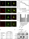
Figure 1
Figure 1
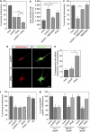
sLIGHT selectively induces death of motoneurons. (ad) Hb9∷GFP motoneurons were cultured for 24 h and immunostained with anti-LT-βR (a), anti-HVEM (b) and anti-LIGHT (c) antibodies. Goat (d) or rabbit (not shown) irrelevant IgGs were used as control. Scale bar, 10 μm. (e) Mouse motoneurons were cultured for 24 h and then incubated with increasing concentrations of human or mouse sLIGHT. Motoneuron survival was determined 48 h later and expressed relative to non-treated cells. Henceforth, sLIGHT will refer to the human form of the recombinant protein. The distance between the x-axis values is arbitrary. (f) After 24 h in culture, motoneurons were treated (or not) with agonistic anti-LT-βR antibodies (100 ng/ml), antagonistic anti-HVEM antibodies (100 ng/ml) or irrelevant goat IgG (100 ng/ml) in combination (or not) with sLIGHT (100 ng/ml). Cell survival was determined 48 h later. (g) Twenty four hours after plating, cortical, hippocampal, sensory and striatal neurons were treated (or not) with sLIGHT (100 ng/ml). Neuron survival was determined 48 h later. (h) Protein extracts from cortical, hippocampal, sensory, striatal and motoneurons cultured for 24 h were resolved by sodium dodecyl sulfate polyacrylamide gel electrophoresis, followed by Western blotting with specific antibodies to LT-βR and LIGHT. The loading control was α-tubulin. Lysates from Cos-7 cells transfected (+) or not (−) with LT-βR or LIGHT expression vectors were used as controls. Graphs in (e), (f) and table in (g) show means values of three independent experiments, each done in triplicate
Figure 2
Figure 2
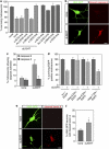
sLIGHT and sFasL show additive killing effect but LIGHT in contrary to FasL functions independently of mitochondrial cytochrome c release. (a) Mouse motoneurons were cultured for 24 h and treated (or not) with sLIGHT (100 ng/ml), recombinant human soluble FasL (sFasL, 100 ng/ml in the presence of 1 μg/ml enhancer antibody), or a combination of both. Motoneuron survival was assessed 48 h after treatment. Note that the enhancer antibody used to cross-link sFasL had no effect on motoneuron survival when added alone or with sLIGHT (not shown)(n=4, ***P<0.001). (b) Quantification of phospho-p38 kinase fluorescence in Hb9∷GFP motoneurons treated (or not) with sLIGHT in the presence or the absence of SB203580 (10 μM) for 1 h. Treatment with sFasL served as a control. Confocal fluorescence imaging and ImageJ image analysis were used to determine the nuclear mean fluorescence intensity of p38 in Hb9∷GFP neurons (a.u, arbitrary unit, *P<0.05, **P<0.01). (c) Motoneurons were maintained in culture for 24 h and treated or not with sLIGHT (100 ng/ml) and SB203580 (10 μM). Survival was determined 48 h later and expressed relative to survival in the absence of any treatment. (d and e) Hb9∷GFP motoneurons were incubated or not with 100 ng/ml of sLIGHT or sFasL, fixed and immunostained with anti-cytochrome c antibody 30 h later. sLIGHT-treated Hb9∷GFP motoneuron shows the same punctuate labeling of mitochondria as non-treated motoneurons (not shown), whereas following Fas activation a proportion of motoneurons show diffuse labeling of cytochrome c. Scale bar, 10 μm. (e) The percentage of Hb9∷GFP motoneurons showing diffuse versus punctuated cytochrome c labeling was determined by direct counting under fluorescence microscope (n=4). (f) Survival assay was done as in (c). The Bax inhibitory peptide V5 was used at the concentration of 50 μM. (g) Mouse motoneurons were co-electroporated with an equimolar ratio of a vector coding for EGFP and a vector coding for either Bcl-Xl, a dominant negative form of Bid (tn-Bid) or an empty vector (e.v) and cultured for 24 h before being treated or not with sLIGHT or sFasL. The survival of EGFP-positive motoneurons for each combination of vectors was expressed as a percentage of surviving motoneurons in the absence of sLIGHT or sFasL. Histograms show mean values±S.D. of at least three independent experiments, each done in triplicate
Figure 3
Figure 3
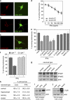
sLIGHT triggers motoneuron death through an unconventional caspase cascade. (a) Motoneurons were incubated with z-IETD-fmk (10 μM), Ac-LEHD-cmk (1 μM), z-DEVD-fmk (10 μM) or z-VEID-fmk (10 μM) added together with sLIGHT (100 ng/ml). Motoneuron survival was determined 48 h later and expressed relative to non-treated condition. (b) Activation of caspase-9 was visualized in sLIGHT-treated Hb9∷GFP motoneurons with antibodies specific to the cleaved form of caspase-9. (c) The percentage of Hb9∷GFP motoneurons immunopositive for active caspase-9 (b) or -3 (see Supplementary Figure 3b) was determined 30 h later following addition of sLIGHT by direct counting using fluorescence microscopy. Statistical attribute is shown for none versus sLIGHT-treated cells (n=3, **P<0.01). (d) Motoneurons were co-electroporated with a combination of expression vectors encoding EGFP and catalytically inactive mutants of caspase-9 (casp-9 DN), -6 (casp-6 DN) or -7 (casp-7 DN), and incubated in the presence or absence of sLIGHT. The effect of the dominant negative caspases on motoneuron survival was determined by counting the EGFP-positive motoneurons 48 h after treatment. e.v, empty vector. (e) The experimental procedure was performed as in (b) only that the percentage of motoneurons positive for cleaved lamin A was determined 38 h after treatment using an anti-cleaved lamin A antibody. Scale bars, 10 μm. (f) The percentage of cleaved lamin-A-positive Hb9∷GFP motoneurons for LIGHT-treated or untreated conditions was determined by counting under fluorescent microscope (n=3, *P<0.05). All values are expressed as the means±S.D. of three independent experiments
Figure 4
Figure 4
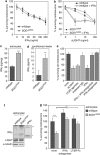
IFNγ selectively kills motoneurons in a LIGHT-dependent manner. (ac) Isolated Hb9∷GFP motoneurons were immunostained 24 h after seeding with antibodies directed against IFNγR1 (a), IFNγR2 (b) or with hamster (c) or mouse (not shown) irrelevant IgG as control. Scale bar, 10 μm. (d) Motoneurons were cultured for 24 h and incubated with increasing concentrations of soluble mouse recombinant IFNγ from two different sources. The percentage of surviving motoneurons was determined 48 h later. The distance between the x-axis values is arbitrary. (e) Motoneuron survival was determined 48 h following treatment or not with LT-βR-Fc (100 ng/ml), HVEM-Fc (10 ng/ml), Fas-Fc (1 μg/ml) or TNFR1-Fc (100 ng/ml) in combination or not with 250 ng/ml of IFNγ. The number of surviving motoneurons is expressed as a percentage of the number of motoneurons in the control condition (none). (f) Motoneurons were isolated from E12.5 embryos of indicated genotype and cultured for 24 h before being treated with 250 ng/ml of IFNγ. Motoneuron survival was determined 48 h later and expressed relative to the non-treated condition for each genotype. (g) Immunoblot analysis of cortical, hippocampal, sensory, striatal and motoneurons proteins separated by sodium dodecyl sulfate polyacrylamide gel electrophoresis. Actin was used as a loading control. (h) Cell survival assay was performed as in (d), with neuronal cells being treated with 250 ng/ml of IFNγ. (i and j) Motoneurons were cultured for 24 h and incubated with 250 ng/ml of IFNγ. Eight hours later, cells were lysed and expression levels of LIGHT (i) and LT-βR (j) were determined by western blotting with indicated antibodies. Fold-increase of LIGHT and LT-βR over non-treated conditions was determined by densitometric analysis of immunoreactive bands, normalized to their respective actin signals (n=3, means±S.D.). Results shown in (d, e, f and h) are the mean values±S.D. of three independent experiments performed in triplicate
Figure 5
Figure 5
SOD1 mutant astrocytes kill motoneurons in an IFNγ/LIGHT-dependent pathway. (a) Twenty four hours after plating, indicated concentrations of mouse recombinant IFNγ were added to motoneurons isolated from SOD1G93A or wild-type embryos of the same littermate. Motoneuron survival was determined 48 h later and expressed relative to non-treated condition of corresponding genotype. (b) Mutant SOD1G93A and wild-type motoneurons were treated after 24 h in culture with increasing concentrations of sLIGHT in combination or not with IFNγ (10 ng/ml). Motoneuron survival was determined 48 h after treatment. (c and d) IFNγ levels in extracts (c) and conditioned media (d) of astrocytes of indicated genotype were quantified by ELISA (n=4, means±S.D.). (e) Immunopurified E14 rat motoneurons were treated or not with sLIGHT (100 ng/ml), agonistic anti-LT-βR antibodies (100 ng/ml), irrelevant IgG (100 ng/ml), recombinant soluble rat IFNγ (250 ng/ml) in combination or not with antagonistic anti-IFNγ (500 ng/ml) or anti-IFNγ antibodies alone. Survival of motoneurons was determined 48 h after treatment by direct counting. (f) Total protein extract of wild-type and SOD1G93A rat astrocytes were subjected to sodium dodecyl sulfate polyacrylamide gel electrophoresis followed by immunoblotting with anti-IFNγ specific antibodies. Recombinant rat IFNγ was used as a control. Asterisks indicate the monomeric (*) form of IFNγ and the apparent stable dimeric (**) biologically active form of IFNγ. (g) Wild-type motoneurons were plated on astrocyte monolayer of indicated genotype (wild type, SOD1G93A) and incubated or not with function-blocking anti-IFNγ antibodies (500 ng/ml) or LT-βR-Fc (100 ng/ml) for 48 h. Survival of motoneurons is expressed as the percentage of the number of motoneurons surviving on wild-type astrocyte monolayer in the absence of any treatment. The graphs show the mean values±S.D. of at least three independent experiments performed in triplicate
Figure 6
Figure 6
IFNγ is upregulated in spinal cords of ALS mice. (a) The percentage of motoneurons, as identified with VAChT immunostaining on adjacent sections (Supplementary Figure 5a and b), immunoreactive for LIGHT and LT-βR was determined in lumbar spinal cord of wild-type and SOD1G93A mice at 75, 90 and 110 days (d) of age. (b) Total protein extracts from lumbar spinal cords of wild-type and SOD1G93A mice at indicated age were resolved by sodium dodecyl sulfate polyacrylamide gel electrophoresis and probed with antibodies to IFNγ and actin (Supplementary Figure 5c). IFNγ signals were quantified, normalized to actin signals and expressed as the ratio of SOD1G93A to wild-type values. (c) ELISA quantification of IFNγ levels in dissociated spinal cord of 110-day-old wild-type and SOD1G93A mice and 365-day-old SOD1WT mice. Values in (ac) are means±S.D., n=3. (d and f) Immunostainings of wild-type and SOD1G93A mice lumbar spinal cord sections at 90 days of age using antibody against IFNγ in combination with either GFAP (d), Iba1 (e) or SMI32 (f). Scale bar, 30 μm. (g) Lumbar spinal cord sections of wild-type and SOD1G93A mice were immunostained as in (f) at 75, 90 and 110 days of age and the percentage of IFNγ immunoreactive motoneurons was determined by counting under fluorescent microscope (75 days, n=3; 90 days, n=4; 110 days, n=5, values are means±S.D.)
Figure 7
Figure 7
Targeted deletion of Light in ALS mice delays progression but not onset of the disease. (a) The cumulative probability of onset of SOD1G93A/Light+/+ (n=12) and SOD1G93A; Light−/− (n=16) was determined by the peak of weight curve. (b) The progressive motor deficit of SOD1G93A/Light+/+, SOD1G93A/Light−/−, Light+/+ and Light−/− was determined by evaluating weekly the swimming performance of mice (values are means±S.E.M.). Statistical attributes are shown only for SOD1G93A/Light+/+ versus SOD1G93A/Light−/− (Supplementary Table). (c) Kaplan–Meier survival curves for SOD1G93A/Light+/+ (n=12) and SOD1G93A/Light−/− (n=14) mice. (d) The mean motoneuron survival was determined by counting the number of VAChT-immunostained motoneurons in 28 sections of lumbar spinal cord from 120-day-old Light+/+ (n=3), Light−/− (n=3), SOD1G93A/Light+/+ (n=4) and SOD1G93A/Light−/− (n=4) (values are means±S.D.)
Figure 8
Figure 8
Model for the non-cell-autonomous effect of mutant SOD1 on the activation of motoneuron selective death pathways. IFNγ produced by mutant astrocytes can selectively trigger death of some motoneurons through activation of the LIGHT-LT-βR pathway. IFNγ from circulating blood (serum) and the cerebrospinal fluid (CSF) may also contribute to this neurodegenerative process. Motoneurons might represent an additional source of IFNγ that could participate to the inflammatory process. In grey, neurotoxic factors, including NO, or members of the TNF family, such as FasL, produced by mutant microglial cells and/or astrocytes, could also participate in the elimination of motoneurons in the disease., Vulnerable motoneurons, having distinct intrinsic features, might be differentially susceptible to these non-cell-autonomous death triggers

Similar articles

Cited by

References

    1. Kanning KC, Kaplan A, Henderson CE. Motor neuron diversity in development and disease. Annu Rev Neurosci. 2010;33:409–440. - PubMed
    1. Boillee S, Yamanaka K, Lobsiger CS, Copeland NG, Jenkins NA, Kassiotis G, et al. Onset and progression in inherited ALS determined by motor neurons and microglia. Science. 2006;312:1389–1392. - PubMed
    1. Yamanaka K, Chun SJ, Boillee S, Fujimori-Tonou N, Yamashita H, Gutmann DH, et al. Astrocytes as determinants of disease progression in inherited amyotrophic lateral sclerosis. Nat Neurosci. 2008;11:251–253. - PMC - PubMed
    1. Vargas MR, Pehar M, Cassina P, Beckman JS, Barbeito L. Increased glutathione biosynthesis by Nrf2 activation in astrocytes prevents p75NTR-dependent motor neuron apoptosis. J Neurochem. 2006;97:687–696. - PubMed
    1. Di Giorgio FP, Boulting GL, Bobrowicz S, Eggan KC. Human embryonic stem cell-derived motor neurons are sensitive to the toxic effect of glial cells carrying an ALS-causing mutation. Cell Stem Cell. 2008;3:637–648. - PubMed

Publication types

MeSH terms

LinkOut - more resources