. 2010 Oct 28;6(10):e1001176.
doi: 10.1371/journal.pgen.1001176.

Actin depolymerizing factors cofilin1 and destrin are required for ureteric bud branching morphogenesis

Affiliations

Actin depolymerizing factors cofilin1 and destrin are required for ureteric bud branching morphogenesis

Satu Kuure et al. PLoS Genet. .

Abstract

The actin depolymerizing factors (ADFs) play important roles in several cellular processes that require cytoskeletal rearrangements, such as cell migration, but little is known about the in vivo functions of ADFs in developmental events like branching morphogenesis. While the molecular control of ureteric bud (UB) branching during kidney development has been extensively studied, the detailed cellular events underlying this process remain poorly understood. To gain insight into the role of actin cytoskeletal dynamics during renal branching morphogenesis, we studied the functional requirements for the closely related ADFs cofilin1 (Cfl1) and destrin (Dstn) during mouse development. Either deletion of Cfl1 in UB epithelium or an inactivating mutation in Dstn has no effect on renal morphogenesis, but simultaneous lack of both genes arrests branching morphogenesis at an early stage, revealing considerable functional overlap between cofilin1 and destrin. Lack of Cfl1 and Dstn in the UB causes accumulation of filamentous actin, disruption of normal epithelial organization, and defects in cell migration. Animals with less severe combinations of mutant Cfl1 and Dstn alleles, which retain one wild-type Cfl1 or Dstn allele, display abnormalities including ureter duplication, renal hypoplasia, and abnormal kidney shape. The results indicate that ADF activity, provided by either cofilin1 or destrin, is essential in UB epithelial cells for normal growth and branching.


Conflict of interest statement

The authors have declared that no competing interests exist.

Figures
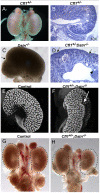
Figure 1
Figure 1. Kidney defects in Dstn−/− and Cfl1+/−;Dstn−/− mutant animals.
Whole mount image (A) and histological section (B) of control kidneys. (C) Double ureter (asterisks) and duplex kidney formation in a Dstn−/− embryo. Arrow points to the cleft between the two lobes of the duplex kidney, and asterisks mark the two ureters. (D) Histology of Cfl1+/−;Dstn−/− kidney reveals irregular surface of the renal cortex. Arrows point to constriction sites, arrowhead to a dilated collecting duct. (E) and (F), Three-dimensional reconstruction from confocal optical sections of control kidney (Hoxb7/myrVenus;Cfl1+/−) (E), showing smooth surface and kidney shape, and Hoxb7/myrVenus;Cfl1+/−;Dstn−/− kidney (F), showing an irregular surface and abnormal cleft (arrow). (G) and (H), reduced kidney size in Cfl1+/−;Dstn−/− (H) compared to Dstn+/− control (G). The dotted curves indicate the outlines of the two control kidneys. All kidneys are at E16.5, except C which is E17.5.
Figure 2
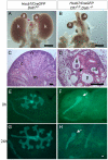
Figure 2. Absence of Cfl1 and Dstn in the ureteric bud of embryonic kidneys results in severe renal hypodysplasia.
(A) Control kidneys at E18.5. (B) Absence of normal kidneys in E18.5 double mutant where two Cfl1F/F alleles are conditionally deleted by HoxB7/CreGFP, specifically in ureteric epithelium, in a Dstn −/− background. (C) and (D), Hematoxylin-eosin staining of E18.5 control kidney (C) and Cfl1;Dstn double mutant kidney (D). Aside from their very small size, the mutant kidneys lack normal patterning, and contain only a few disorganized tubules. There are a few nephrons containing proximal tubules (p) and distal tubules (arrow), and very few tubules resembling collecting ducts (cd). (E–F) ureteric bud branching in double mutant and control kidneys. Control Dstn +/− kidneys show normal branched ureteric bud morphology at E12.5 (E) and continue to branch when cultured for 24 hours (G). In contrast, Cfl1;Dstn double mutants show an unbranched ureteric bud outgrowth (white arrow) at E12 (F), which fails to branch further when cultured for 24 hours (H). Abbreviations: a, adrenal gland; c, cortex; g, glomerulus; k, kidney; m, medulla; n, nephrogenic zone; Scale bar 1 mm for A–B; 200 µm for C–H 100 µm.
Figure 3
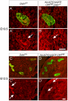
Figure 3. Efficiency of Hoxb7/CreGFP-mediated deletion of Cfl1 as evaluated by Cofilin1 antibody staining.
Calbindin (green) was used to visualize Wolffian duct epithelium in (A–D). (A–B'), cofilin1 (red) and calbindin (green) in E10.5 Wolffian duct (sections through the expanded region that will give rise to the UB). Cofilin1 is expressed in every cell in both ureteric bud and metanephric mesenchyme, and seems slightly enriched in UB epithelial cells. At E10.5, there is no detectable difference in cofilin1 protein amount or localization between control (Dstn+/−) and Hoxb7/CreGFP; Cfl1F/F; Dstn−/− kidneys. (C–D'), At E12.5, cofilin1 localization in the Dstn+/− control is similar to that at the earlier stages. However, Hoxb7/CreGFP has efficiently deleted the gene in the UB, as the protein is absent in ureteric epithelium of Cfl1F/F kidneys. Arrows point to ureteric buds, scale bar 50 µm.
Figure 4
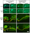
Figure 4. Exogenous GDNF is unable to rescue the ureteric bud branching defect, or induce ectopic budding from Wolffian duct, in Cfl1;Dstn double mutants.
(A–H) Time-lapse images of E11.5 kidney cultures in control medium (A, B, E, F) or with added GDNF (C, D, G, H). Control kidneys start from early T-shape (A, C) and without GDNF end up with secondary branch points at 20 h (B), but when cultured for 20 h with exogenous GDNF, they develop a huge enlargement of the ureteric bud tip (D). Cfl1;Dstn double mutant kidneys show only a small, unbranched ureteric bud at E11.5 (E, G), which during 20 hrs of culture is able to elongate but does not begin branching regardless of GDNF addition (F, H). (I–L), E11.5 urogenital blocks (UGBs) cultured for 48 h without (I, K) or with GDNF (J, L) and stained for Pax2 (green) and pan-cytokeratin (red). Under normal conditions, the control Dstn+/− kidney (I) shows normal branching and Pax2-positive induced metanephric mesenchyme surrounding the ureteric tips, while the Cfl1;Dstn double mutant kidney (K) retains some Pax2-positive mesenchyme around a single outgrowth of ureteric bud. In a control Dstn+/− UGB (J), exogenous GDNF induced massive swelling of the ureteric tip, which still maintains Pax2-positive metanephric mesenchyme around it, and induced extra ureteric budding (arrows) from the Wolffian duct. GDNF is unable to induce normal ureteric bud branching or ectopic ureteric budding in Cfl1;Dstn double mutant UGB (L), but induces slight bulging (arrowhead) in the lower ureteric bud epithelium. Scale bar 200 µm.
Figure 5
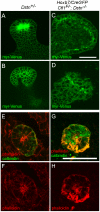
Figure 5. Disruption of actin depolymerization results in irregular ureteric epithelial cell shape and organization due to F-actin accumulation in Cfl;Dstn double mutant ureteric bud.
(A–D), A transgene encoding myristoylated-Venus driven by Hoxb7-promoter was introduced to Cfl1;Dstn mutant mice to visualize ureteric epithelial cell outlines; optical sections through ureteric bud tips of control Dstn+/− (A–B) and double mutant (C–D) kidneys at E12.5. (A) and (C) are optical sections through the lumen of the UB tip, while (B) and (D) are glancing sections through the epithelium. Control ureteric epithelium shows cells that vary in shape, but have smooth outlines and are organized in an orderly pattern along the whole epithelium. (C–D), Double mutant epithelial cells are often abnormal in shape and exhibit a disorganized pattern throughout the ureteric bud. (E–H), Confocal images of phalloidin (red)/calbindin (green) double staining, which visualizes actin filaments (F-actin) in developing kidney. (E–F), In control kidney, actin filaments are enriched in apical side of ureteric epithelium while some localize to the basolateral surfaces. (G–H) Disruption of actin depolymerization in Cfl1;Dstn double mutants results in huge accumulation of F-actin, specifically in the ureteric epithelium where Cfl1F is deleted. Scale bars: 50 µm.
Figure 6
Figure 6. Migration assay for primary ureteric bud cells reveals defects in epithelial cell movement in Cfl1;Dstn double mutant.
A scratch was introduced 2 days after plating the isolated ureteric buds (0 h, A and C) and followed for 24 h (B and D). Control Dstn−/−cells (A–B) had completely filled the gap produced by the scratch (N = 12 cultures), while double mutant cells (C–D) were impaired in their movement (N = 5 cultures). Scale bars 200 µm.
Figure 7
Figure 7. Primary UB epithelial cells lacking Cfl1 but maintaining one wild-type Dstn allele (Hoxb7/CreGFP; Cfl1F/F; Dstn+/−) show a migration delay in vitro.
Examples of wild type control (A–C) and mutant (D–F) primary ureteric bud epithelial cell migration in scratch assay. Already at 8 h after establishment of the scratch, wild-type cells (N = 12) have migrated to almost fill the gap (A–C), while mutant cells (D–F, N = 9) have not migrated as far. (G) Quantification of movements by control and mutant primary epithelial cells at 8 h after introducing the scratch. The percent of gap filled at this time point was calculated by dividing the average width of gap at 8 h by the width of initial scratch. Scale bar 200 µm.
Figure 8
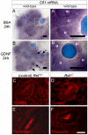
Figure 8. Cfl1 gene expression is not regulated by GDNF/Ret signaling in developing kidney.
(A–A'), Cfl1 in situ hybridization in E11.5 wild-type kidney cultured with control, BSA-soaked bead. A' shows a higher magnification of the boxed area in A. mRNA is detected both in metanephric mesenchyme (m) and ureteric epithelium (u). (B–B'), Similar expression levels of Cfl1 in kidneys cultured with GDNF-soaked beads, which induce enlargement of ureteric tips and extra budding from Wolffian duct (arrows). B' shows a higher magnification of the boxed area in B. (C), Cofilin1 antibody staining of Ret +/− (control) E10.5 embryo, where Wolffian duct shows epithelial thickening as a hallmark of early ureteric bud outgrowth. (D), Normal expression of Cofilin1 protein (red) in Ret −/− Wolffian duct epithelium at E10.5. (E,F) F-actin localization in control and Ret −/− kidneys at E10.5. Scale bars 100 µm for A–B, 50 µm for C–F.

Similar articles

Cited by

References

    1. Van Troys M, Huyck L, Leyman S, Dhaese S, Vandekerkhove J, et al. Ins and outs of ADF/cofilin activity and regulation. Eur J Cell Biol. 2008;87:649–667. - PubMed
    1. Insall RH, Machesky LM. Actin dynamics at the leading edge: from simple machinery to complex networks. Dev Cell. 2009;17:310–322. - PubMed
    1. Lenart P, Bacher CP, Daigle N, Hand AR, Eils R, et al. A contractile nuclear actin network drives chromosome congression in oocytes. Nature. 2005;436:812–818. - PubMed
    1. Gurniak CB, Perlas E, Witke W. The actin depolymerizing factor n-cofilin is essential for neural tube morphogenesis and neural crest cell migration. Dev Biol. 2005;278:231–241. - PubMed
    1. Bellenchi GC, Gurniak CB, Perlas E, Middei S, Ammassari-Teule M, et al. N-cofilin is associated with neuronal migration disorders and cell cycle control in the cerebral cortex. Genes Dev. 2007;21:2347–2357. - PMC - PubMed

Publication types

MeSH terms