Semen protects CD4+ target cells from HIV infection but promotes the preferential transmission of R5 tropic HIV
- PMID: 21059891
- PMCID: PMC3071682
- DOI: 10.4049/jimmunol.1002846
Semen protects CD4+ target cells from HIV infection but promotes the preferential transmission of R5 tropic HIV
Abstract
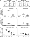
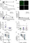
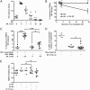
Sexual intercourse is the major means of HIV transmission, yet the impact of semen on HIV infection of CD4(+) T cells remains unclear. To resolve this conundrum, we measured CD4(+) target cell infection with X4 tropic HIV IIIB and HC4 and R5 tropic HIV BaL and SF162 after incubation with centrifuged seminal plasma (SP) from HIV-negative donors and assessed the impact of SP on critical determinants of target cell susceptibility to HIV infection. We found that SP potently protects CD4(+) T cells from infection with X4 and R5 tropic HIV in a dose- and time-dependent manner. SP caused a diminution in CD4(+) T cell surface expression of the HIVR CD4 and enhanced surface expression of the HIV coreceptor CCR5. Consequently, SP protected CD4(+) T cells from infection with R5 tropic HIV less potently than it protected CD4(+) T cells from infection with X4 tropic HIV. SP also reduced CD4(+) T cell activation and proliferation, and the magnitude of SP-mediated suppression of target cell CD4 expression, activation, and proliferation correlated closely with the magnitude of the protection of CD4(+) T cells from infection with HIV. Taken together, these data show that semen protects CD4(+) T cells from HIV infection by restricting critical determinants of CD4(+) target cell susceptibility to HIV infection. Further, semen contributes to the selective transmission of R5 tropic HIV to CD4(+) target cells.
Figures


Similar articles
-
Comparison of the effect of semen from HIV-infected and uninfected men on CD4+ T-cell infection.AIDS. 2016 May 15;30(8):1197-208. doi: 10.1097/QAD.0000000000001048. AIDS. 2016. PMID: 26854806 Free PMC article.
-
Human immunodeficiency virus type 1 strains R5 and X4 induce different pathogenic effects in hu-PBL-SCID mice, depending on the state of activation/differentiation of human target cells at the time of primary infection.J Virol. 1999 Aug;73(8):6453-9. doi: 10.1128/JVI.73.8.6453-6459.1999. J Virol. 1999. PMID: 10400739 Free PMC article.
-
Differential effects of R5 and X4 human immunodeficiency virus type 1 infection on CD4+ cell proliferation and activation.J Gen Virol. 2005 Apr;86(Pt 4):1171-1179. doi: 10.1099/vir.0.80674-0. J Gen Virol. 2005. PMID: 15784911
-
Selective transmission of R5 HIV-1 variants: where is the gatekeeper?J Transl Med. 2011 Jan 27;9 Suppl 1(Suppl 1):S6. doi: 10.1186/1479-5876-9-S1-S6. J Transl Med. 2011. PMID: 21284905 Free PMC article. Review.
-
Evolution of Host Target Cell Specificity During HIV-1 Infection.Curr HIV Res. 2018;16(1):13-20. doi: 10.2174/1570162X16666171222105721. Curr HIV Res. 2018. PMID: 29268687 Review.
Cited by
-
HIV-1 subverts the complement system in semen to enhance viral transmission.Mucosal Immunol. 2021 May;14(3):743-750. doi: 10.1038/s41385-021-00376-9. Epub 2021 Feb 10. Mucosal Immunol. 2021. PMID: 33568786 Free PMC article.
-
Immune biomarkers and anti-HIV activity in the reproductive tract of sexually active and sexually inactive adolescent girls.Am J Reprod Immunol. 2018 Jun;79(6):e12846. doi: 10.1111/aji.12846. Epub 2018 Mar 13. Am J Reprod Immunol. 2018. PMID: 29533494 Free PMC article.
-
Characterization of the Influence of Semen-Derived Enhancer of Virus Infection on the Interaction of HIV-1 with Female Reproductive Tract Tissues.J Virol. 2015 May;89(10):5569-80. doi: 10.1128/JVI.00309-15. Epub 2015 Mar 4. J Virol. 2015. PMID: 25740984 Free PMC article.
-
X4 tropic multi-drug resistant quasi-species detected at the time of primary HIV-1 infection remain exclusive or at least dominant far from PHI.PLoS One. 2011;6(8):e23301. doi: 10.1371/journal.pone.0023301. Epub 2011 Aug 24. PLoS One. 2011. PMID: 21887243 Free PMC article.
-
Development of HIV-1 rectal-specific microbicides and colonic tissue evaluation.PLoS One. 2014 Jul 15;9(7):e102585. doi: 10.1371/journal.pone.0102585. eCollection 2014. PLoS One. 2014. PMID: 25025306 Free PMC article.
References
-
- Granich RM, Gilks CF, Dye C, De Cock KM, Williams BG. Universal voluntary HIV testing with immediate antiretroviral therapy as a strategy for elimination of HIV transmission: a mathematical model. Lancet. 2009;373:48–57. - PubMed
-
- Galvin SR, Cohen MS. The role of sexually transmitted diseases in HIV transmission. Nat. Rev. Microbiol. 2004;2:33–42. - PubMed
-
- Ball JK, Curran R, Irving WL, Dearden AA. HIV-1 in semen: determination of proviral and viral titres compared to blood, and quantification of semen leukocyte populations. J. Med. Virol. 1999;59:356–363. - PubMed
-
- Marcelin AG, Tubiana R, Lambert-Niclot S, Lefebvre G, Dominguez S, Bonmarchand M, Vauthier-Brouzes D, Marguet F, Mousset-Simeon N, Peytavin G, Poirot C. Detection of HIV-1 RNA in seminal plasma samples from treated patients with undetectable HIV-1 RNA in blood plasma. AIDS. 2008;22:1677–1679. - PubMed
Publication types
MeSH terms
Grants and funding
LinkOut - more resources
Full Text Sources
Medical
Research Materials

