. 2010 Nov;22(11):863-73.
doi: 10.1093/intimm/dxq440. Epub 2010 Nov 8.

A panel of human cell-based artificial APC enables the expansion of long-lived antigen-specific CD4+ T cells restricted by prevalent HLA-DR alleles

Affiliations

A panel of human cell-based artificial APC enables the expansion of long-lived antigen-specific CD4+ T cells restricted by prevalent HLA-DR alleles

Marcus O Butler et al. Int Immunol. 2010 Nov.

Abstract

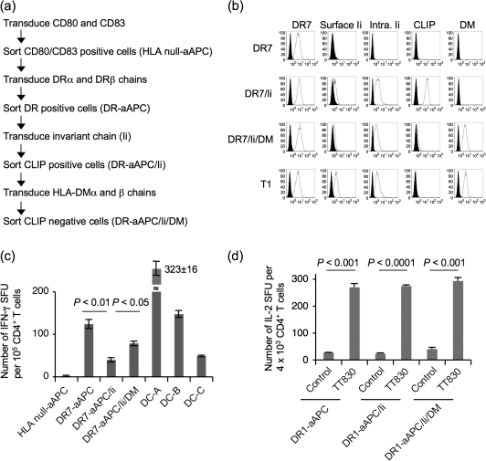
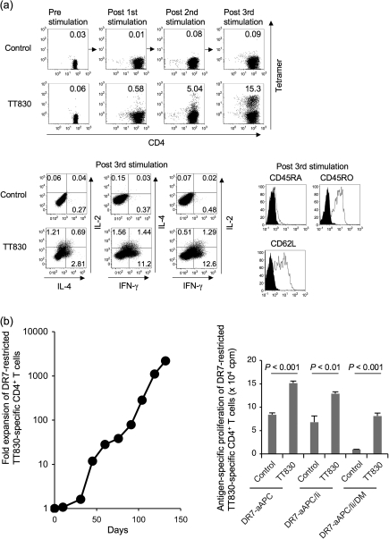
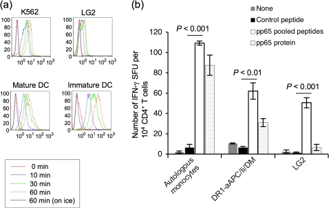
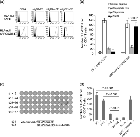
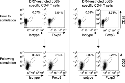
Many preclinical experiments have attested to the critical role of CD4(+) T cell help in CD8(+) cytotoxic T lymphocyte (CTL)-mediated immunity. Recent clinical trials have demonstrated that reinfusion of CD4(+) T cells can induce responses in infectious diseases and cancer. However, few standardized and versatile systems exist to expand antigen-specific CD4(+) T(h) for clinical use. K562 is a human erythroleukemic cell line, which lacks expression of HLA class I and class II, invariant chain and HLA-DM but expresses adhesion molecules such as intercellular adhesion molecule-1 and leukocyte function-associated antigen-3. With this unique immunologic phenotype, K562 has been tested in clinical trials of cancer immunotherapy. Previously, we created a K562-based artificial antigen-presenting cell (aAPC) that generates ex vivo long-lived HLA-A2-restricted CD8(+) CTL with a central/effector memory phenotype armed with potent effector function. We successfully generated a clinical version of this aAPC and conducted a clinical trial where large numbers of anti-tumor CTL are reinfused to cancer patients. In this article, we shifted focus to CD4(+) T cells and developed a panel of novel K562-derived aAPC, where each expresses a different single HLA-DR allele, invariant chain, HLA-DM, CD80, CD83 and CD64; takes up soluble protein by endocytosis and processes and presents CD4(+) T-cell peptides. Using this aAPC, we were able to determine novel DR-restricted CD4(+) T-cell epitopes and expand long-lived CD4(+) T-cells specific for multiple antigens without growing bystander Foxp3(+) regulatory T cells. Our results suggest that K562-based aAPC may serve as a translatable platform to generate both antigen-specific CD8(+) CTL and CD4(+) T(h).


Figures

Fig. 1.
Fig. 1.
Generation of K562-based aAPC that expresses DR7 as a single HLA allele. (a) The experimental protocol to generate K562-based aAPC that expresses DR7 as a single HLA allele is shown. K562 was sequentially transduced with CD80, CD83, HLA-DR7αβ heterodimers, Ii and HLA-DMαβ heterodimers. Each molecule was retrovirally infected, and transduced cells were isolated using specific mAb and flow cytometry-guided sorting. (b) HLA null aAPC was serially transduced with DR7, Ii and HLA-DM to generate stable cell lines, DR7-aAPC (DR7), DR7-aAPC/Ii (DR7/Ii) and DR7-aAPC/Ii/DM (DR7/Ii/DM). T1 cells that hemizygously express DR7 served as a control. The expression of cell surface DR7, cell surface and intracellular Ii, cell surface CLIP and intracellular HLA-DM was studied by flow cytometric analysis using specific mAb. (c) DR7-transduced aAPC were immunogenic and able to induce allogeneic responses. CD4+ T cells purified from DR7 healthy donors were stimulated with DR7-aAPC, DR7-aAPC/Ii, DR7-aAPC/Ii/DM and monocyte-derived mature DC derived from three different allogeneic donors (DC-A, B and C). IFN-γ secretion by allogeneic CD4+ T cells was measured by IFN-γ ELISPOT. Data shown represent means ± SDs of quadricates. (d) DR1-restricted tetanus TT830 peptide-specific CD4+ T cells were stimulated with DR1-aAPC, DR1-aAPC/Ii and DR1-aAPC/Ii/DM pulsed with pan-DR PADRE (control) or TT830 peptide. TT830-specific IL-2 secretion by CD4+ T cells were evaluated by IL-2 ELISPOT on day 28. Data shown represent means ± SDs of triplicates.
Fig. 2.
Fig. 2.
DR-aAPC/Ii/DM can generate long-lived antigen-specific CD4+ T cells. (a) DR1-aAPC/Ii/DM can expand DR1-restricted antigen-specific Th1-biased CD4+ T cells with a central memory phenotype in the presence of low dose IL-2 and IL-15. Purified DR1+ CD4+ T cells were stimulated every 10 days with TT830 peptide-pulsed and -irradiated DR1-aAPC/Ii/DM. Following each stimulation, IL-2 (10 IU ml−1) and IL-15 (10 ng ml−1) were added to the T-cell cultures every 3 days. The percentage of DR1-restricted TT830-specific CD4+ T cells was determined by TT830 tetramer staining (top). DR1-restricted CLIP-specific tetramer was employed as a negative control. Following three stimulations (day 28), IL-2, IL-4 and IFN-γ secretion was also analyzed by intracellular cytokine staining (bottom left). DR1-transduced T2 cells were pulsed with TT830 or pan-DR PADRE (control) peptide and were used for stimulation. Cells were co-stained with anti-CD45RA, CD45RO and CD62L mAbs along with TT830-tetramer (bottom right). Expression on gated tetramer-staining cells is depicted. Isotype staining was used as a control. (b) DR-aAPC/Ii/DM was able to generate long-lived antigen-specific CD4+ T cells. Purified CD4+ T cells isolated from DR7-positive healthy donors were repeatedly stimulated with TT830-pulsed and -irradiated DR7-aAPC/Ii/DM every 14 days. Between stimulations, 10 IU ml−1 IL-2 and 10 ng ml−1 IL-15 were added to the T-cell cultures. To confirm the antigen specificity of the long-lived DR7-restricted TT830-specific T cells, antigen-specific proliferation was measured on day 103 using pan-DR PADRE (control) or TT830 peptide-pulsed and mitomycin-C treated DR7-aAPC, DR7-aAPC/Ii or DR7-aAPC/II/DM as a stimulator. Data shown represents means ± SDs of triplicates. A TT830-specific proliferation assay was also performed on days 77, 91 and 110 with similar results (data not shown). In addition, TT830 specificity was confirmed by IFN-γ ELISPOT on day 150 (data not shown).
Fig. 3.
Fig. 3.
K562-based aAPC possesses pinocytic activity and processes protein to present DR-restricted peptides to CD4+ T cells. (a) K562 cells, the aAPC backbone, possess pinocytic activity. K562, LG2 (EBV-transformed lymphoblastoid cells) and immature and mature DC ex vivo generated using peripheral blood monocytes were incubated with FITC–dextran (1 mg ml−1; molecular weight 70 000; Invitrogen) at 37°C for 0, 10, 30 and 60 min. As a control experiment, cells were similarly incubated with FITC–dextran on ice for 60 min. After incubation, the cells were washed twice with cold PBS to stop pinocytosis and to remove excess dextran. Pinocytosis was measured as the cellular uptake of FITC–dextran and was quantified by flow cytometry. Data shown are representative of three similar experiments. (b) DR-aAPC/Ii/DM can process and present CD4+ T-cell peptides derived from pinocytosed protein. CD4+ T cells isolated from DR1+ and CMV Ab (+) healthy donors were stimulated once with autologous DC pulsed with recombinant full-length pp65 protein. IFN-γ ELISPOT was conducted using autologous monocytes, DR1-aAPC/Ii/DM and LG2 mixed with 10 nM control pan-DR PADRE peptide, pp65 peptide mix or full-length pp65 protein as a stimulator. Data shown represent means ± SDs of triplicates.
Fig. 4.
Fig. 4.
Transduction of CD64 enables K562-based aAPC to effectively endocytose via Fcγ receptor and process protein for presentation of DR-restricted peptides to CD4+ T cells. (a) HLA-null aAPC (K562/CD80/CD83) was transduced with CD64 and common Fcγ receptor to generate HLA-null aAPC/CD64. The surface expression of CD64 was confirmed by specific mAb staining. HLA-null aAPC and HLA-null aAPC/CD64 cultured in the presence of 10% bovine IgG-depleted FCS were incubated with PE-conjugated mouse IgG1, 2a or 2b at 1 μg ml−1 for 24 h at 37°C. Cells were trypsinized and washed intensively to remove membrane-bound mIgG. Endocytosed PE–mIgG was measured in the FL2 channel by flow cytometry analysis. (b) DR7-restricted pp65-specific CD4+ T cells were generated using autologous monocytes pulsed with pp65 protein from DR7+ CMV Ab (+) healthy donors. On day 19, cells were subjected to an IL-2 ELISPOT assay using DR7-aAPC/Ii/DM or DR7-aAPC/Ii/DM/CD64 as an APC. Unlike in Fig. 3(b), the APC was pulsed for only 30 min with 10 nM pp65 protein or pp65 IC, washed and then used. Pan-DR PADRE peptide and pp65 peptide mix were used as negative and positive controls, respectively. Data shown represent means ± SDs of triplicates. (c) CD4+ T cells purified from DR7-positive CMV Ab (+) healthy donors were stimulated with DR7-aAPC/Ii/DM/CD64 mixed with 10 nM pp65 IC. IL-2 ELISPOT was conducted on day 48 using DBB (EBV-transformed lymphoblastoid cells homozygous for DR7) pulsed with each of 57 20-mer pp65 peptides overlapping by 10 amino acids (#1∼#57). Two adjacent wells (#34 and #35) that were positive were boxed in a solid line. Three wells boxed in a dotted line served as a control. The overlapping sequence, QYDPVAALFP, between #34 and #35 peptides is boxed. (d) CD4+ T cells purified from DR4-positive CMV Ab (+) healthy donors were stimulated with DR4-aAPC/Ii/DM/CD64 mixed with 10 nM pp65 IC. On day 28, IL-2 ELISPOT was conducted using Preiss (EBV-transformed lymphoblastoid cells homozygous for DR4) pulsed with each of 57 20-mer pp65 peptides overlapping by 10 amino acids (#1∼#57) or IC. Data for three positive peptides, #6, #16 and 19, and IC is depicted. Pan-DR PADRE peptide was used as a negative control.
Fig. 5.
Fig. 5.
K562-based DR-aAPC/Ii/DM does not grow Foxp3+ CD4+ T cells as a bystander. DR7-restricted pp65-specific CD4+ T cells (day 48) and DR4-restricted pp65-specific CD4+ T cells (day 38) were analyzed for the intracellular expression of Foxp3. Total CD4+ T cells in culture were gated. Purified CD4+ CD25+ T cells from the same donors prior to stimulation were also studied.

Similar articles

Cited by

References

    1. June CH. Adoptive T cell therapy for cancer in the clinic. J. Clin. Invest. 2007;117:1466. - PMC - PubMed
    1. Leen AM, Rooney CM, Foster AE. Improving T cell therapy for cancer. Annu. Rev. Immunol. 2007;25:243. - PubMed
    1. Rosenberg SA, Dudley ME. Adoptive cell therapy for the treatment of patients with metastatic melanoma. Curr. Opin. Immunol. 2009;21:233. - PMC - PubMed
    1. Berger C, Turtle CJ, Jensen MC, Riddell SR. Adoptive transfer of virus-specific and tumor-specific T cell immunity. Curr. Opin. Immunol. 2009;21:224. - PMC - PubMed
    1. Bollard CM, Gottschalk S, Leen AM, et al. Complete responses of relapsed lymphoma following genetic modification of tumor-antigen presenting cells and T-lymphocyte transfer. Blood. 2007;110:2838. - PMC - PubMed

Publication types

Substances