. 2011 Feb;9(1):58-68.
doi: 10.1089/adt.2010.0303. Epub 2010 Nov 4.

Use of base modifications in primers and amplicons to improve nucleic acids detection in the real-time snake polymerase chain reaction

Affiliations

Use of base modifications in primers and amplicons to improve nucleic acids detection in the real-time snake polymerase chain reaction

Igor V Kutyavin. Assay Drug Dev Technol. 2011 Feb.

Abstract

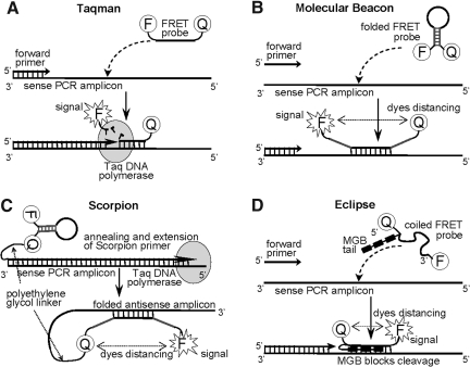
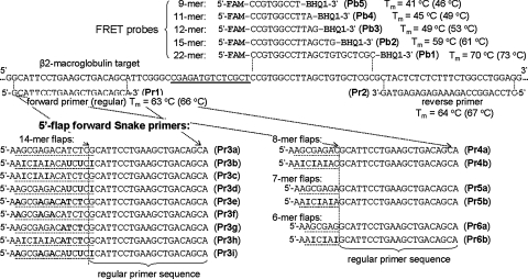
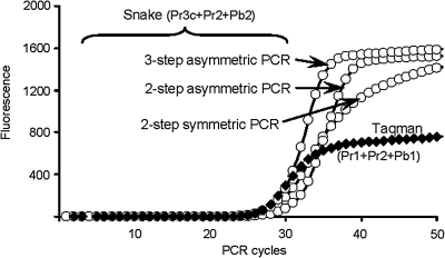
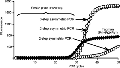
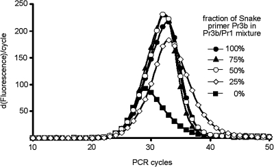
The addition of relatively short flap sequence at the 5'-end of one of the polymerase chain reaction (PCR) primers considerably improves performance of real-time assays based on 5'-nuclease activity. This new technology, called Snake, was shown to supersede the conventional methods like TaqMan, Molecular Beacons, and Scorpions in the signal productivity and discrimination of target polymorphic variations as small as single nucleotides. The present article describes a number of reaction conditions and methods that allow further improvement of the assay performance. One of the identified approaches is the use of duplex-destabilizing modifications such as deoxyinosine and deoxyuridine in the design of the Snake primers. This approach was shown to solve the most serious problem associated with the antisense amplicon folding and cleavage. As a result, the method permits the use of relatively long-in this study-14-mer flap sequences. Investigation also revealed that only the 5'-segment of the flap requires the deoxyinosine/deoxyuridine destabilization, whereas the 3'-segment is preferably left unmodified or even stabilized using 2-amino deoxyadenosine d(2-amA) and 5-propynyl deoxyuridine d(5-PrU) modifications. The base-modification technique is especially effective when applied in combination with asymmetric three-step PCR. The most valuable discovery of the present study is the effective application of modified deoxynucleoside 5'-triphosphates d(2-amA)TP and d(5-PrU)TP in Snake PCR. This method made possible the use of very short 6-8-mer 5'-flap sequences in Snake primers.


Figures

Fig. 1.
Fig. 1.
The diagrams show the systems' design, the key reaction stages, and the mechanism of the fluorescence signal generation in the probe-based technologies most commonly used in real-time PCR. TaqMan (A) is a rare example of the methods wherein FRET is abolished due to the probe cleavage. In the competing hybridization-triggered assays such as Molecular Beacons (B), Scorpion primers (C), and Eclipse probes (D), the fluorescence signal is produced by the distancing of a reporter dye from a quencher when the probe is annealed to its target. Molecular Beacons are known for the relatively low fluorescence background—efficient FRET in the unhybridized state—that is achieved by intramolecular stem formation (shown in gray), bringing the dyes in close proximity. In Scorpion primers the 5′-end of a PCR primer is conjugated to the 3′-end of a molecular beacon through a long, polyethylene glycol linker, precluding extension over the beacon sequence. Unlike for Molecular Beacons, the DNA detection stage in Scorpions becomes a rapid intramolecular reaction. Eclipse probes (D) are yet another example of hybridization-based FRET probes that have a low fluorescence background. These probes carry an MGB moiety at their 5′-end in addition to two FRET dyes. Due to the strong DNA-duplex stabilizing effect of the MGB moiety, the probes can be designed as short as 12-18-mers. Placing the MGB-tail at the 5′-end of the probes completely blocks 5′-nuclease cleavage and fluorescent signal is generated solely due to the hybridization-triggered dye separation. Unlike Molecular Beacons and Scorpions, the FRET effect in unhybridized Eclipse probes is achieved through a collisional quenching of the dyes in a coiled single-stranded oligonucleotide. In the diagrams, F is the abbreviation of a reporter dye and Q is a quencher. PCR, polymerase chain reaction; FRET, Förster resonance energy transfer; MGB, minor groove binder.
Fig. 2.
Fig. 2.
The scheme shows the sequences of FRET probes and PCR primers used in the TaqMan and Snake assays of the present study. Wherever it is possible, the primers and probes are aligned with a detected sequence of 96-mer target oligodeoxyribonucleotide. The forward Snake primers comprised the regular primer (Pr1) bearing 5′-flap sequences of variable length. The primer segments that participate in the intramolecular stem formation of the folded Snake amplicons and their binding site within the target sequence are underlined. The bolded characters in the flap sequences correspond to the following modifications: A – 2-amino deoxyadenosine, T – 5-propynyl deoxyuridine, I – deoxyinosine, and U – deoxyuridine. Tms were calculated for reactions with 2 mM MgCl2. The values in the parentheses were, respectively, calculated for the 5 mM MgCl2 conditions. FAM is 6-fluorescein and BHQ1 is a Black Hole Quencher from Biosearch Technologies. Tm, melting temperatures.
Fig. 3.
Fig. 3.
Illustrated are the reaction pathways of an antisense Snake amplicon that can have a negative effect on PCR efficiency. Because of the flap sequence, the Snake amplicon folds into a stem-loop secondary structure in stage A. During the extension of a reverse primer hybridized to the antisense amplicon in stage B, DNA polymerase reaches the amplicon secondary structure (shown in gray) and partially cleaves its 5′-end (stage C) while displacing the rest of the sequence and completing the replication. This results in a truncated double-stranded amplicon (stage D) that, after strand separation at 95°C, provides a 3′-excised single-stranded sense amplicon (stage E). This 3′-excised sense amplicon does not contribute to the Snake assay signal productivity because it does not form an optimal cleavage structure for Taq 5′-nuclease when hybridized to a FRET probe in stage G. However, if a 5′-flap forward primer hybridizes to this amplicon, then the original sequence integrity of the sense amplicon along with its catalytic function can be restored as shown in stage F. Troublesome is the reaction pathway E→H→I. Unlike the normal Snake amplicon shown in Figure 4, this curtailed amplicon can form a self-priming intramolecular duplex with no 3′-terminal mismatched nucleotide (stage H). Extension of such a duplex by DNA polymerase leads to a long hairpin-like structure (stage I) that does not participate in PCR due to it exceptional thermal stability. The pathway E→H→I can actually be signal contributing but only through the regular TaqMan mechanism, and, for this reason, the FRET probes have to have elevated hybridization properties.
Fig. 4.
Fig. 4.
The scheme shows reaction pathways of a sense Snake amplicon during PCR. Folding of this particular amplicon catalyzes the FRET probe cleavage in stages A→B→C→D, resulting in an exceptionally strong fluorescence signal that is typical for the Snake technology. The same amplicon, however, needs to replicate in PCR, and that is where its stable secondary structure may become a problem. At least a small fraction of this sense amplicon is always in a linear form A and accessible for a forward 5′-flap primer to hybridize and extend in stage E. In theory, the strand replication can be solely accomplished through this passive hybridization pathway A→E. On the other hand, active hybridization of the forward primer shown in stage F followed by strand displacement in stage G can contribute and substantially accelerate the replication process. The 5′-flap sequence in the forward primer and its replica in the sense amplicon are shown in gray color.
Fig. 5.
Fig. 5.
Real-time PCR detection of the β2-microglobulin target sequence in TaqMan and Snake assays. Shown are the reactions using one of the best-performing Snake primers (Pr3c) carrying a 14-mer 5′-flap sequence. Experiments with other analogs of this 14-mer flap sequence are not shown, but they exhibited similar responses to the changes in PCR protocol. The structures of the primers and FRET probes can be found in Figure 2. The experiments were performed in 2 mM MgCl2 and at an annealing temperature of 64°C. Other reaction details, for example, PCR time/temperature profiles, reagent compositions, and concentrations, are described in Materials and Methods. The TaqMan real-time curve was obtained using regular two-step symmetric PCR.
Fig. 6.
Fig. 6.
Effect of changes in PCR protocol on real-time performance of Snake assays employing very short 6-mer 5′-flap sequence in the forward primer Pr6a. The primers and FRET probes used are indicated for each assay, and their structures are shown Figure 2. All Snake experiments were performed in 5 mM MgCl2 and at an annealing temperature of 56°C. Other reaction details, including the PCR time/temperature profiles, are described in Materials and Methods. The TaqMan real-time curve is the same experiment shown in Figure 5, and it was conducted using regular two-step symmetric PCR.
Fig. 7.
Fig. 7.
Performance of exceptionally short 9 and 11-mer FRET probes in real-time Snake and TaqMan PCR. The TaqMan detection experiments were conducted using two-step symmetric PCR, regular forward and reverse primers (Pr1 and Pr2) and 9 or 11-mer FRET probe as indicated. An asymmetric three-step PCR protocol was used in the case of the Snake experiments and the reactions comprised a 6-mer 5′-flap forward primer Pr6a, a regular reverse primer Pr2 and one of the short probes Pb4 or Pb5. All shown detection tests of TaqMan and Snake were performed at an annealing temperature of 56°C in 5 mM MgCl2 reaction mixtures with the 2-amino deoxyadenosine 5′-triphosphate analog completely substituted for dATP. Other reaction details are described in Materials and Methods. The structures of primers and FRET probes used are shown Figure 2.
Fig. 8.
Fig. 8.
This experiment illustrates the resistance of the Snake detection system to loss of the 5′-flap sequence in the forward primer. The real-time curves are shown in a differential format for better detection of minor differences. The reactions were conducted using three-step asymmetric PCR, 15-mer FRET probe Pb2, reverse Pr2, and forward 5′-flap primer Pr3b in 2 mM MgCl2 reaction buffer and at the annealing temperature of 64°C. During the experiment, the overall concentration of forward primer was maintained at 100 nM, but the Snake primer Pr3b was gradually replaced with a regular no-flap forward primer Pr1. The final fractions of the flap-incorporating Snake primer are indicated for each curve. Other reaction details are described in Materials and Methods. The structures of primers and FRET probes used are shown in Figure 2.

Similar articles

Cited by

References

    1. Higuchi R. Dollinger G. Walsh PS. Griffith R. Simultaneous amplification and detection of specific DNA sequences. Biotechnology. 1992;10:413–417. - PubMed
    1. Higuchi R. Fockler C. Dollinger G. Watson R. Kinetic PCR: real time monitoring of DNA amplification reactions. Biotechnology. 1993;11:1026–1030. - PubMed
    1. Förster T. Delocalized excitation, excitation transfer. In: Sinanoglu O, editor. Modern Quantum Chemistry, Istanbul Lectures, Part III. Academic Press; New York: 1965. pp. 93–137.
    1. Clegg RM. Fluorescence resonance energy transfer and nucleic acids. Methods Enzymol. 1992;211:353–388. - PubMed
    1. Didenko VV. DNA probes using fluorescence resonance energy transfer (FRET): design and application. Biotechniques. 2001;31:1106–1121. - PMC - PubMed

Publication types