. 2010 Oct;5(4):201-16.
doi: 10.4103/1817-1737.69106.

Imaging lung manifestations of HIV/AIDS

Affiliations

Imaging lung manifestations of HIV/AIDS

Carolyn M Allen et al. Ann Thorac Med. 2010 Oct.

Abstract

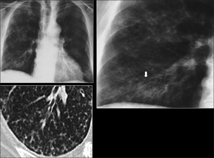
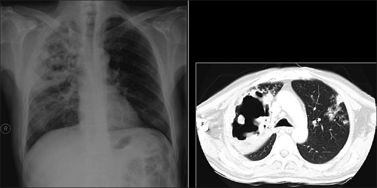
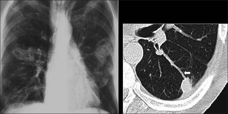
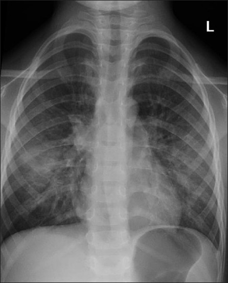
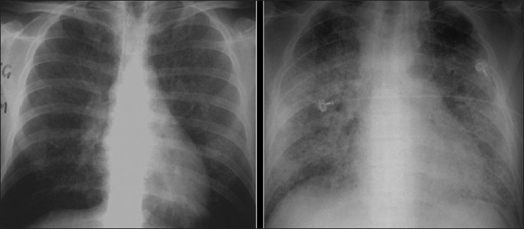
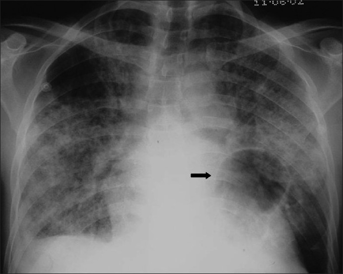
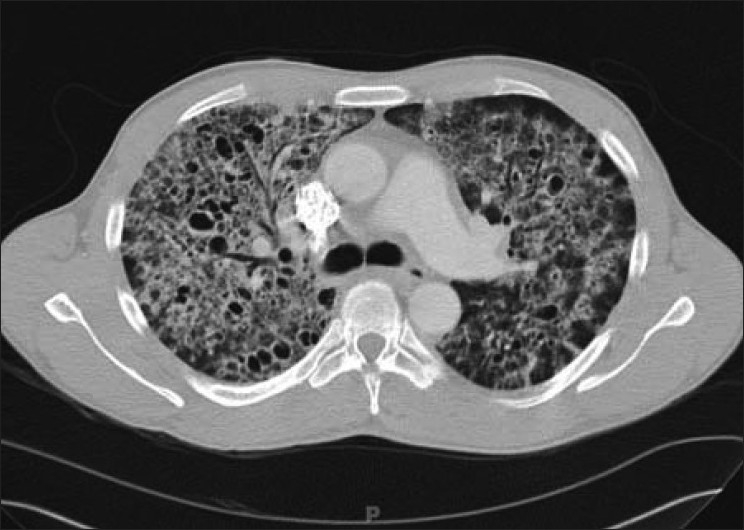
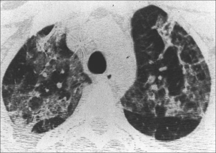
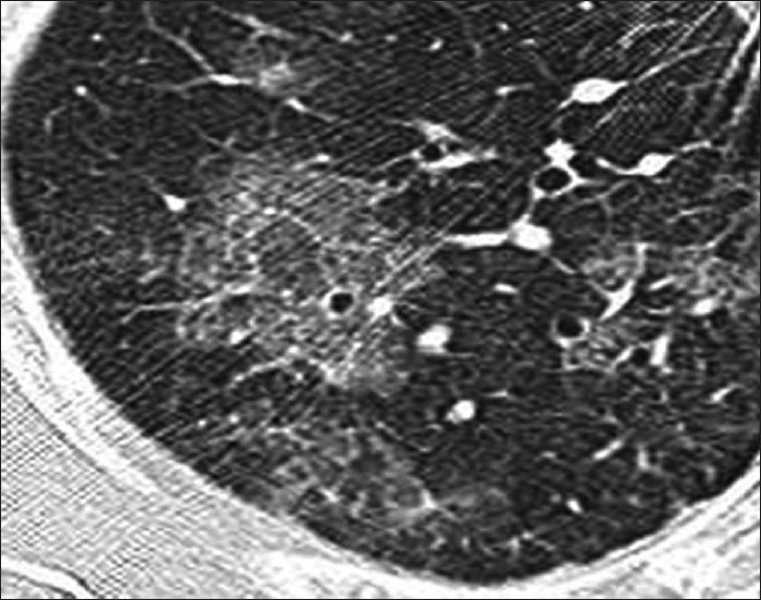
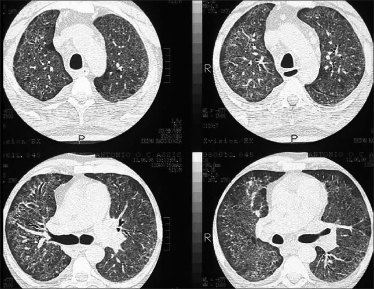
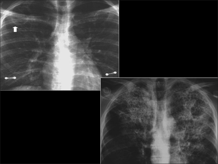
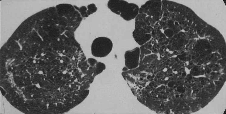
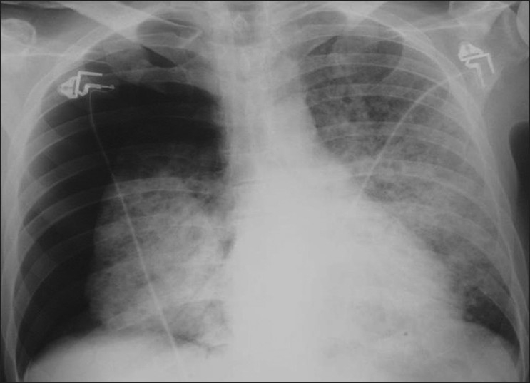
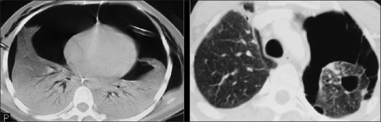
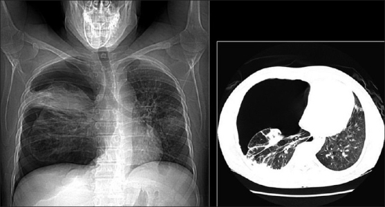
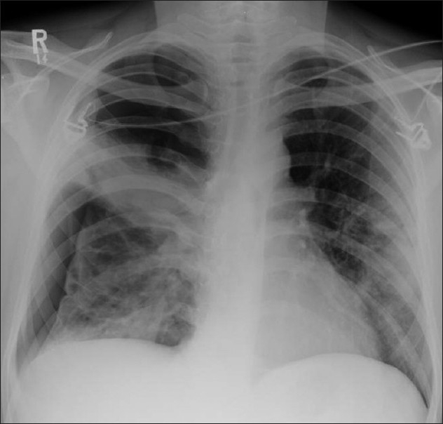
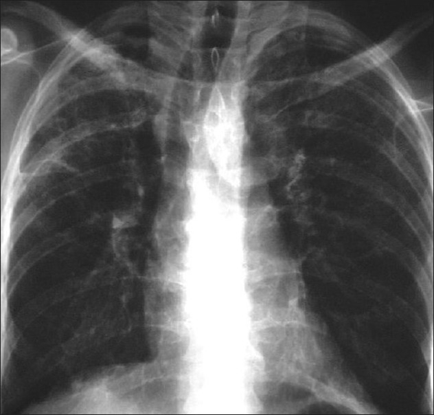
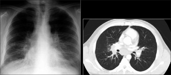
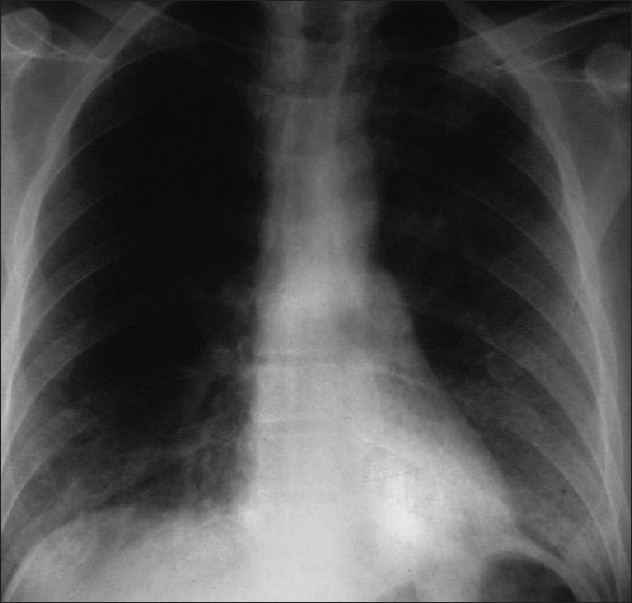
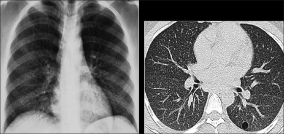
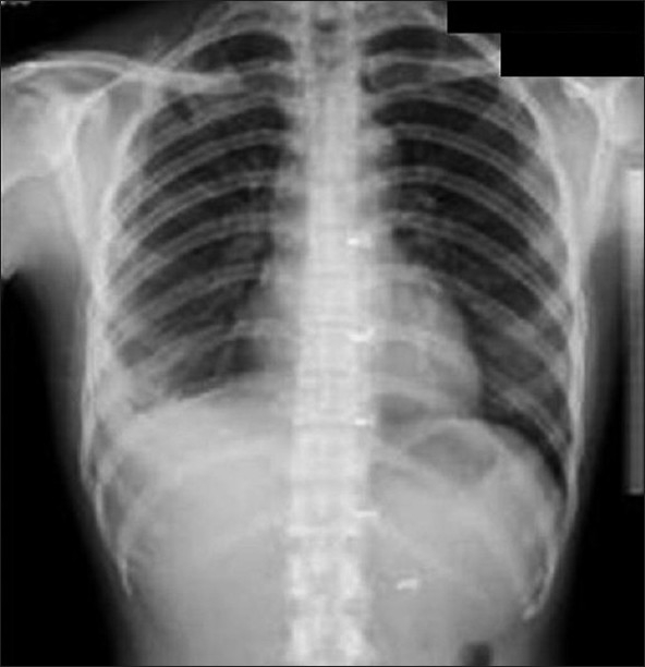
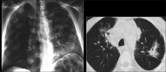
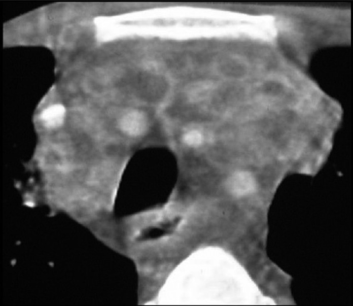
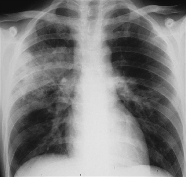
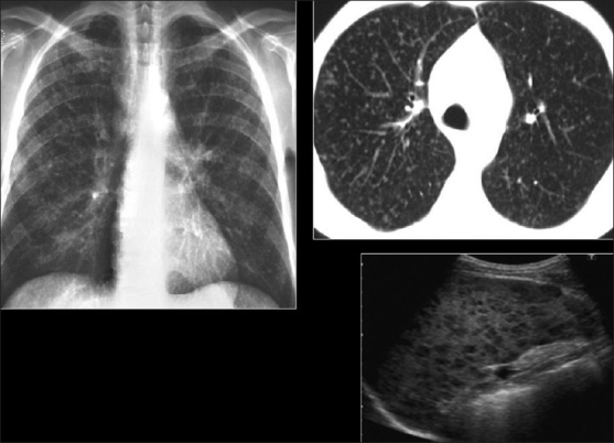
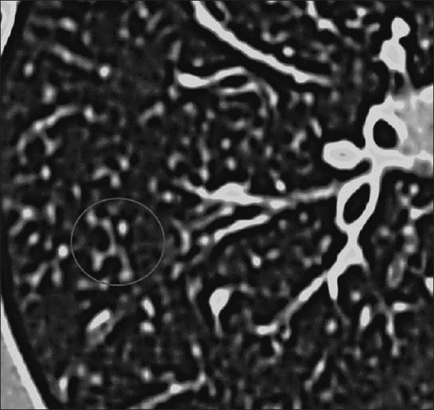
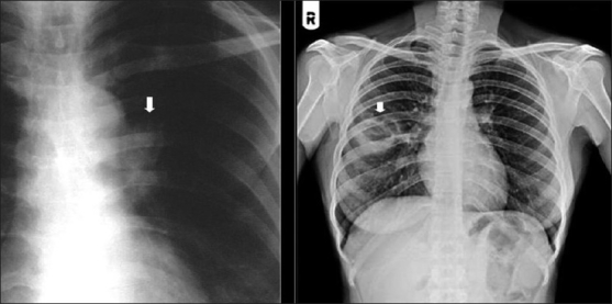
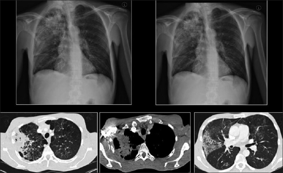
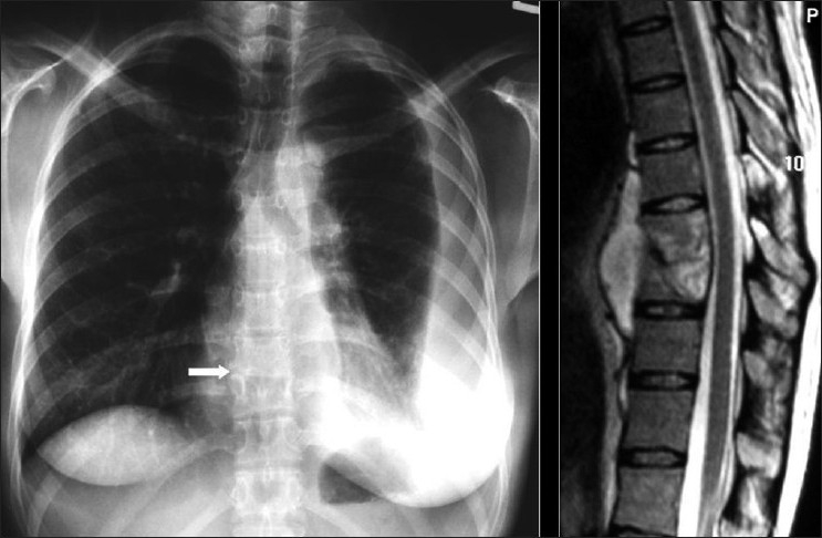
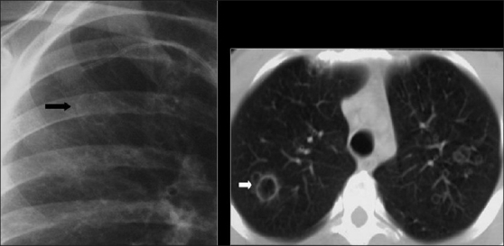
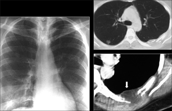
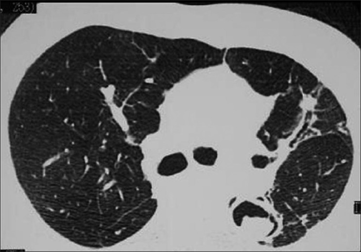
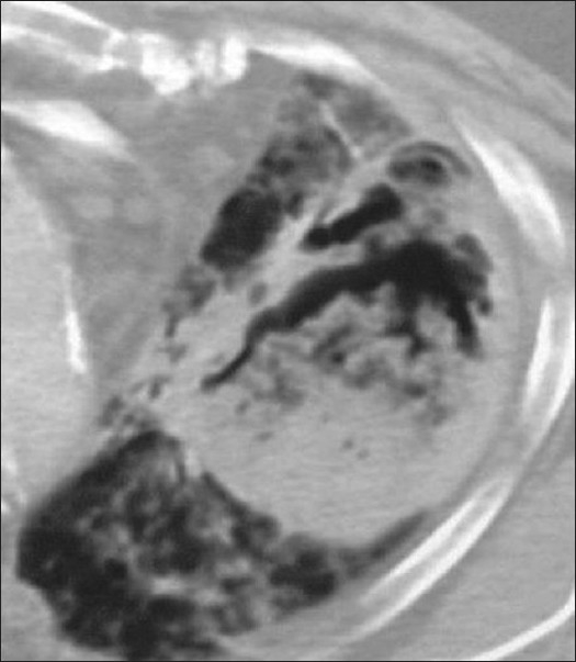
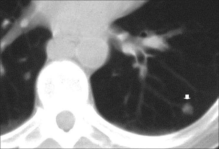
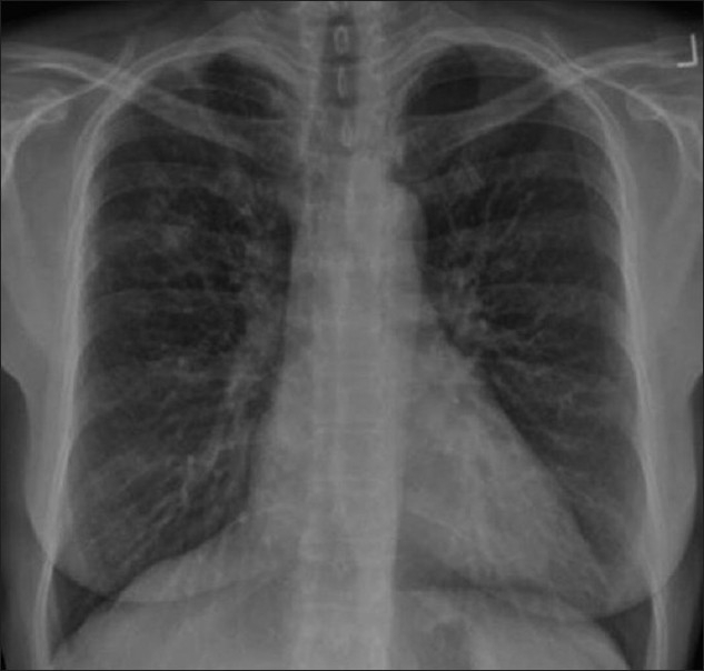
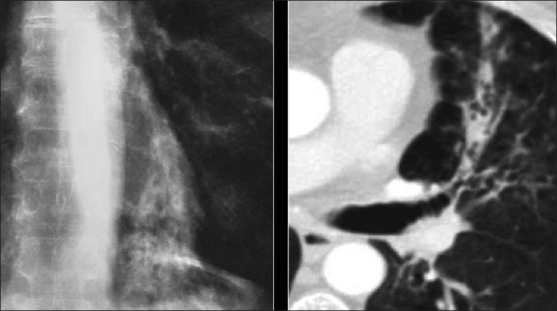
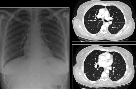
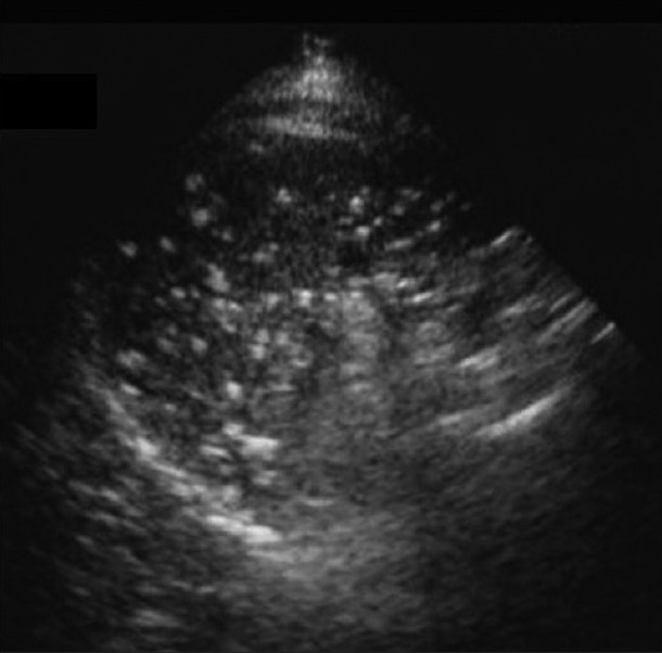
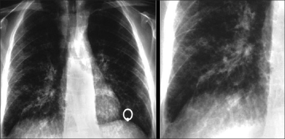
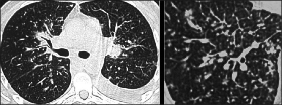
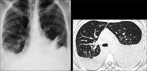
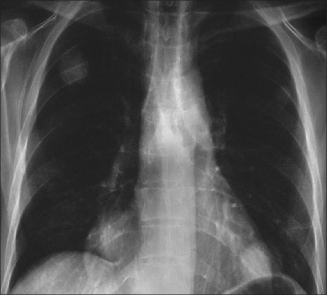
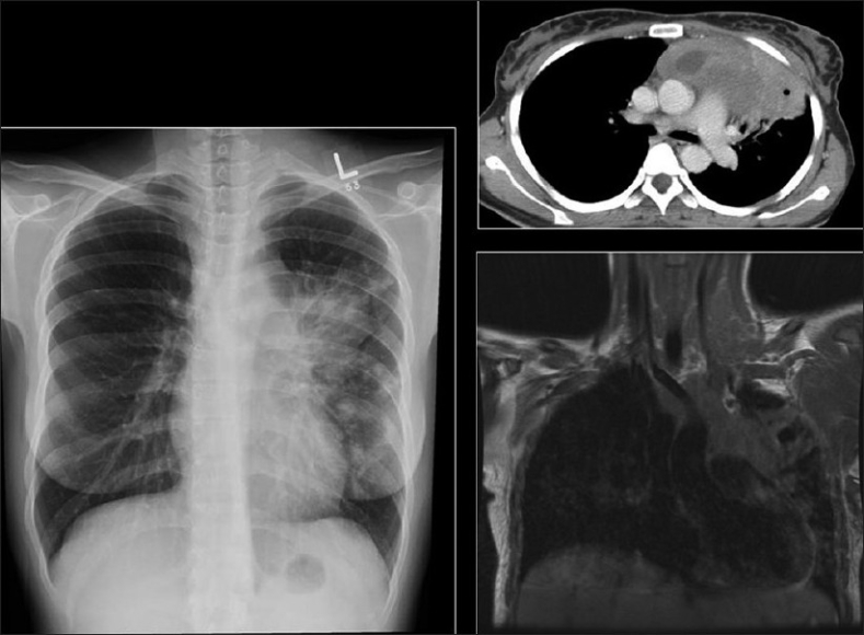
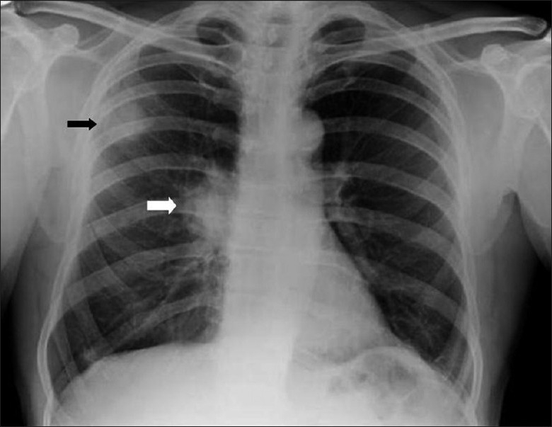
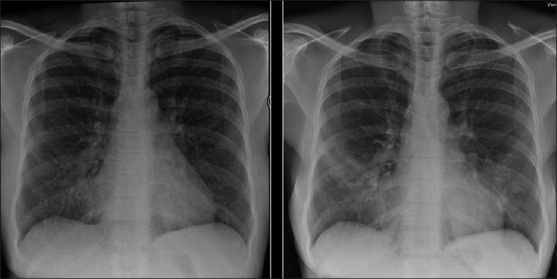
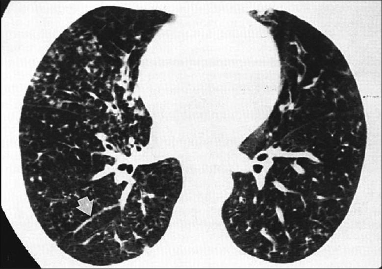
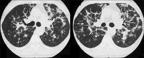
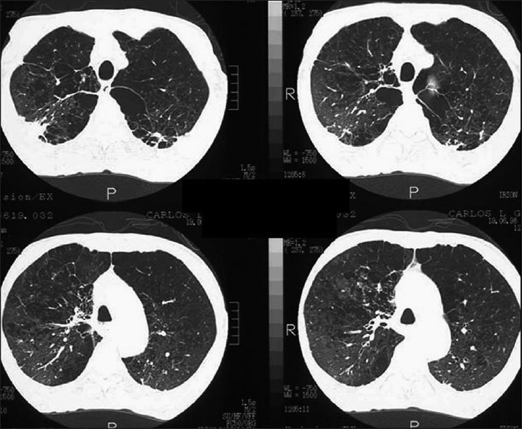
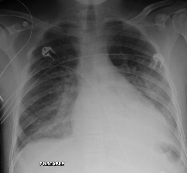
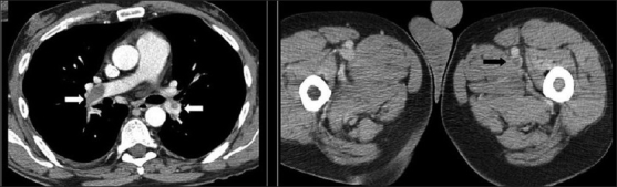
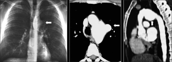
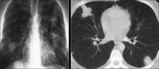
Advances in our understanding of human immunodeficiency virus (HIV) infection have led to improved care and incremental increases in survival. However, the pulmonary manifestations of HIV/acquired immunodeficiency syndrome (AIDS) remain a major cause of morbidity and mortality. Respiratory complaints are not infrequent in patients who are HIV positive. The great majority of lung complications of HIV/AIDS are of infectious etiology but neoplasm, interstitial pneumonias, Kaposi sarcoma and lymphomas add significantly to patient morbidity and mortality. Imaging plays a vital role in the diagnosis and management of lung of complications associated with HIV. Accurate diagnosis is based on an understanding of the pathogenesis of the processes involved and their imaging findings. Imaging also plays an important role in selection of the most appropriate site for tissue sampling, staging of disease and follow-ups. We present images of lung manifestations of HIV/AIDS, describing the salient features and the differential diagnosis.

Keywords: HIV/AIDS; imaging lung; mediastinal manifestations.


Conflict of interest statement

Conflict of Interest: None declared.

Figures

Figure 1
Figure 1
Bacterial bronchiolitis. Chest X-ray of a 36-year-old human immunodeficiency virus-positive male with a CD4 count of >400 presenting with productive cough. Note (left) the peribronchial thickening. The right hand film is a blown-up image showing the peribronchial thickening more clearly (arrow). The high-resolution computed tomography (lower left) depicts bronchiectasis, centrilobular nodularity/“tree-in-bud” mosaic attenuation in the same patient
Figure 2
Figure 2
Necrotizing cavitating pneumonia. Chest X-ray and computed tomography depicting necrotizing cavitating pneumonia due to Staphylococcus aureus in a 29-year-old male with acquired immunodeficiency syndrome
Figure 3
Figure 3
Septic emboli. A chest X-ray (CXR) and high-resolution computed tomography (HRCT) on a human immunodeficiency virus-positive patient with gram negative septicemia and septic emboli. Note the wedge-shaped pleural-based opacity and the feeding vessel on HRCT (arrow) and multiple cavitating nodules on the CXR
Figure 4
Figure 4
A chest X-ray of a patient with a CD4 count <200/mm3 showing perihilar ground-glass appearance in the shape of bats-wings
Figure 5
Figure 5
Pneumocystis carinii pneumonia. These chest radiographs are of two patients. Both show -ground glass appearance. The left chest X-ray (CXR) shows a much more subtle ground-glass appearance while the right CXR shows a much more gross ground-glass appearance mimicking pulmonary edema
Figure 6
Figure 6
Pneumocystis carinii pneumonia. If left untreated, chest X-ray may progress to alveolar consolidation in 3 or 4 days. Infiltrates clear within 2 weeks, but in a proportion infection will be followed by coarse reticular opacification and fibrosis. Note the large cyst (arrow)
Figure 7
Figure 7
Pneumocystis carinii pneumonia. Computed tomography (CT) in a subacute phase showing foci of consolidation and interlobular septal thickening due to organized inflammatory infiltrate on high-resolution CT
Figure 8
Figure 8
Pneumocystis carinii pneumonia (PCP). High-resolution computed tomography showing the hallmark of PCP in a clinical setting of immune compromise. Note the ground-glass attenuation with a geographic or mosaic distribution
Figure 9
Figure 9
Pneumocystis carinii pneumonia. Computed tomography (CT) in a subacute phase showing foci of consolidation and interlobular septal thickening due to organized inflammatory infiltrate on high-resolution CT
Figure 10
Figure 10
Pneumocystis carinii pneumonia (PCP). High-resolution computed tomography showing the hallmark of PCP in a clinical setting of immune compromise. Note the ground-glass attenuation with a geographic or mosaic distribution
Figure 11
Figure 11
Pneumocystis carinii pneumonia. High-resolution computed tomography shows diffuse ground-glass attenuation with a geographic distribution
Figure 12
Figure 12
Pneumocystis carinii pneumonia. Chest X-ray (R) shows a thin-walled cyst in the right upper lobe (arrow). The left image shows multiple cysts in the apical regions related to pentamidine inhalation in a human immunodeficiency virus patient
Figure 13
Figure 13
Pneumocystis carinii pneumonia. Lung cysts are usually multiple and bilateral, but range in size, shape and distribution. They are more commonly appreciated on computed tomography (CT)/high-resolution CT
Figure 14
Figure 14
Pneumocystis carinii pneumonia. Chest X-ray showing an approximately 50% right-sided spontaneous pneumothorax
Figure 15
Figure 15
Pneumocystis carinii pneumonia. Computed tomography showing bilateral pneumothoraces in conjunction with lung cysts
Figure 16
Figure 16
Pneumocystis carinii pneumonia. Chest X-ray and computed tomography show a left-sided ground-glass pattern and a right-sided large tension pneumothorax. Note the mediastinal shift
Figure 17
Figure 17
Pneumocystis carinii pneumonia. Pneumothoraces are often refractory to conventional chest tube drainage, becoming chronic, requiring pleurodesis or surgical intervention as in this patient
Figure 18
Figure 18
Pneumocystis carinii pneumonia. Chest X-ray shows atypical features of upper lobe focal reticulation associated with minor ground-glass appearance
Figure 19
Figure 19
Pneumocystis carinii pneumonia. Perihilar haze associated with hilar lymphadenopathy mimicking sarcoidosis
Figure 20
Figure 20
Pneumocystis carinii pneumonia atypical features. Chest X-ray shows focal consolidation of the left lung base
Figure 21
Figure 21
Pneumocystis carinii pneumonia atypical features. Chest X-ray shows finely granular/miliary appearance better depicted on high-resolution computed tomography
Figure 22
Figure 22
Pneumocystis carinii pneumonia atypical features. Chest X-ray shows diffuse, vague, ill-defined lung nodules associated with a right-sided pleural effusion (confirmed by ultrasound)
Figure 23
Figure 23
Chest X-ray and computed tomography showing features of reactivation Mycobacterium tuberculosis as patchy consolidation, including involvement at unusual sites, e.g. lower lobes as seen here, cavitation, nodularity and adenopathy
Figure 24
Figure 24
Mycobacterium tuberculosis (TB). TB mediastinal lymph nodes are typically markedly enlarged and of low attenuation on computed tomography, often demonstrating rim enhancement following contrast administration, as in this case
Figure 25
Figure 25
“Primary” Mycobacterium tuberculosis. Chest X-ray shows right upper lobe and left midzone consolidation and adenopathy. Note lack of cavitation in this patient with a low CD4 count
Figure 26
Figure 26
“Primary” Mycobacterium tuberculosis (MTB). Chest X-ray and high-resolution computed tomography show diffuse randomly distributed miliary nodules. Ultrasound of the spleen of the same patient shows splenomegaly and multiple hypoechoic nodules due to MTB granulomas
Figure 27
Figure 27
Mycobacterium tuberculosis. High-resolution computed tomography shows a tree-in-bud appearance
Figure 28
Figure 28
Nontuberculous/atypical Mycobacterium (NTMB). The chest X-ray (CXR) abnormalities resemble Mycobacterium tuberculosis, with focal consolidation CXR (left), diffuse patchy infiltrates and cavities (right). The features are, however, nonspecific and the diagnosis is often delayed
Figure 29
Figure 29
Mycobacterium xenopi in a human immunodeficiency virus, 36-year old, male patient with a CD4 count of 80 with four positive sputum samples and a bronchoalveolar lavage for acid-fast bacilli. The chest X-ray and computed tomography scans show cavitating consolidation, loss of volume, traction bronchiectasis and ground-glass appearances in the right apical region superimposed on bullous disease of the lungs. There is no associated lymphadenopathy
Figure 30
Figure 30
A 26-year-old human immunodeficiency virus-positive female presented with shortness of breath. The chest X-ray shows a large left-sided pleural effusion and loss of height and erosion of the articular plates between the 9th and 10th vertebral bodies associated with soft tissue swelling. A sagittal T2-weigted magnetic resonance scan of the dorsal spine shows complete obliteration of the disc between the 9th and 10th dorsal vertebral bodies associated with fluid collection anterior to the spine representing pus. Acid-fast bacilli were identified in the aspirated pus
Figure 31
Figure 31
Cryptococcosis. Imaging findings are varied and nonspecific. Reticular chest X-ray or reticulonodular infiltrates are the most common pattern as in this case where a reticulonodular infiltrate involved the left costophrenic angle
Figure 32
Figure 32
Cryptococcosis. Chest X-ray and computed tomography show a solitary pulmonary nodule. The diagnosis of Cryptococcosis was confirmed on biopsy
Figure 33
Figure 33
Cryptococcosis. Chest X-ray shows a less-frequently seen cavitation due to Cryptococcosis in an acquired immunodeficiency syndrome patient
Figure 34
Figure 34
Cryptococcosis. Chest X-ray (L) shows a thin-walled cavity (arrow) associated with patchy consolidation. Computed tomography section at the level of the lower trachea (R) shows multiple cavities of varying sizes associated with subtle ground-glass opacification
Figure 35
Figure 35
Cryptococcosis. Chest X-ray and computed tomography show lessfrequent manifestations of lung nodules and a chest wall abscess (arrow)
Figure 36
Figure 36
Aspergillosis. Mycetomas are the least common, but can complicate cavitary Mycobacterium tuberculosis or Pneumocystis carinii pneumonia. The computed tomography here represents a mycetoma in a tuberculous cavity. Note the traction bronchiectasis and loss of volume in the left upper zone
Figure 37
Figure 37
Aspergillosis. Angioinvasive disease chest X-ray is the most common, manifesting as thick-walled cavitary lesions predominating in the upper lobes, with air-crescents surrounding areas of desquamated infarcted lung. Here, we see all the described features on computed tomography
Figure 38
Figure 38
Aspergillosis. Less-common patterns include computed tomography nodules (arrow) with a peripheral halo of ground-glass attenuation (not shown)
Figure 39
Figure 39
Aspergillosis. ABPA chest X-ray (CXR), manifesting as bilateral consolidation, mostly lower lobe, bronchiectasis and airway impaction or “finger in glove.” The consolidation in the CXR shown is much more diffuse
Figure 40
Figure 40
Aspergillosis. ABPA a chest X-ray/computed tomography on the same patient shows bilateral lower lobe consolidation, bronchiectasis and airway impaction or “finger in glove” appearance
Figure 41
Figure 41
Cytomegalovirus (CMV). Human immunodeficiency virus patient with a CD4 count of 40 presented with fever and dry cough. The chest X-ray shows ill defined diffuse small nodules. High-resolution computed tomography scans confirm scattered bilateral centrilobular nodule-associated interlobular septal thickening. Imaging appearances of CMV pneumonia are nonspecific and may mimic other opportunistic infections
Figure 42
Figure 42
Ultrasound of the spleen on the same patient as in Figure 41 showing multiple microabscesses due to systemic cytomegalovirus infection
Figure 43
Figure 43
Kaposi’s sarcoma is the most common acquired immunodeficiency syndrome-related malignancy. The chest X-ray shows bilateral perihilar/lower zone reticulonodular infiltrates
Figure 44
Figure 44
Kaposi’s sarcoma. High-resolution computed tomography shows thickening of the bronchovascular bundles reflecting bronchocentric disease. Interlobular septal thickening due to lymphatic obstruction because of tumor invasion is also seen
Figure 45
Figure 45
Kaposi sarcoma. Pleural effusions chest X-ray/computed tomography are common. They may be unilateral or bilateral and may be large and characteristically hemorrhagic on aspiration
Figure 46
Figure 46
Lymphoma. Chest X-ray (CXR) on a human immunodeficiency virus patient that presented with multiple lung masses, which grew rapidly mimicking infection. Note that there is no associated lymphadenopathy. Well-defined solitary or multiple parenchymal nodules CXR are common. A percutaneous biopsy revealed a non-Hodgkin’s lymphoma
Figure 47
Figure 47
NHL in a 23-year-old human immunodeficiency virus female. The chest radiograph shows multiple well-defined lung nodules within the left lung associated with mediastinal lymphadenopathy. Lymphadenopathy is a less common feature in acquired immunodeficiency disease-related NHL and nodes are rarely significant according to size criteria unlike as in the case shown here, where there is significant lymphadenopathy as confirmed by computed tomography (right upper frame). Magnetic resonance imaging is the imaging of choice to detect vascular encasement
Figure 48
Figure 48
Bronchogenic carcinoma. Radiographic appearances are similar to ordinary lung cancer, except that lesions tend to be more peripheral and in the upper lobes as in this case (black arrow). Note the right hilar lymphadenopathy (white arrow)
Figure 49
Figure 49
Lymphocytic interstitial pneumonitis in a human immunodeficiency virus patient. Chest X-ray showing bilateral reticulonodular interstitial infiltrates. Diagnosis was confirmed by a transbronchial biopsy
Figure 50
Figure 50
Lymphocytic interstitial pneumonitis in a human immunodeficiency virus patient. High-resolution computed tomography shows smooth and nodular thickening of bronchovascular bundles, centrilobular and subpleural nodularity, ground-glass opacification and interlobular septal thickening. Cysts and mediastinal adenopathy may also be seen (not shown here)
Figure 51
Figure 51
Bronchiolitis obliterans with or without organizing pneumonia in the absence of infection can be a feature of acquired immunodeficiency syndrome (AIDS). This is an infrequent imaging diagnosis, although focal air trapping on expiratory computed tomography, consistent with bronchiolitis obliterans, has been demonstrated in two-thirds of human immunodeficiency virus-positive patients without AIDS, the severity increasing with the duration of infection
Figure 52
Figure 52
High-resolution computed tomography of a 46-year-old man with acquired immunodeficiency syndrome-related emphysema. This patient was also a smoker and experimented with cocaine and marijuana. A biopsy revealed destruction of the lung parenchyma distal to the terminal bronchioles accompanied by various degrees of inflammation
Figure 53
Figure 53
Anterioposterior chest radiograph on a 43-year-old man treated with highly active antiretroviral therapy for over 8 years who presented with increasing shortness of breath over the past 3 months that had suddenly worsened, prompting hospital admission. The chest X-ray shows features of pulmonary edema. Subsequent investigations revealed congestive cardiomyopathy
Figure 54
Figure 54
Thromboembolism. Computed tomography (CT) images from a 38-year old human immunodeficiency virus-positive man with a CD count of 400 presented with acute tightness in the chest and low O2 saturation. The CT scans show pulmonary emboli within the lower lobe pulmonary arteries on both sides (white arrows). Note the thrombus in the left femoral artery (black arrow)
Figure 55
Figure 55
This human immunodeficiency virus patient suffered from gram negative septicemia, which was successfully treated. However, routine physical examination revealed an audible bruit on thoracic auscultation. The chest X-ray shows a prominent hump over the proximal descending aorta due to mycotic aortic aneurysm. The axial computed tomography (CT) and coronal CT reconstruction elegantly demonstrate the abnormality
Figure 56
Figure 56
This hemophilic patient was infected with human immunodeficiency virus (HIV) from factor 8 before HIV testing became compulsory. The images show pulmonary hemorrhage on the background of Pneumocystis carinii pneumonia

Similar articles

  • HIV-related Malignancies and Mimics: Imaging Findings and Management.
    Javadi S, Menias CO, Karbasian N, Shaaban A, Shah K, Osman A, Jensen CT, Lubner MG, Gaballah AH, Elsayes KM. Javadi S, et al. Radiographics. 2018 Nov-Dec;38(7):2051-2068. doi: 10.1148/rg.2018180149. Epub 2018 Oct 19. Radiographics. 2018. PMID: 30339518 Review.
  • Human immunodeficiency virus infection in childhood.
    Blokzijl ML. Blokzijl ML. Ann Trop Paediatr. 1988 Mar;8(1):1-17. doi: 10.1080/02724936.1988.11748530. Ann Trop Paediatr. 1988. PMID: 2456715 Review.
  • Is HIV involved in the pathogenesis of non-infectious pulmonary complications in infected patients?
    Kanmogne GD, Kennedy RC, Grammas P. Kanmogne GD, et al. Curr HIV Res. 2003 Oct;1(4):385-93. doi: 10.2174/1570162033485177. Curr HIV Res. 2003. PMID: 15049425 Review.
  • Tuberculosis.
    Bloom BR, Atun R, Cohen T, Dye C, Fraser H, Gomez GB, Knight G, Murray M, Nardell E, Rubin E, Salomon J, Vassall A, Volchenkov G, White R, Wilson D, Yadav P. Bloom BR, et al. In: Holmes KK, Bertozzi S, Bloom BR, Jha P, editors. Major Infectious Diseases. 3rd edition. Washington (DC): The International Bank for Reconstruction and Development / The World Bank; 2017 Nov 3. Chapter 11. In: Holmes KK, Bertozzi S, Bloom BR, Jha P, editors. Major Infectious Diseases. 3rd edition. Washington (DC): The International Bank for Reconstruction and Development / The World Bank; 2017 Nov 3. Chapter 11. PMID: 30212088 Free Books & Documents. Review.
  • Noninfectious pulmonary complications of HIV/AIDS.
    Kanmogne GD. Kanmogne GD. Curr Opin Pulm Med. 2005 May;11(3):208-12. doi: 10.1097/01.mcp.0000159833.28271.00. Curr Opin Pulm Med. 2005. PMID: 15818181 Review.

Cited by

References

    1. The latest statistics on the world epidemic of HIV and AIDS were published by UNAIDS/WHO in July 2008, and refer to the end of 2007
    1. Beck JM, Rosen MJ, Peavy HH. Pulmonary complications of HIV infection: Report of the Fourth NHLBI Workshop. Am J Respir Crit Care Med. 2001;164:2120–6. - PubMed
    1. Taylor IK, Coker RJ, Clarke J, Moss FM, Nieman R, Evans DJ, et al. Pulmonary complications of HIV disease: 10-year retrospective evaluation of yields from bronchoalveolar lavage, 1983–93. Thorax. 1995;50:1240–5. - PMC - PubMed
    1. Rosen MJ. Overview of pulmonary complications. Clin Chest Med. 1996;17:621–31. - PubMed
    1. Poirier CD, Inhaber N, Lalonde RG, Ernst P. Prevalence of bronchial hyperrsponsiveness among HIV-infected men. Am J Respir Crit Care Med. 2001;164:542–5. - PubMed