. 2010 Oct 27;30(43):14411-9.
doi: 10.1523/JNEUROSCI.3537-10.2010.

Amyloid beta-protein dimers rapidly form stable synaptotoxic protofibrils

Affiliations

Amyloid beta-protein dimers rapidly form stable synaptotoxic protofibrils

Brian O'Nuallain et al. J Neurosci. .

Abstract

Nonfibrillar, water-soluble low-molecular weight assemblies of the amyloid β-protein (Aβ) are believed to play an important role in Alzheimer's disease (AD). Aqueous extracts of human brain contain Aβ assemblies that migrate on SDS-polyacrylamide gels and elute from size exclusion as dimers (∼8 kDa) and can block long-term potentiation and impair memory consolidation in the rat. Such species are detected specifically and sensitively in extracts of Alzheimer brain suggesting that SDS-stable dimers may be the basic building blocks of AD-associated synaptotoxic assemblies. Consequently, understanding the structure and properties of Aβ dimers is of great interest. In the absence of sufficient brain-derived dimer to facilitate biophysical analysis, we generated synthetic dimers designed to mimic the natural species. For this, Aβ(1-40) containing cysteine in place of serine 26 was used to produce disulphide cross-linked dimer, (AβS26C)2. Such dimers had no detectable secondary structure, produced an analytical ultracentrifugation profile consistent for an ∼8.6 kDa protein, and had no effect on hippocampal long-term potentiation (LTP). However, (AβS26C)2 aggregated more rapidly than either AβS26C or wild-type monomers and formed parastable β-sheet rich, thioflavin T-positive, protofibril-like assemblies. Whereas wild-type Aβ aggregated to form typical amyloid fibrils, the protofibril-like structures formed by (AβS26C)2 persisted for prolonged periods and potently inhibited LTP in mouse hippocampus. These data support the idea that Aβ dimers may stabilize the formation of fibril intermediates by a process distinct from that available to Aβ monomer and that higher molecular weight prefibrillar assemblies are the proximate mediators of Aβ toxicity.


Figures
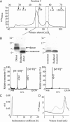
Figure 1.
Figure 1.
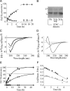
Isolation and initial characterization of AβS26C dimers. Disulfide cross-linked Aβ dimers were generated by atmospheric oxidation of 20 μm AβS26C in 10 mm sodium bicarbonate, pH 8.5, for 5 d at room temperature. A, The cross-linked Aβ dimer product was isolated by SEC using a HiLoad 16/60 Superdex 75 column equilibrated with 25 mm ammonium acetate, pH 8.5. Arrows indicate elution of dextran standards. B, SDS-PAGE and MALDI-ToF MS analysis of the two low molecular weight major SEC peaks confirms the presence of a disulfide cross-link in the dimer but not monomer fractions. C, Analytical ultracentrifugation analysis confirmed the predominant species to have a predicted mass of 10 ± 2 kDa. D, The same dimer used for AUC was incubated at room temperature for 16 h and when rechromatographed on SEC revealed a significant peak in the void volume (gray line) that was not detected in the t = 0 sample (black line).
Figure 2.
Figure 2.
Quiescent aggregation of AβS26C dimers. A, Progress curves for the formation of ThT-positive material are shown as percentage of the maximum fluorescence detected from freshly isolated (AβS26C)2 (•), S26C (○) and wild-type (▴) monomers. ThT fluorescence was monitored in real time at 37°C. Reactions contained 10 μm (AβS26C)2 or 20 μm wild-type/S26C monomer in 20 mm sodium phosphate, pH 7.4, plus 10 μm ThT. B, Freshly isolated and 1 d aggregated dimer samples were analyzed by SDS-PAGE. Dimer supernatant (sup) was generated by centrifuging the sample at 16,000 × g for 20 min at room temperature. C, D, Circular dichroism spectra for solutions of freshly isolated (AβS26C)2 (10 μm, —), S26C (20 μm, – –) or wild-type (20 μm, · · ·), monomers at 0 h (C) and after 24 h at 37°C (D). E, Concentration dependency of S26C dimer aggregation: 10 μm (•); 5 μm (○); 2.5 μm (▴), and 1. 25 μm (□) were incubated at 37°C for up to 48 h. F, Dimer aggregation follows pseudo first-order kinetics. The data from E were plotted as the natural log of the difference between the maximum ThT fluorescence and the observed signal versus time.
Figure 3.
Figure 3.
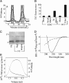
Biophysical analysis of AβS26C dimer aggregates. A, (AβS26C)2 (10 μm) was incubated in 20 mm sodium phosphate, pH 7.4, without agitation and at intervals samples removed and chromatographed on a Superdex 75 10/300 GL column equilibrated with 20 mm sodium phosphate, pH 7.4. The chromatographs show the conversion of Aβ dimers into high molecular weight conformers that eluted in the void volume (dashed vertical line). Freshly prepared Aβ dimers after 4 h at 4°C (—), and Aβ dimer preparation after 6 h (– –), 24 h (· · ·), and 3 d (—· ·) at 37°C. The gray bars show the peak fractions that were collected for subsequent experiments. B, ThT fluorescence of SEC-isolated 3 d aggregated (AβS26C)2 compared with the same concentration (0.09 mg/ml) of the unaggregated peptide and WT Aβ conformers. C, SDS-PAGE analysis of time 0 and 3 d incubated dimer before (unspun) and after centrifugation (sup). D, Circular dichroism spectra obtained using SEC isolated dimers (2.5 μm, – –) and the void component of SEC fractionated 3 d aggregated dimers (3 μm, —). E, Multi-angle light scattering indicated that the void component of SEC fractionated 3 d aggregated dimers (—) had a size distribution of ∼1–4 MDa (· · ·).
Figure 4.
Figure 4.
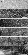
Morphology of aggregates formed by AβS26C dimers. A–E, Negative contrast EM was performed on freshly isolated 10 μm (AβS26C)2 in 25 mm ammonium acetate, pH 8.5 (A), and on aliquots of the (AβS26C)2 reaction after incubation at 37°C in 20 mm sodium phosphate, pH 7.4, for 1 d (B), 3 d (C), or 1 month (D). When the 1 month sample was centrifuged at 16,000 × g and room temperature for 20 min and the supernatant examined protofibrils were still detected (E), but on average these protofibrils were shorter than those detected in the unspun sample. F, Fibrils formed from 30 μm monomeric wild-type Aβ after a 2 week incubation. Images are representative of at least 6–8 fields from duplicate grids for each time point. Scale bar, 100 nm.
Figure 5.
Figure 5.
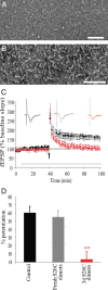
Protofibrils formed from AβS26C dimers potently inhibit LTP. A–C, Freshly SEC-isolated (AβS26C)2 was immediately diluted to 17 μm with 25 mm ammonium acetate, pH 8.5, and used to prepare samples for negative contrast electron microscopy (A). As in the prior figure, micrographs are representative of at least 6–8 fields from duplicate grids for each time point. Size bar = 100 nm. The remaining solution was held on ice for 1–4 h, then diluted to 10 μm with 20 mm phosphate, pH 7.4, and used for electrophysiology (C) or incubated at 37°C for a further 72 h (B). C, Perfusion of mouse hippocampal slices with nACSF containing 3 d aggregated (AβS26C)2 (red triangles), but not vehicle (ammonium acetate/phosphate buffer) (black squares) or an equivalent amount (50 nm) of freshly isolated (AβS26C)2 (gray circles) blocked LTP (p < 0.001). Values are mean ± SEM percentage of baseline, n = 7 (aggregated dimer), n = 7 (fresh dimer) and n = 7 (vehicle). The horizontal bar represents the time during which the vehicle or peptide was present in the recording solution. Insets show typical fEPSP 5 min pre- and post-TBS. Calibration: 5 ms, 0.5 mV. D, The histogram shows the magnitude of LTP between 55 and 60 min post-TBS for all 3 groups; *p < 0.01 (ANOVA).
Figure 6.
Figure 6.
A model for dimer-mediated protofibril toxicity. In vitro, wild-type Aβ monomer is known to assemble into amyloid fibrils by a process that appears to require the transient formation of prefibrillar structures referred to as protofibrils. The steady-state level of protofibrils is controlled by four key reactions: (1) formation of protofibrils, (2) disassembly of protofibrils (3), formation of fibrils and (4) disassembly of fibrils. The rate of protofibril formation and the time period over which protofibrils persist is strongly influenced by Aβ primary sequence. Specifically, the population of protofibrils is greater for Aβ1-42 and Aβ1-40E22G than for wild-type Aβ1-40 (Walsh et al., 1997; Nilsberth et al., 2001). Similarly, covalent cross-linking of Aβ by either 4-hydroxynonenal (HNE) or transglutaminase (TGase) accelerates formation of protofibrils while inhibiting fibril formation (Siegel et al., 2007; Hartley et al., 2008). Here we demonstrate that pure (AβS26C)2 also increases the rate of protofibril, but not fibril formation. This suggests that formation of a stable dimer (either covalently cross-linked as shown in the current study or non-covalently cross-linked as seen in human brain) may better facilitate protofibril formation and persistence than Aβ monomer. It has also been demonstrated that certain lipids can destabilize mature fibrils and liberate protofibrils (Johansson et al., 2007; Martins et al., 2008) and that such “reverse” protofibrils, like “forward” protofibrils are potent synaptotoxins (Martins et al., 2008). Since SDS-stable Aβ dimers appear specific for AD it seems plausible that the presence of dimers and abundance of protofibrils are linked. That is, dimers exert toxicity as a consequence of their ability to form relatively stable protofibrils.

Similar articles

Cited by

References

    1. Betts V, Leissring MA, Dolios G, Wang R, Selkoe DJ, Walsh DM. Aggregation and catabolism of disease-associated intra-Abeta mutations: reduced proteolysis of AbetaA21G by neprilysin. Neurobiol Dis. 2008;31:442–450. - PMC - PubMed
    1. Glabe CG. Structural classification of toxic amyloid oligomers. J Biol Chem. 2008;283:29639–29643. - PMC - PubMed
    1. Hardy J, Selkoe DJ. The amyloid hypothesis of Alzheimer's disease: progress and problems on the road to therapeutics. Science. 2002;297:353–356. - PubMed
    1. Harmeier A, Wozny C, Rost BR, Munter LM, Hua H, Georgiev O, Beyermann M, Hildebrand PW, Weise C, Schaffner W, Schmitz D, Multhaup G. Role of amyloid-beta glycine 33 in oligomerization, toxicity, and neuronal plasticity. J Neurosci. 2009;29:7582–7590. - PMC - PubMed
    1. Harper JD, Lansbury PT., Jr Models of amyloid seeding in Alzheimer's disease and scrapie: mechanistic truths and physiological consequences of the time-dependent solubility of amyloid proteins. Annu Rev Biochem. 1997;66:385–407. - PubMed

Publication types

MeSH terms

LinkOut - more resources