

IP(3) receptors: toward understanding their activation
- PMID: 20980441
- PMCID: PMC2982166
- DOI: 10.1101/cshperspect.a004010
IP(3) receptors: toward understanding their activation
Abstract
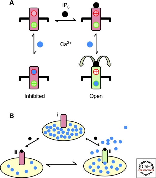
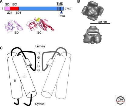
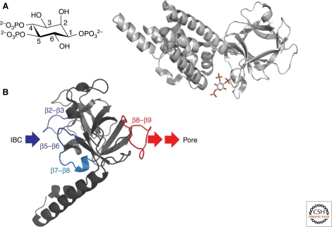
Inositol 1,4,5-trisphosphate receptors (IP(3)R) and their relatives, ryanodine receptors, are the channels that most often mediate Ca(2+) release from intracellular stores. Their regulation by Ca(2+) allows them also to propagate cytosolic Ca(2+) signals regeneratively. This brief review addresses the structural basis of IP(3)R activation by IP(3) and Ca(2+). IP(3) initiates IP(3)R activation by promoting Ca(2+) binding to a stimulatory Ca(2+)-binding site, the identity of which is unresolved. We suggest that interactions of critical phosphate groups in IP(3) with opposite sides of the clam-like IP(3)-binding core cause it to close and propagate a conformational change toward the pore via the adjacent N-terminal suppressor domain. The pore, assembled from the last pair of transmembrane domains and the intervening pore loop from each of the four IP(3)R subunits, forms a structure in which a luminal selectivity filter and a gate at the cytosolic end of the pore control cation fluxes through the IP(3)R.
Figures
Similar articles
-
Structural and functional conservation of key domains in InsP3 and ryanodine receptors.Nature. 2012 Jan 29;483(7387):108-12. doi: 10.1038/nature10751. Nature. 2012. PMID: 22286060 Free PMC article.
-
IP(3) receptors: the search for structure.Trends Biochem Sci. 2004 Apr;29(4):210-9. doi: 10.1016/j.tibs.2004.02.010. Trends Biochem Sci. 2004. PMID: 15082315 Review.
-
The contribution of inositol 1,4,5-trisphosphate and ryanodine receptors to agonist-induced Ca(2+) signaling of airway smooth muscle cells.Am J Physiol Lung Cell Mol Physiol. 2009 Aug;297(2):L347-61. doi: 10.1152/ajplung.90559.2008. Epub 2009 May 22. Am J Physiol Lung Cell Mol Physiol. 2009. PMID: 19465516 Free PMC article.
-
Regulation of Ca2+-release-activated Ca2+ current (Icrac) by ryanodine receptors in inositol 1,4,5-trisphosphate-receptor-deficient DT40 cells.Biochem J. 2001 Nov 15;360(Pt 1):17-22. doi: 10.1042/0264-6021:3600017. Biochem J. 2001. PMID: 11695987 Free PMC article.
-
Ca(2+) signalling by IP(3) receptors.Subcell Biochem. 2012;59:1-34. doi: 10.1007/978-94-007-3015-1_1. Subcell Biochem. 2012. PMID: 22374086 Review.
Cited by
-
The signatures of organellar calcium.Plant Physiol. 2021 Dec 4;187(4):1985-2004. doi: 10.1093/plphys/kiab189. Plant Physiol. 2021. PMID: 33905517 Free PMC article. Review.
-
Update on vascular endothelial Ca(2+) signalling: A tale of ion channels, pumps and transporters.World J Biol Chem. 2012 Jul 26;3(7):127-58. doi: 10.4331/wjbc.v3.i7.127. World J Biol Chem. 2012. PMID: 22905291 Free PMC article.
-
Recovirus NS1-2 Has Viroporin Activity That Induces Aberrant Cellular Calcium Signaling To Facilitate Virus Replication.mSphere. 2019 Sep 18;4(5):e00506-19. doi: 10.1128/mSphere.00506-19. mSphere. 2019. PMID: 31533997 Free PMC article.
-
Amyloid-β disrupts unitary calcium entry through endothelial NMDA receptors in mouse cerebral arteries.J Cereb Blood Flow Metab. 2022 Jan;42(1):145-161. doi: 10.1177/0271678X211039592. Epub 2021 Aug 31. J Cereb Blood Flow Metab. 2022. PMID: 34465229 Free PMC article.
-
Synthesis of dimeric analogs of adenophostin A that potently evoke Ca2+ release through IP3 receptors.RSC Adv. 2016 Nov 3;6(89):86346-86351. doi: 10.1039/c6ra19413c. Epub 2016 Sep 5. RSC Adv. 2016. PMID: 28066549 Free PMC article.
References
-
- Adkins CE, Taylor CW 1999. Lateral inhibition of inositol 1,4,5-trisphosphate receptors by cytosolic Ca2+. Curr Biol 9: 1115–1118 - PubMed
Publication types
MeSH terms
Substances
Grants and funding
LinkOut - more resources
Full Text Sources
Molecular Biology Databases
Miscellaneous