

αβγ-Synuclein triple knockout mice reveal age-dependent neuronal dysfunction
- PMID: 20974939
- PMCID: PMC2984188
- DOI: 10.1073/pnas.1005005107
αβγ-Synuclein triple knockout mice reveal age-dependent neuronal dysfunction
Abstract
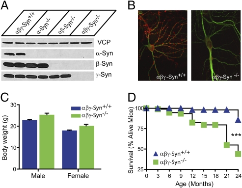
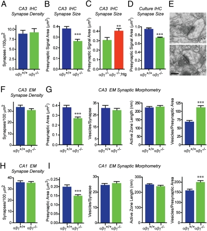
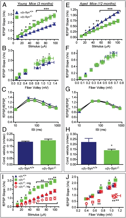
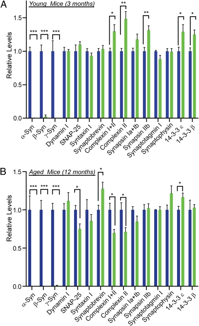
Synucleins are a vertebrate-specific family of abundant neuronal proteins. They comprise three closely related members, α-, β-, and γ-synuclein. α-Synuclein has been the focus of intense attention since mutations in it were identified as a cause for familial Parkinson's disease. Despite their disease relevance, the normal physiological function of synucleins has remained elusive. To address this, we generated and characterized αβγ-synuclein knockout mice, which lack all members of this protein family. Deletion of synucleins causes alterations in synaptic structure and transmission, age-dependent neuronal dysfunction, as well as diminished survival. Abrogation of synuclein expression decreased excitatory synapse size by ∼30% both in vivo and in vitro, revealing that synucleins are important determinants of presynaptic terminal size. Young synuclein null mice show improved basic transmission, whereas older mice show a pronounced decrement. The late onset phenotypes in synuclein null mice were not due to a loss of synapses or neurons but rather reflect specific changes in synaptic protein composition and axonal structure. Our results demonstrate that synucleins contribute importantly to the long-term operation of the nervous system and that alterations in their physiological function could contribute to the development of Parkinson's disease.
Conflict of interest statement
The authors declare no conflict of interest.
Figures

Similar articles
-
Functional alterations to the nigrostriatal system in mice lacking all three members of the synuclein family.J Neurosci. 2011 May 18;31(20):7264-74. doi: 10.1523/JNEUROSCI.6194-10.2011. J Neurosci. 2011. PMID: 21593311 Free PMC article.
-
Monomeric synucleins generate membrane curvature.J Biol Chem. 2013 Jan 18;288(3):1829-40. doi: 10.1074/jbc.M112.418871. Epub 2012 Nov 26. J Biol Chem. 2013. PMID: 23184946 Free PMC article.
-
Localization of synucleins in the mammalian cochlea.J Assoc Res Otolaryngol. 2008 Dec;9(4):452-63. doi: 10.1007/s10162-008-0134-y. Epub 2008 Jul 30. J Assoc Res Otolaryngol. 2008. PMID: 18665422 Free PMC article.
-
Synucleins and their relationship to Parkinson's disease.Cell Tissue Res. 2004 Oct;318(1):163-74. doi: 10.1007/s00441-004-0921-7. Epub 2004 Jul 24. Cell Tissue Res. 2004. PMID: 15503152 Review.
-
The synucleins: a family of proteins involved in synaptic function, plasticity, neurodegeneration and disease.Trends Neurosci. 1998 Jun;21(6):249-54. doi: 10.1016/s0166-2236(97)01213-7. Trends Neurosci. 1998. PMID: 9641537 Review.
Cited by
-
Monomeric Alpha-Synuclein Exerts a Physiological Role on Brain ATP Synthase.J Neurosci. 2016 Oct 12;36(41):10510-10521. doi: 10.1523/JNEUROSCI.1659-16.2016. J Neurosci. 2016. PMID: 27733604 Free PMC article.
-
Current insights and assumptions on α-synuclein in Lewy body disease.Acta Neuropathol. 2024 Aug 14;148(1):18. doi: 10.1007/s00401-024-02781-3. Acta Neuropathol. 2024. PMID: 39141121 Free PMC article. Review.
-
An extracellular mechanism that can explain the neurotoxic effects of α-synuclein aggregates in the brain.Front Physiol. 2012 Jul 26;3:297. doi: 10.3389/fphys.2012.00297. eCollection 2012. Front Physiol. 2012. PMID: 22934048 Free PMC article.
-
The Close Encounter Between Alpha-Synuclein and Mitochondria.Front Neurosci. 2018 Jun 7;12:388. doi: 10.3389/fnins.2018.00388. eCollection 2018. Front Neurosci. 2018. PMID: 29930495 Free PMC article. Review.
-
α-Synuclein Dimers Impair Vesicle Fission during Clathrin-Mediated Synaptic Vesicle Recycling.Front Cell Neurosci. 2017 Dec 11;11:388. doi: 10.3389/fncel.2017.00388. eCollection 2017. Front Cell Neurosci. 2017. PMID: 29321725 Free PMC article.
References
-
- Clayton DF, George JM. Synucleins in synaptic plasticity and neurodegenerative disorders. J Neurosci Res. 1999;58:120–129. - PubMed
-
- Chandra S. Synucleins. In: Squire L, editor. The New Encyclopedia of Neuroscience. Oxford: Academic; 2009. pp. 833–837.
-
- Iwai A, et al. The precursor protein of non-A beta component of Alzheimer's disease amyloid is a presynaptic protein of the central nervous system. Neuron. 1995;14:467–475. - PubMed
-
- Petersen K, Olesen OF, Mikkelsen JD. Developmental expression of alpha-synuclein in rat hippocampus and cerebral cortex. Neuroscience. 1999;91:651–659. - PubMed
-
- Abeliovich A, et al. Mice lacking alpha-synuclein display functional deficits in the nigrostriatal dopamine system. Neuron. 2000;25:239–252. - PubMed
Publication types
MeSH terms
Substances
Grants and funding
LinkOut - more resources
Full Text Sources
Molecular Biology Databases
Research Materials
