Review
. 2011 Apr 1;14(7):1315-35.
doi: 10.1089/ars.2010.3356.

High-mobility group box 1, oxidative stress, and disease

Affiliations
Review

High-mobility group box 1, oxidative stress, and disease

Daolin Tang et al. Antioxid Redox Signal. .

Abstract

Oxidative stress and associated reactive oxygen species can modify lipids, proteins, carbohydrates, and nucleic acids, and induce the mitochondrial permeability transition, providing a signal leading to the induction of autophagy, apoptosis, and necrosis. High-mobility group box 1 (HMGB1) protein, a chromatin-binding nuclear protein and damage-associated molecular pattern molecule, is integral to oxidative stress and downstream apoptosis or survival. Accumulation of HMGB1 at sites of oxidative DNA damage can lead to repair of the DNA. As a redox-sensitive protein, HMGB1 contains three cysteines (Cys23, 45, and 106). In the setting of oxidative stress, it can form a Cys23-Cys45 disulfide bond; a role for oxidative homo- or heterodimerization through the Cys106 has been suggested for some of its biologic activities. HMGB1 causes activation of nicotinamide adenine dinucleotide phosphate oxidase and increased reactive oxygen species production in neutrophils. Reduced and oxidized HMGB1 have different roles in extracellular signaling and regulation of immune responses, mediated by signaling through the receptor for advanced glycation end products and/or Toll-like receptors. Antioxidants such as ethyl pyruvate, quercetin, green tea, N-acetylcysteine, and curcumin are protective in the setting of experimental infection/sepsis and injury including ischemia-reperfusion, partly through attenuating HMGB1 release and systemic accumulation.


Figures

FIG. 1.
FIG. 1.
Generation of free radicals. Superoxide can be generated by specialized enzymes, such as the nicotinamide adenine dinucleotide phosphate (NADPH) oxidases or as a byproduct of cellular metabolism, particularly the mitochondrial electron transport chain (METC). During mitochondrial respiration some electrons go directly to oxygen-forming superoxide anion (O2•−) that leads to the production of other free radicals such as peroxynitrite (ONOO) through reaction with nitric oxide (NO), hydrogen peroxide (H2O2) through enzymatic dismutation by superoxide dismutase (SOD), and hydroxyl radical (OH) by Fenton reaction. Singlet oxygen (1O2) is the diamagnetic form of molecular oxygen (O2). Redox potentials related to these cellular processes are adapted (189).
FIG. 2.
FIG. 2.
Pathways activated in response to different degree of oxidative stress. When low oxidative stress is just enough to lead to the mitochondrial permeability transition (MPT), and autophagy and mitophagy are likely induced (261). As the oxidative damage increases, molecules such as cytochrome c may be released from mitochondria activating the caspase cascade and triggering apoptosis. At the extreme, oxidative stress causes severe MPT or even the rupture of the mitochondrial membrane, and neither autophagy nor apoptosis can provide an adequate response. The Bcl-2 proteins are a family of proteins involved in the response to autophagy and apoptosis, and play important roles in regulating MPT. (For interpretation of the references to color in this figure legend, the reader is referred to the web version of this article at www.liebertonline.com/ars).
FIG. 3.
FIG. 3.
Cystine is composed of two cysteines linked by a disulfide bond. Rarely is it found as a free amino acid, but rather as a consequence of proteolysis.
FIG. 4.
FIG. 4.
Examples of redox sensors in prokaryotic cells, yeast, and mammal cells. There are significant interactions between zinc (Zn), iron (Fe), calcium (Ca), and redox species.
FIG. 5.
FIG. 5.
Functions of high-mobility group box 1 (HMGB1). HMGB1 is present in almost all metazoans and plants. As a DNA chaperone, HMGB1 participates in DNA replication, recombination, transcription, and repair. HMGB1 is passively released from necrotic cells and is actively secreted by inflammatory cells, mediating the response to inflammation, immunity, chemotaxis, and tissue regeneration. (For interpretation of the references to color in this figure legend, the reader is referred to the web version of this article at www.liebertonline.com/ars).
FIG. 6.
FIG. 6.
Structure of the HMGB1 protein. (A) HMGB1 is a conserved chromosomal protein composed of two similar DNA binding domains (A and B box) linked by a short basic stretch to an acidic C-terminal tail of 30 residues. There are oxidation-sensitive unpaired cysteines at positions 23, 45, and 106. (B) Helical secondary structure of A box domain and B box domain (226). Green shades are neutral amino acids; red-shaded amino acids are basic and blue-shaded amino acids are acidic residues. (For interpretation of the references to color in this figure legend, the reader is referred to the web version of this article at www.liebertonline.com/ars).
FIG. 7.
FIG. 7.
Extracellular HMGB1 functions as a damage-associated molecular pattern molecule signal. HMGB1 is passively released from injury and necrotic cells and is actively secreted by inflammatory cells, binding with high affinity to several receptors, including the receptor for advanced glycation end products (RAGE), Toll-like receptors (TLR)-2, TLR-4, triggering receptor expressed on myeloid cells-1 (TREM-1), and CD24, mediating the response to cell migration, cell activation, cell proliferation, and cell differentiation. (For interpretation of the references to color in this figure legend, the reader is referred to the web version of this article at www.liebertonline.com/ars).
FIG. 8.
FIG. 8.
Hsp72 regulates oxidative stress-induced HMGB1 cytoplasmic translocation and release. Upon stimulation with oxidative stress (i.e., H2O2), mitogen-activated protein kinase (MAPK) signal pathways are activated to trigger potential acetylation of HMGB1. On the other hand, oxidative stress also induces nuclear translocation of Hsp72, which directly or indirectly interacts with various nuclear proteins (such as HMGB1 and histone deacetylase-1 [HDAC1]) within the nucleus. The Hsp72-facilitated potential recruitment of HDAC1 to HMGB1 may consequently prevent HMGB1 acetylation, cytoplasmic translocation, and subsequent release via the secretory lysosome pathway. Hsp70 inhibits apoptosis downstream of cytochrome c release and with LAMP-2A drives chaperone-mediated autophagy. (For interpretation of the references to color in this figure legend, the reader is referred to the web version of this article at www.liebertonline.com/ars).
FIG. 9.
FIG. 9.
Hsp72 is an important intracellular protein to inhibition of HMGB1 release and proinflammatory function in macrophages (217). Hsp72 attenuates lipopolysaccharide (LPS)- or tumor necrosis factor (TNF)-α-induced HMGB1 release partly through inhibiting chromosome region maintenance 1 (CRM1)-dependent nuclear export pathway of HMGB1. Hsp72 also inhibits HMGB1-induced cytokine (i.e., TNF-α and interleukin [IL]-1β) expression and release potentially by inhibiting MAPKs and nuclear factor (NF)-κB activation. (For interpretation of the references to color in this figure legend, the reader is referred to the web version of this article at www.liebertonline.com/ars).
FIG. 10.
FIG. 10.
TLR-4 mediates HMGB1-induced reactive oxygen species (ROS) production. The integral membrane of the phagocyte consists of two subunits: p22phox and gp91phox, which, respectively, produce the smaller and larger chain of the cytochrome-b558. Two cytosolic subunits (p67phox and p47phox), a p40phox accessory protein, and an Rac-GTP binding protein then translocate to the cell membrane upon cell activation to form the NADPH oxidase complex, which generates a respiratory burst. Hemorrhagic shock/resuscitation-induced HMGB1 release. HMGB1 increases neutrophil NADPH oxidase activation and subsequent ROS production by TLR-4-MyD88-IRAK4 signaling. (For interpretation of the references to color in this figure legend, the reader is referred to the web version of this article at www.liebertonline.com/ars).
FIG. 11.
FIG. 11.
Redox of HMGB1 mediated its immunogenic activity. In necrotic cells, HMGB1 is released from dying cells possessing full immunogenic activity, including dendritic cell (DC). Binding of HMGB1 to RAGE and/or TLRs induces activation and/or maturation of DCs. In contrast, in cells undergoing apoptosis, activated caspase-3 and caspase-7 cleave the complex I component p75 NDUFS1 and thus halt the respiratory chain, leading to production of ROS, which oxidize cysteine 106 in HMGB1. Oxidized HMGB1 cannot fully activate DCs and has tolerogenic activities. (For interpretation of the references to color in this figure legend, the reader is referred to the web version of this article at www.liebertonline.com/ars).
FIG. 12.
FIG. 12.
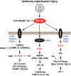
Signaling pathway of HMGB1-mediated ischemia reperfusion injury. HMGB1 is an early mediator of injury and inflammation in liver, kidney, heart, and brain ischemia reperfusion injury and implicates TLR-4, TLR-9, and RAGE as receptors that are involved in the process. HMGB1's extracellular function partly through activation of MAPKs and NF-κB pathway. (For interpretation of the references to color in this figure legend, the reader is referred to the web version of this article at www.liebertonline.com/ars).
FIG. 13.
FIG. 13.
HMGB1 induces endothelial dysfunction and promotes atherosclerotic plaque formation. Activated vascular smooth muscle cells (SMCs) are the source of HMGB1 in human advanced atherosclerotic lesions. HMGB1 activates vascular endothelial cells to express and the secretion of intercellular adhesion molecule 1 (ICAM-1), vascular cell adhesion molecule 1 (VCAM-1), RAGE, TNF-α, IL-8, monocyte chemotactic protein-1 (MCP-1), plasminogen activator inhibitor 1 (PAI-1), and tissue plasminogen activator (tPA). Moreover, HMGB1 activates macrophagy, promotes neutrophil adherence, and increases ROS production. Atherosclerosis develops from low-density lipoprotein (LDL) molecules becoming oxidized by ROS in artery wall. (For interpretation of the references to color in this figure legend, the reader is referred to the web version of this article at www.liebertonline.com/ars).
FIG. 14.
FIG. 14.
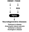
The HMGB1/RAGE pathway is involving in aging. Aging is a major risk factor for several common neurodegenerative diseases, including Parkinson's disease, amyotrophic lateral sclerosis, Alzheimer's disease, and Huntington's disease. ROS is one of the primary determinants of aging.
FIG. 15.
FIG. 15.
Several agents such as ethyl pyruvate, quercetin, green tea, N-acetylcysteine, and curcumin have not only antioxidative properties, but also antiinflammatory properties. They are protective in the setting of experimental inflammation, partly through attenuating systemic HMGB1 accumulation.

Similar articles

Cited by

References

    1. Abd El-Gawad HM. Khalifa AE. Quercetin, coenzyme Q10, and L-canavanine as protective agents against lipid peroxidation and nitric oxide generation in endotoxin-induced shock in rat brain. Pharmacol Res. 2001;43:257–263. - PubMed
    1. Abraham E. Arcaroli J. Carmody A. Wang H. Tracey KJ. HMG-1 as a mediator of acute lung inflammation. J Immunol. 2000;165:2950–2954. - PubMed
    1. Agresti A. Lupo R. Bianchi ME. Muller S. HMGB1 interacts differentially with members of the Rel family of transcription factors. Biochem Biophys Res Commun. 2003;302:421–426. - PubMed
    1. Andersson U. Wang H. Palmblad K. Aveberger AC. Bloom O. Erlandsson-Harris H. Janson A. Kokkola R. Zhang M. Yang H. Tracey KJ. High mobility group 1 protein (HMG-1) stimulates proinflammatory cytokine synthesis in human monocytes. J Exp Med. 2000;192:565–570. - PMC - PubMed
    1. Andrassy M. Volz HC. Igwe JC. Funke B. Eichberger SN. Kaya Z. Buss S. Autschbach F. Pleger ST. Lukic IK. Bea F. Hardt SE. Humpert PM. Bianchi ME. Mairbaurl H. Nawroth PP. Remppis A. Katus HA. Bierhaus A. High-mobility group box-1 in ischemia-reperfusion injury of the heart. Circulation. 2008;117:3216–3226. - PubMed

MeSH terms