. 2011 Jan;85(1):568-81.
doi: 10.1128/JVI.01611-10. Epub 2010 Oct 20.

The alphaherpesvirus US3/ORF66 protein kinases direct phosphorylation of the nuclear matrix protein matrin 3

Affiliations

The alphaherpesvirus US3/ORF66 protein kinases direct phosphorylation of the nuclear matrix protein matrin 3

Angela Erazo et al. J Virol. 2011 Jan.

Abstract

The protein kinase found in the short region of alphaherpesviruses, termed US3 in herpes simplex virus type 1 (HSV-1) and pseudorabies virus (PRV) and ORF66 in varicella-zoster virus (VZV), affects several viral and host cell processes, and its specific targets remain an area of active investigation. Reports suggesting that HSV-1 US3 substrates overlap with those of cellular protein kinase A (PKA) prompted the use of an antibody specific for phosphorylated PKA substrates to identify US3/ORF66 targets. HSV-1, VZV, and PRV induced very different substrate profiles that were US3/ORF66 kinase dependent. The predominant VZV-phosphorylated 125-kDa species was identified as matrin 3, one of the major nuclear matrix proteins. Matrin 3 was also phosphorylated by HSV-1 and PRV in a US3 kinase-dependent manner and by VZV ORF66 kinase at a novel residue (KRRRT150EE). Since VZV-directed T150 phosphorylation was not blocked by PKA inhibitors and was not induced by PKA activation, and since PKA predominantly targeted matrin 3 S188, it was concluded that phosphorylation by VZV was PKA independent. However, purified VZV ORF66 kinase did not phosphorylate matrin 3 in vitro, suggesting that additional cellular factors were required. In VZV-infected cells in the absence of the ORF66 kinase, matrin 3 displayed intranuclear changes, while matrin 3 showed a pronounced cytoplasmic distribution in late-stage cells infected with US3-negative HSV-1 or PRV. This work identifies phosphorylation of the nuclear matrix protein matrin 3 as a new conserved target of this kinase group.


Figures
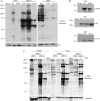
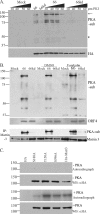
FIG. 1.
FIG. 1.
Anti-PKAps profiles for cells infected with alphaherpesviruses (VZV, HSV-1, or PRV) with or without expression of their US3 kinases. MeWo cells were either mock infected, infected at an MOI of 0.1 with VZV.GFP-66 or VZV.GFP-66kd (expressing kinase-inactivated ORF66 protein), or infected at an MOI of 1 with HSV-1 (US3), HSV-EGFP-US3.5 (US3.5), HSV-EGFP-ΔUS3 (ΔUS3), PRV (US3), or PRV.ΔUS3 (ΔUS3). Cell extracts were harvested 20 h postinfection and were separated on a 7% SDS-PAGE gel. (A) Immunoblot analysis of proteins is shown following antibody probing with a rabbit α-p-PKA substrate antibody (top) or a mouse α-tubulin (VZV and PRV) or α-Ku-86 (HSV-1) antibody (bottom) as a loading control (LC). (B) Immunoblot analysis of viral proteins to show equal infections (rabbit α-VZV ORF9, mouse α-HSV gB, or goat α-PRV UL34 antibody). (C) Comparisons of the anti-PKAps profiles of MeWo cells or Vero cells infected with HSV-1 or PRV, either with (+) or deleted for (−) the US3 kinase. Profiles of cells infected at two different multiplicities of infection (1 and 5) for each virus, harvested at 18 h postinfection, are shown. The same blot was simultaneously probed with antibodies to tubulin for a loading control. Antibodies in panels A and B were detected by use of secondary antibodies bound to horseradish peroxidase, followed by a fluorescent substrate and collection by film exposure. Antibodies in panel C were simultaneously detected using secondary antibodies linked to IRDye 800CW (anti-mouse) or IRDye 680 (anti-rabbit) and were captured by a LI-COR Odyssey infrared imager. Protein size markers (in kilodaltons) are specified to the left of each panel.
FIG. 2.
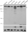
FIG. 2.
The 125-kDa ORF66-specific PKA substrate protein is a host cell protein. MRC-5 cells were coinfected with replication-deficient adenoviruses: Ad.Toff, which expresses a tetracycline-controlled transactivator, at an MOI of 2.5, and either mock infection or Ad.GFP-66 (expressing functional ORF66) (Ad.66), Ad.GFP-66kd (expressing kinase-inactive ORF66) (Ad.66kd), or Ad.Vector, at an MOI of 5. Doxycycline was added (+) to turn off ORF66/66kd gene expression. Cells were harvested 1 day postinfection, and immunoblot analysis was performed on cell extracts with a rabbit α-p-PKA substrate antibody. The blot was stripped and reprobed with mouse α-HA to analyze for ORF66/66kd expression. Mouse α-tubulin was used to evaluate equal amounts of total protein levels. The star marks a nonspecific protein species that was present under all conditions. The arrow indicates the ORF66-dependent 125-kDA p-PKA substrate band. Protein size markers (in kilodaltons) are specified to the left.
FIG. 3.
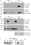
FIG. 3.
VZV ORF66 kinase activity induces specific matrin 3 phosphorylation. (A) Hek 293 cells were either infected with Ad.GFP-66 or Ad.GFP-66kd at an MOI of 0.3 or mock infected and were then incubated for 24 h. Soluble fractions (Sol) and immunoprecipitation (IP) products were separated on a 7% gel and analyzed by immunoblotting (Western blotting [WB]). IPs were performed using rabbit α-matrin 3, followed by probing of membranes with the rabbit α-PKA substrate antibody (top), or IPs were performed using the rabbit α-PKA substrate antibody, followed by probing of membranes with the rabbit α-matrin 3 antibody (bottom). (B) MRC-5 cells either were infected with VZV.GFP-66 (66), VZV.GFP-66kd (66kd), or VZV.GFP-66s (66s) at an MOI of 0.01 or were mock infected; then they were harvested 2 days postinfection. Soluble extracts (Sol) or immunoprecipitations were separated on a 7% SDS-PAGE gel and were immunoblotted (top and center) as described above. The top membrane was then stripped and reprobed for matrin 3 to analyze levels of matrin 3 in IP fractions (bottom). Protein size markers (in kilodaltons) are specified to the left. (C) MRC-5 cells either were infected with VZV expressing ORF66 or ORF66kd at an MOI of 0.01 or were mock infected; they were harvested at 2 days postinfection. Cleared lysates were then immunoprecipitated using a rabbit α-matrin 3 antibody in addition to matrin 3 blocking peptide (BP) where indicated. Proteins were separated on a 6% SDS-PAGE gel, and membranes were then probed either with a rabbit α-matrin 3 antibody (left), to test for the efficiency of IPs and the blocking peptide, or with a rabbit α-PKA substrate antibody, to analyze for phosphorylation of matrin 3 (right).
FIG. 4.
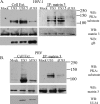
FIG. 4.
Matrin 3 is a conserved phosphorylation target for the US3 kinases during infection. MRC-5 cells were either infected with HSV-1 (US3), HSV-EGFP-US3.5 (US3.5), or HSV-EGFP-ΔUS3 (ΔUS3) at an MOI of 1 (A) or with PRV (US3) or PRV.ΔUS3 (ΔUS3) at an MOI of 1 (B) or were mock infected; then they were harvested 1 day postinfection. Cleared lysates were immunoprecipitated using the rabbit α-matrin 3 antibody, and whole-cell extracts (Cell Ext.) or immunoprecipitation (IP) products were separated on a 7% (HSV) or 6% (PRV) SDS-PAGE gel and analyzed by immunoblotting. Membranes were probed with a rabbit α-PKA substrate antibody and were then stripped and reprobed using a rabbit α-matrin 3 antibody as indicated. Cell extracts were also analyzed for viral protein expression using a mouse α-HSV gB (A) or a goat α-PRV UL34 (B) antibody. Protein size markers (in kilodaltons) are specified to the right.
FIG. 5.
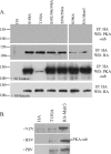
FIG. 5.
Matrin 3 amino acid T150 is the major phosphorylation site for VZV ORF66 and for HSV-1 and PRV US3 kinases. (A) The top two panels show immunoblot analysis of Hek 293T cells transfected with plasmids expressing HA alone, HA-tagged full-length matrin 3 (HA-Matr3), or one of the following five HA-tagged matrin 3 serine/threonine-to-alanine mutants: the S188A, T150A, S592/596/598A, S596/598A, or S596A mutant. The lower two panels are similar analyses of cells cotransfected with one of the HA-tagged proteins and EGFP.66. The next day, cells were harvested, and cleared lysates were immunoprecipitated using a mouse α-HA antibody, followed by separation of IP products on a 6% SDS-PAGE gel and transfer to PVDF membranes. Membranes were probed with a rabbit α-PKA substrate antibody to analyze matrin 3 phosphorylation (top panel of each pair) or with a mouse α-HA antibody to analyze equivalent HA immunoprecipitation (bottom panel of each pair). Protein size markers (in kilodaltons) are specified to the left. (B) Immunoblot analysis of a MeWo cell transfection/infection in which cells were transfected with plasmids expressing either a vector (HA), HA-tagged matrin 3 containing a T150A mutation (T150A), or HA-tagged matrin 3 (HA-Matr3). At 24 h posttransfection, cells either were overlaid with VZV (top) at an MOI of 0.3 or were infected with HSV-1 or PRV at an MOI of 5. Immunoprecipitates from cleared lysates using a mouse α-HA antibody were separated on a 6% SDS-PAGE gel and analyzed by immunoblotting using a rabbit α-PKA substrate.
FIG. 6.
FIG. 6.
The 125-kDa matrin 3 is phosphorylated through a non-PKA pathway. (A) MRC-5 cells were pretreated with a PKA substrate inhibitor (PKI), 14-22 amide, 2 h prior to infection. Then cells either were infected with Ad.Toff at an MOI of 2.5 and with Ad.GFP-66 (66) or Ad.GFP-66kd (66kd) at an MOI of 5, with or without PKI, or were mock infected. Doses of PKI at increasing micromolar concentrations are given above the gel. Whole-cell extracts were immunoblotted. The membrane was first probed with a mouse α-HA antibody, stripped, and then reprobed with a rabbit α-PKA substrate antibody. The arrow indicates the 125-kDa PKA substrate (PKA-sub)/matrin 3 protein. The star indicates an undefined protein whose phosphorylation is PKI resistant. (B) MRC-5 cells either were infected with VZV.GFP-66 (66) or VZV.GFP-66kd (66kd) at an MOI of 0.1 or were mock infected. Where indicated, cells were treated with 10 μM forskolin or DMSO at the time of infection. Cells were harvested 1 day postinfection, analyzed by immunoblotting, and probed with a rabbit α-PKA substrate antibody. Viral protein expression was analyzed using a rabbit α-VZV ORF4 antibody. Cleared lysates were immunoprecipitated for matrin 3 and were subsequently analyzed by immunoblotting using rabbit α-PKA substrate; the blot was stripped and reprobed with a rabbit matrin 3 antibody. (C) In vitro kinase assay of immunoprecipitated HA alone, HA-tagged matrin 3 (HA-Matr3), or the HA-tagged S188A, T150A, or S596A matrin 3 mutant, incubated with PKA where specified (+ PKA). HA proteins were derived from transfected Hek 293T cells and were purified by immunoprecipitation using a mouse α-HA antibody. Following the kinase assay, proteins were separated on an SDS-PAGE gel, transferred to a PVDF membrane, and exposed to film. These membranes were then analyzed for HA fusion protein expression by probing with a mouse α-HA antibody. Protein size markers (in kilodaltons) are specified to the left.
FIG. 7.
FIG. 7.
ORF66 does not phosphorylate matrin 3 under the conditions required for phosphorylation of IE62. Purified MBP fusion proteins and GST kinase were incubated in optimal kinase buffer, separated on a 7% SDS-PAGE gel, transferred to PVDF membranes, and exposed to film for autoradiography. Lanes 1 to 4 contain GST, GST.66, GST66kd, or MBP incubated alone, respectively. Lanes 5 to 7 contain MBP incubated with GST, GST.66, or GST.66kd, respectively. Lane 8 contains MBP62p alone, and lanes 9 to 11 contain MBP62p incubated with GST, GST.66, or GST.66kd, respectively. Lane 12 contains MBP-matrin 3 (MBPmatr3) alone, and lanes 13 to 15 contain MBPmatr3 incubated with GST, GST.66, or GST.66kd, respectively. Images of lanes 12 to 15 are 4 times more exposed in order to show MBPmatr3 proteins. Lane 16 also contains MBPmatr3 alone, and lanes 17 and 18 contain equivalent levels of PKA incubated with MBPmatr3 in PKA reaction buffer (lane 17) or optimal ORF66 kinase buffer (lane 18). Protein levels of substrate and CRF66 protein were analyzed by immunoblotting using rabbit α-MBP and goat α-GST antibodies (lanes 1 to 15). Protein size markers (in kilodaltons) are specified to the left of autoradiographs.
FIG. 8.
FIG. 8.
The ORF66 kinase influences matrin 3 nuclear distribution in VZV infections. Shown are immunofluorescence analyses of mock-infected MRC-5 cells (A) and of MRC-5 cells infected with VZV.GFP-66 (B) or VZV.GFP-66kd (C) at an MOI of 0.003. Cells were fixed with 4% paraformaldehyde 3 days postinfection and were immunostained either with a rabbit α-matrin 3 antibody (A to C, panels i, and D), detected with α-rabbit Alexa Fluor 546, or with mouse α-IE62 (A to C, panels iii), detected with α-mouse Alexa Fluor 647. ORF66 expression was determined by GFP autofluorescence (A to C, panels ii). The merge images (A to C, panels iv) are overlays of matrin 3 (gray), GFP (green), IE62 (red), and nuclei stained with Hoechst dye (blue). (D) Representative single-cell images depicting matrin 3 localization. Infection conditions are given on the images. Fluorescence images were taken using a 60× objective.
FIG. 9.
FIG. 9.
HSV-1 and PRV US3 kinases drive nuclear retention of matrin 3 during infection. Shown are immunofluorescence analyses of MRC-5 cells that were either mock infected (A) or infected with HSV-1 (B), HSV.US3.5 (C), HSV.ΔUS3 (D), PRV (E), or PRV.ΔUS3 (F) at an MOI of 5. Cells were fixed with 4% paraformaldehyde 18 h (HSV) or 11 h (PRV) postinfection; then they were immunostained either with a rabbit α-matrin 3 antibody (A to F, panels i), detected with α-rabbit Alexa Fluor 546 (HSV) or α-rabbit Alexa Fluor 555 (PRV); with mouse α-HSV ICP4 (A to D, panels iii) (magenta), detected with α-mouse Alexa Fluor 647; or with goat α-PRV UL34 (E and F, panels iii) (magenta), detected with α-goat Alexa Fluor 647. GFP-US3.5 expression or US3 promoter activity in HSV.ΔUS3- and PRV.ΔUS3-infected cells was determined using GFP autofluorescence (A to F, panels ii) (green). The merge images (A to F, panels iv) are overlays of matrin 3 (gray) and nuclei stained with Hoechst dye (blue). Fluorescence images were taken using a 60× objective.

Similar articles

Cited by

References

    1. Abendroth, A., I. Lin, B. Slobedman, H. Ploegh, and A. M. Arvin. 2001. Varicella-zoster virus retains major histocompatibility complex class I proteins in the Golgi compartment of infected cells. J. Virol. 75:4878-4888. - PMC - PubMed
    1. Anachkova, B., V. Djeliova, and G. Russev. 2005. Nuclear matrix support of DNA replication. J. Cell. Biochem. 96:951-961. - PubMed
    1. Beausoleil, S. A., M. Jedrychowski, D. Schwartz, J. E. Elias, J. Villen, J. Li, M. A. Cohn, L. C. Cantley, and S. P. Gygi. 2004. Large-scale characterization of HeLa cell nuclear phosphoproteins. Proc. Natl. Acad. Sci. U. S. A. 101:12130-12135. - PMC - PubMed
    1. Belgrader, P., R. Dey, and R. Berezney. 1991. Molecular cloning of matrin 3. A 125-kilodalton protein of the nuclear matrix contains an extensive acidic domain. J. Biol. Chem. 266:9893-9899. - PubMed
    1. Benetti, L., and B. Roizman. 2004. Herpes simplex virus protein kinase US3 activates and functionally overlaps protein kinase A to block apoptosis. Proc. Natl. Acad. Sci. U. S. A. 101:9411-9416. - PMC - PubMed

Publication types

MeSH terms

LinkOut - more resources