

CT enterography with polyethylene glycol solution vs CT enteroclysis in small bowel disease
- PMID: 20959377
- PMCID: PMC3473850
- DOI: 10.1259/bjr/71649888
CT enterography with polyethylene glycol solution vs CT enteroclysis in small bowel disease
Abstract
Objective: The aim of the study is to compare CT enterography with polyethylene glycol solution (PEG-CT) with CT enteroclysis (CT-E) in patients with suspected small bowel disease.
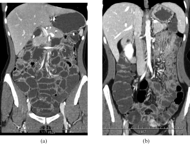
Methods: 145 patients underwent abdominal contrast-enhanced 16-row multidetector CT after administration of 2000 ml of PEG by mouth (n = 75) or after administration of 2000 ml of methylcellulose by nasojejunal tube (n = 70). Small bowel distension, luminal and extraluminal findings were evaluated and compared with small bowel follow-through examination in 60 patients, double contrast enema in 50, surgery in 25 and endoscopy in 35. Statistical evaluation was carried out by χ² testing. For both techniques we have also calculated the effective dose and the equivalent dose in a standard patient.
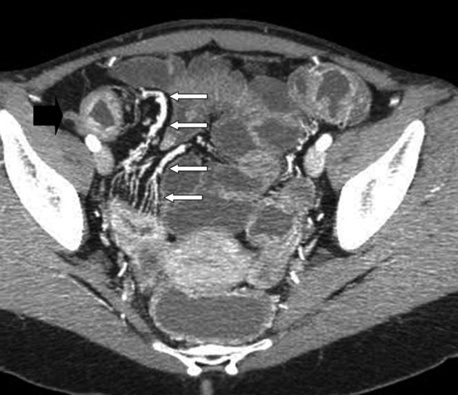
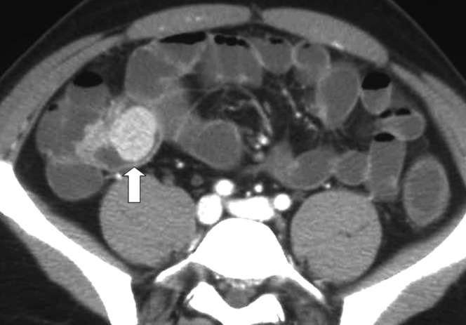
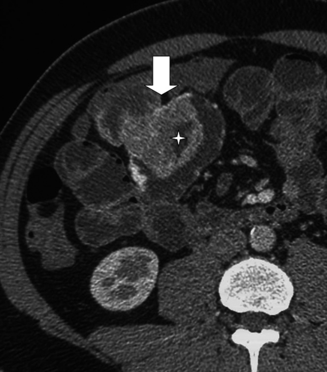
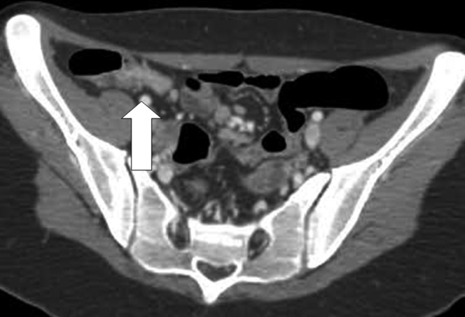
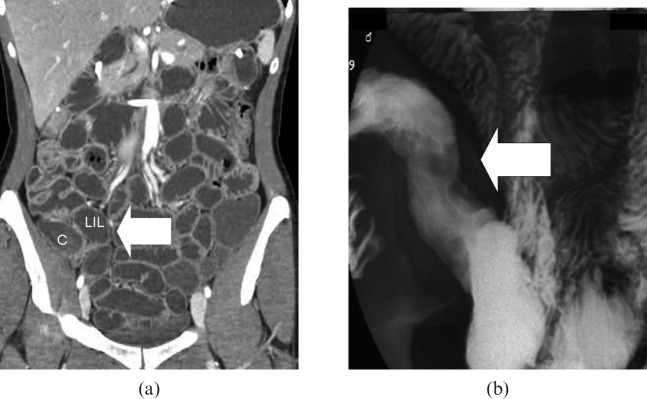
Results: Crohn's disease was diagnosed in 64 patients, neoplasms in 16, adhesions in 6. Distension of the jejunum was better with CT-E than PEG-CT (p<0.05: statistically significant difference). No significant difference was present for others sites (p>0.05). Evaluation of pathological ileal loops was good with both techniques. The values of sensitivity, specificity and diagnostic accuracy were respectively 94%, 100% and 96% with CT-E, and 93%, 94% and 93% with PEG-CT. The effective dose for PEG-CT was less than the dose for the CT-E (34.7 mSv vs 39.91 mSv).
Conclusion: PEG-CT shows findings of Crohn's disease as well as CT-E does, although CT-E gives better bowel distension, especially in the jejunum, and has higher specificity than PEG-CT.
Figures
Similar articles
-
Enterography CT without and with water enema in patients with Crohn's disease: Results from a comparative observational study in comparison with endoscopy.Eur J Radiol. 2016 Feb;85(2):404-13. doi: 10.1016/j.ejrad.2015.11.028. Epub 2015 Nov 26. Eur J Radiol. 2016. PMID: 26781146
-
Computed tomographic enterography and enteroclysis: pearls and pitfalls.Curr Probl Diagn Radiol. 2008 Nov-Dec;37(6):279-87. doi: 10.1067/j.cpradiol.2007.08.007. Curr Probl Diagn Radiol. 2008. PMID: 18823868 Review.
-
Enteroclysis CT and PEG-CT in patients with previous small-bowel surgical resection for Crohn's disease: CT findings and correlation with endoscopy.Eur Radiol. 2009 Oct;19(10):2432-40. doi: 10.1007/s00330-009-1423-5. Epub 2009 May 5. Eur Radiol. 2009. PMID: 19415289 Clinical Trial.
-
Accuracy of small-intestine contrast ultrasonography, compared with computed tomography enteroclysis, in characterizing lesions in patients with Crohn's disease.Clin Gastroenterol Hepatol. 2013 Aug;11(8):950-5. doi: 10.1016/j.cgh.2013.01.015. Epub 2013 Jan 29. Clin Gastroenterol Hepatol. 2013. PMID: 23375998
-
The role of nasoenteric intubation in the MR study of patients with Crohn's disease: our experience and literature review.Radiol Med. 2011 Apr;116(3):389-406. doi: 10.1007/s11547-010-0605-1. Epub 2010 Oct 27. Radiol Med. 2011. PMID: 20981501 Review. English, Italian.
Cited by
-
Crohn's Disease: Radiological Answers to Clinical Questions and Review of the Literature.J Clin Med. 2024 Jul 16;13(14):4145. doi: 10.3390/jcm13144145. J Clin Med. 2024. PMID: 39064186 Free PMC article. Review.
-
Advanced imaging techniques for small bowel Crohn's disease: what does the future hold?Therap Adv Gastroenterol. 2018 Feb 12;11:1756283X18757185. doi: 10.1177/1756283X18757185. eCollection 2018. Therap Adv Gastroenterol. 2018. PMID: 29467827 Free PMC article. Review.
-
Prospective evaluation of magnetic resonance enterography for the detection of mesenteric small bowel tumours.Eur Radiol. 2013 Jul;23(7):1901-10. doi: 10.1007/s00330-013-2800-7. Epub 2013 Mar 12. Eur Radiol. 2013. PMID: 23479221
-
Imaging of Strictures in Crohn's Disease.Life (Basel). 2023 Nov 29;13(12):2283. doi: 10.3390/life13122283. Life (Basel). 2023. PMID: 38137884 Free PMC article. Review.
-
Evaluation of neutral oral contrast agents for assessment of the small bowel at abdominal staging CT.PLoS One. 2019 Nov 14;14(11):e0225160. doi: 10.1371/journal.pone.0225160. eCollection 2019. PLoS One. 2019. PMID: 31725763 Free PMC article.
References
-
- Birnbaum BA. Computed tomography of the small bowel. Technique and principles of interpretation. Herlinger H, Maglinte DDT, Birnbaum BA, editors. Clinical imaging of the small intestine. Berlin, Germany: Springer, 1999: 153–66
-
- Bender GN, Timmons JH, Williard WC, Carter J. Computed tomographic enteroclysis. One methodology. Invest Radiol 1996;31:43–9 - PubMed
-
- Engin G. Computed tomography enteroclysis in the diagnosis of intestinal diseases. J Comput Assist 2008;32:9–16 - PubMed
-
- Maglinte DDT, Sandrasegaran K, Lappas JC, Chiorean M. CT enteroclysis. Radiology 2007;245:661–71 - PubMed
-
- Makó EK, Mester AR, Tarján Z, Karlinger K, Tóth G. Enteroclysis and spiral CT examination in diagnosis and evaluation of small bowel Crohn's disease. Eur J Radiol 2000;35:168–75 - PubMed
