

Enhanced neuronal expression of major histocompatibility complex class I leads to aberrations in neurodevelopment and neurorepair
- PMID: 20950866
- PMCID: PMC5776042
- DOI: 10.1016/j.jneuroim.2010.09.009
Enhanced neuronal expression of major histocompatibility complex class I leads to aberrations in neurodevelopment and neurorepair
Abstract
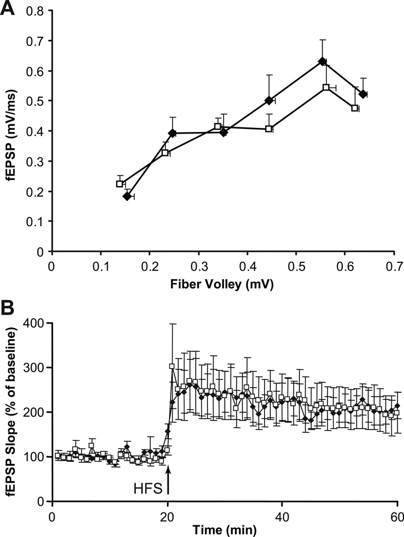
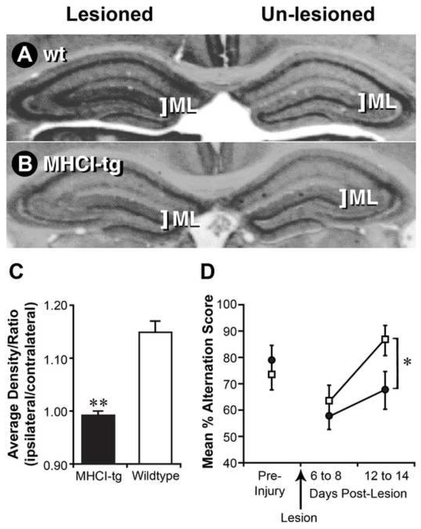
Mice deficient in classical major histocompatibility complex class I (MHCI) have aberrations in neurodevelopment. The consequences of upregulated neuronal MHCI expression have not been examined. We found that transgenic C57Bl/6 mice that are engineered to express higher levels of self-D(b) on their CNS neurons have alterations in their hippocampal morphology and retinogeniculate projections, as well as impaired neurorepair responses. Thus, enhanced neuronal classical MHCI expression can lead to aberrations in neural circuitry and neurorepair. These findings complement a growing body of knowledge concerning the neurobiological activities of MHCI and may have potential clinical relevance.
Copyright © 2010 Elsevier B.V. All rights reserved.
Conflict of interest statement
All authors declare that there are no conflicts of interest
Figures


Similar articles
-
Transgenic mice with enhanced neuronal major histocompatibility complex class I expression recover locomotor function better after spinal cord injury.J Neurosci Res. 2011 Mar;89(3):365-72. doi: 10.1002/jnr.22557. Epub 2010 Dec 22. J Neurosci Res. 2011. PMID: 21259323 Free PMC article.
-
MHC class I limits hippocampal synapse density by inhibiting neuronal insulin receptor signaling.J Neurosci. 2014 Aug 27;34(35):11844-56. doi: 10.1523/JNEUROSCI.4642-12.2014. J Neurosci. 2014. PMID: 25164678 Free PMC article.
-
Regulation of CNS synapses by neuronal MHC class I.Proc Natl Acad Sci U S A. 2007 Apr 17;104(16):6828-33. doi: 10.1073/pnas.0702023104. Epub 2007 Apr 9. Proc Natl Acad Sci U S A. 2007. PMID: 17420446 Free PMC article.
-
Major histocompatibility complex class I proteins in brain development and plasticity.Trends Neurosci. 2012 Nov;35(11):660-70. doi: 10.1016/j.tins.2012.08.001. Epub 2012 Aug 30. Trends Neurosci. 2012. PMID: 22939644 Free PMC article. Review.
-
Major histocompatibility complex I in brain development and schizophrenia.Biol Psychiatry. 2014 Feb 15;75(4):262-8. doi: 10.1016/j.biopsych.2013.10.003. Epub 2013 Oct 10. Biol Psychiatry. 2014. PMID: 24199663 Free PMC article. Review.
Cited by
-
Major histocompatibility complex class I molecules modulate embryonic neuritogenesis and neuronal polarization.J Neuroimmunol. 2012 Jun 15;247(1-2):1-8. doi: 10.1016/j.jneuroim.2012.03.008. Epub 2012 Apr 12. J Neuroimmunol. 2012. PMID: 22503373 Free PMC article.
-
Major histocompatibility complex class I-mediated inhibition of neurite outgrowth from peripheral nerves.Immunol Lett. 2011 Mar 30;135(1-2):118-23. doi: 10.1016/j.imlet.2010.10.011. Epub 2010 Oct 23. Immunol Lett. 2011. PMID: 20974178 Free PMC article.
-
Genomic, transcriptomic, and metabolomic profiles of hiPSC-derived dopamine neurons from clinically discordant brothers with identical PRKN deletions.NPJ Parkinsons Dis. 2022 Jun 29;8(1):84. doi: 10.1038/s41531-022-00346-3. NPJ Parkinsons Dis. 2022. PMID: 35768426 Free PMC article.
-
Major Histocompatibility Complex I Expression by Motor Neurons and Its Implication in Amyotrophic Lateral Sclerosis.Front Neurol. 2016 Jun 13;7:89. doi: 10.3389/fneur.2016.00089. eCollection 2016. Front Neurol. 2016. PMID: 27379008 Free PMC article. Review.
-
A potential role for shed soluble major histocompatibility class I molecules as modulators of neurite outgrowth.PLoS One. 2011 Mar 31;6(3):e18439. doi: 10.1371/journal.pone.0018439. PLoS One. 2011. PMID: 21483793 Free PMC article.
References
-
- Bai A, Broen J, Forman J. The pathway for processing leader-derived peptides that regulate the maturation and expression of Qa-1b. Immunity. 1998;9:413–421. - PubMed
-
- Ballabh P, Braun A, Nedergaard M. The blood-brain barrier: an overview: structure, regulation, and clinical implications. Neurobiol Dis. 2004;16:1–13. - PubMed
-
- Banks WA, Kastin AJ, Broadwell RD. Passage of cytokines across the blood-brain barrier. Neuroimmunomodulation. 1995;2:241–248. - PubMed
-
- Barco A, Alarcon JM, Kandel ER. Expression of constitutively active CREB protein facilitates the late phase of long-term potentiation by enhancing synaptic capture. Cell. 2002;108:689–703. - PubMed
-
- Bauer A, Huttinger R, Staffler G, Hansmann C, Schmidt W, Majdic O, Knapp W, Stockinger H. Analysis of the requirement for beta 2-microglobulin for expression and formation of human CD1 antigens. Eur J Immunol. 1997;27:1366–1373. - PubMed
Publication types
MeSH terms
Substances
Grants and funding
LinkOut - more resources
Full Text Sources
Other Literature Sources
