

Co-evolution of cancer microenvironment reveals distinctive patterns of gastric cancer invasion: laboratory evidence and clinical significance
- PMID: 20950454
- PMCID: PMC2965128
- DOI: 10.1186/1479-5876-8-101
Co-evolution of cancer microenvironment reveals distinctive patterns of gastric cancer invasion: laboratory evidence and clinical significance
Abstract
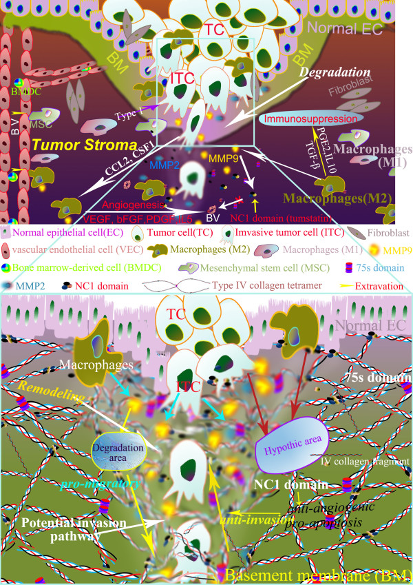
Background: Cancer invasion results from constant interactions between cancer cells and their microenvironment. Major components of the cancer microenvironment are stromal cells, infiltrating inflammatory cells, collagens, matrix metalloproteinases (MMP) and newly formed blood vessels. This study was to determine the roles of MMP-9, MMP-2, type IV collagen, infiltrating macrophages and tumor microvessels in gastric cancer (GC) invasion and their clinico-pathological significance.
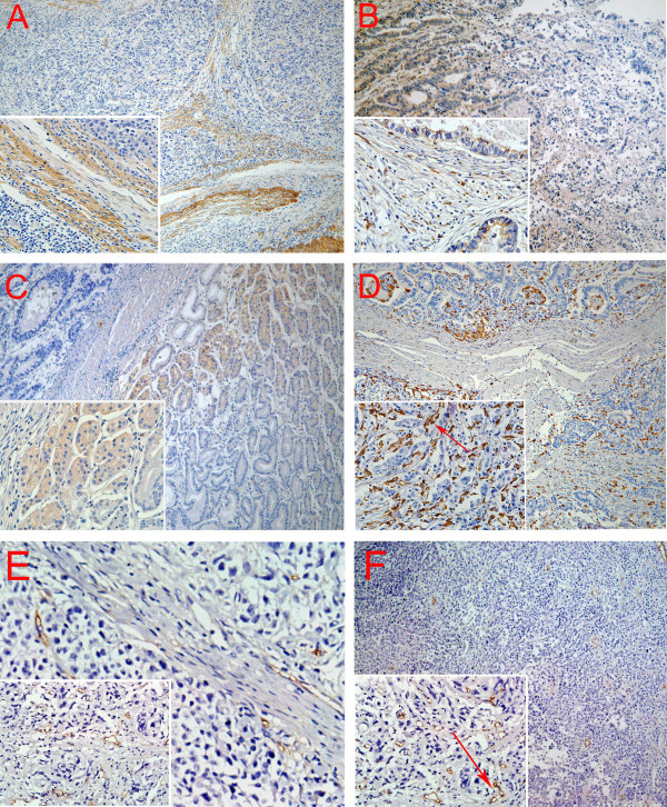
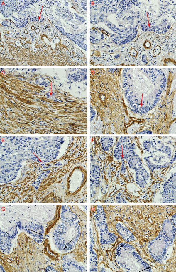
Methods: Paraffin-embedded tissue sections from 37 GC patients were studied by Streptavidin-Peroxidase (SP) immunohistochemical technique to determine the levels of MMP-2, MMP-9, type IV collagen, macrophages infiltration and microvessel density (MVD). Different invasion patterns were delineated and their correlation with major clinico-pathological information was explored.
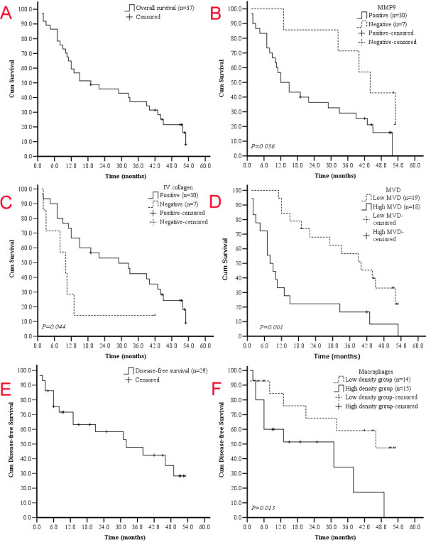
Results: MMP2 expression was higher in malignant gland compared to normal gland, especially nearby the basement membrane (BM). High densities of macrophages at the interface of cancer nests and stroma were found where BM integrity was destroyed. MMP2 expression was significantly increased in cases with recurrence and distant metastasis (P = 0.047 and 0.048, respectively). Infiltrating macrophages were correlated with serosa invasion (P = 0.011) and TNM stage (P = 0.001). MVD was higher in type IV collagen negative group compared to type IV collagen positive group (P = 0.026). MVD was related to infiltrating macrophages density (P = 0.040). Patients with negative MMP9 expression had better overall survival (OS) compared to those with positive MMP9 expression (Median OS 44.0 vs 13.5 mo, P = 0.036). Median OS was significantly longer in type IV collagen positive group than negative group (Median OS 25.5 vs 10.0 mo, P = 0.044). The cumulative OS rate was higher in low macrophages density group than in high macrophages density group (median OS 40.5 vs 13.0 mo, P = 0.056). Median OS was significantly longer in low MVD group than high MVD group (median OS 39.0 vs 8.5 mo, P = 0.001). The difference of disease-free survival (DFS) between low MVD group and high MVD group was not statistically significant (P = 0.260). Four typical patterns of cancer invasion were identified based on histological study of the cancer tissue, including Washing pattern, Ameba-like pattern, Spindle pattern and Linear pattern.
Conclusions: Proteolytic enzymes MMP9, MMP2 and macrophages in stroma contribute to GC progression by facilitating the angiogenesis. Cancer invasion patterns may help predict GC metastasis.
Figures
Similar articles
-
[Relationship between bFGF mRNA and MMP-9 mRNA expression in gastric carcinoma and their clinicopathological features as well as patients survival].Zhonghua Wai Ke Za Zhi. 2005 Feb 1;43(3):169-72. Zhonghua Wai Ke Za Zhi. 2005. PMID: 15842896 Chinese.
-
Overexpression of twist and matrix metalloproteinase-9 with metastasis and prognosis in gastric cancer.Asian Pac J Cancer Prev. 2013;14(9):5055-60. doi: 10.7314/apjcp.2013.14.9.5055. Asian Pac J Cancer Prev. 2013. PMID: 24175775
-
[Matrix metalloproteinase-2 and angiogenesis in gastric cancer].Ai Zheng. 2002 Jan;21(1):25-8. Ai Zheng. 2002. PMID: 12500392 Chinese.
-
[The significance of metalloproteinases and their inhibitors in gastric cancer].Postepy Hig Med Dosw (Online). 2009 Jun 5;63:258-65. Postepy Hig Med Dosw (Online). 2009. PMID: 19502687 Review. Polish.
-
Tumor Microenvironment, Clinical Features, and Advances in Therapy for Bone Metastasis in Gastric Cancer.Cancers (Basel). 2022 Oct 6;14(19):4888. doi: 10.3390/cancers14194888. Cancers (Basel). 2022. PMID: 36230816 Free PMC article. Review.
Cited by
-
The Immune Microenvironment in Gastric Cancer: Prognostic Prediction.Front Oncol. 2022 Apr 28;12:836389. doi: 10.3389/fonc.2022.836389. eCollection 2022. Front Oncol. 2022. PMID: 35574386 Free PMC article. Review.
-
Interleukin-8 is associated with adhesion, migration and invasion in human gastric cancer SCG-7901 cells.Med Oncol. 2012 Mar;29(1):91-9. doi: 10.1007/s12032-010-9780-0. Epub 2010 Dec 30. Med Oncol. 2012. PMID: 21191670
-
Antibody targeting of Cathepsin S induces antibody-dependent cellular cytotoxicity.Mol Cancer. 2011 Dec 14;10:147. doi: 10.1186/1476-4598-10-147. Mol Cancer. 2011. PMID: 22168338 Free PMC article.
-
Application of multispectral imaging in quantitative immunohistochemistry study of breast cancer: a comparative study.Tumour Biol. 2016 Apr;37(4):5013-24. doi: 10.1007/s13277-015-4327-9. Epub 2015 Nov 5. Tumour Biol. 2016. PMID: 26537585 Free PMC article.
-
Expression and clinical significance of matrix metalloproteinase-17 and -25 in gastric cancer.Oncol Lett. 2015 Feb;9(2):671-676. doi: 10.3892/ol.2014.2747. Epub 2014 Dec 1. Oncol Lett. 2015. PMID: 25621036 Free PMC article.
References
Publication types
MeSH terms
Substances
LinkOut - more resources
Full Text Sources
Medical
Miscellaneous
